. 2000 Oct 30;151(3):495-506.
doi: 10.1083/jcb.151.3.495.

DRAL is a p53-responsive gene whose four and a half LIM domain protein product induces apoptosis

Affiliations

DRAL is a p53-responsive gene whose four and a half LIM domain protein product induces apoptosis

F A Scholl et al. J Cell Biol. .

Abstract

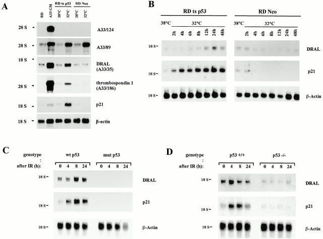
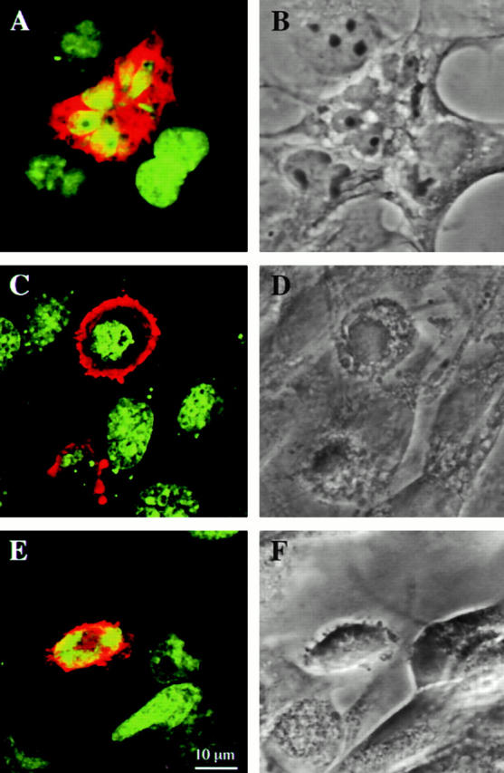
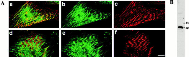
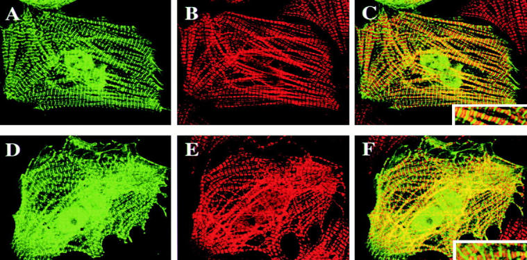
DRAL is a four and a half LIM domain protein identified because of its differential expression between normal human myoblasts and the malignant counterparts, rhabdomyosarcoma cells. In the current study, we demonstrate that transcription of the DRAL gene can be stimulated by p53, since transient expression of functional p53 in rhabdomyosarcoma cells as well as stimulation of endogenous p53 by ionizing radiation in wild-type cells enhances DRAL mRNA levels. In support of these observations, five potential p53 target sites could be identified in the promoter region of the human DRAL gene. To obtain insight into the possible functions of DRAL, ectopic expression experiments were performed. Interestingly, DRAL expression efficiently triggered apoptosis in three cell lines of different origin to the extent that no cells could be generated that stably overexpressed this protein. However, transient transfection experiments as well as immunofluorescence staining of the endogenous protein allowed for the localization of DRAL in different cellular compartments, namely cytoplasm, nucleus, focal contacts, as well as Z-discs and to a lesser extent the M-bands in cardiac myofibrils. These data suggest that downregulation of DRAL might be involved in tumor development. Furthermore, DRAL expression might be important for heart function.


Figures

Figure 1
Figure 1
(A) Screening for p53-inducible genes. 5 μg total RNA from the indicated human cell lines was loaded in each lane and hybridized with the labeled cDNAs A33/124, A33/89, A33/35, A33/186, p21WAF1, and β-actin. RD are RMS cells, A33 GM are primary myoblasts kept in growth medium, RD-tsp53 are RD cells expressing a temperature-sensitive p53 mutant, and RD-Neo are RD cells expressing the vector alone. The mutant p53 protein is inactive at 38°C, but dropping the temperature to 32°C leads to wild-type p53 protein functions. RNA was extracted one day after temperature shift. (B) Time course analyses of p53 target gene induction. Total RNA was extracted from RD-tsp53 or RD-Neo cells either grown at 38° or 32°C for the indicated time. 3 μg total RNA was loaded in each lane. The blot was hybridized with the labeled cDNAs coding for DRAL, p21WAF1, and β-actin. (C) Induction of DRAL expression after γ-irradiation. Normal human myoblasts (wtp53) and RD RMS cells (mutp53) were treated with 20 Gy, and afterwards fed with fresh medium. Total RNA was isolated at the time points indicated and subjected to Northern blot analysis (3 μg/lane). Inserts from the indicated cDNAs were isolated, labeled by random priming, and used for hybridization. The blot was also reprobed for β-actin as a control for equal loading and transfer. (D) Same as in C except that p53+/+ or p53−/− mouse fibroblasts were irradiated.
Figure 4
Figure 4
Time course of DRAL expression in COS-1 cells. Cells were transfected with either DRAL-CF (A–C), DRAL-NF (D–F), or the control plasmid pFLAG–CMV-2–BAP (G–I). After 32, 48, and 72 h cells were fixed and stained with the monoclonal anti-FLAG antibody. Pictures were taken from representative areas. The scale bar line is valid for all the pictures and indicates 100 μm. Arrows point to antibody-labeled remnants, which were absent in the transfections with the control pFLAG–CMV-2–BAP construct.
Figure 2
Figure 2
Partial genomic organization of the human DRAL gene. (A) The 7.8-kb HindIII fragment of the P1 clone 21280 contains the first two noncoding exons (exon 1: nt 1 to nt 63, exon 2: nt 64 to nt 114 of the DRAL cDNA). Letters correspond to the restriction sites: H, HindIII; P, PstI; S, SstI; E, EcoRI; and B, BamHI. Putative transcription factor binding sites in the promoter region are indicated. The five putative p53-binding sites within the 7.8-kb subclone are designated as sites p53 I–p53 V, sequences are listed separately in C. (B) Southern blot analysis using either human genomic DNA or the P1:21280 clone digested with HindIII (H) and PstI (P). The blot was hybridized with the 2.5-kb PstI fragment of the 7.8-kb subclone containing exon II. The sequence of the 7.8-kb HindIII fragment is available from Genbank/EMBL/DDBJ under accession number AF211174.
Figure 3
Figure 3
Expression pattern of DRAL in normal human fetal and adult tissues. A commercially available human RNA dot blot containing normalized quantities of mRNA per dot was hybridized with a labeled DRAL cDNA probe. The tissues used for mRNA extraction are indicated in the lower panel.
Figure 5
Figure 5
Induction of apoptosis by ectopic expression of DRAL. COS-1 (A and B), NIH 3T3 (C and D), and RD (E and F) cells were transiently transfected with FLAG-tagged DRAL. A, C, and E are composite confocal microscopy images of DRAL (red) and DNA (green) staining. B, D, and F represent the corresponding phase contrast picture. Bar: (A–F) 10 μm.
Figure 6
Figure 6
Quantitative analysis of the events following ectopic expression of DRAL. (A) COS-1, RD, and NIH 3T3 cells were transfected with DRAL-CF (circle), DRAL-NF (square), or the control plasmid pFLAG–CMV-2–BAP (cross). After 24, 32, 48, and 72 h cells were fixed and stained for FLAG and Hoechst. Data are shown as mean values with standard error bars representing three independent transfection experiments. (% apoptotic cells) Refers to rounded FLAG-positive cells displaying nuclear condensation and fragmentation in percent of the total number of FLAG positive cells. In the right hand panels, transfected cells were counted as FLAG-positive cells and indicated as percentage of total cell number during time course. (B) COS-1 cells were stained with annexin V 42 h after transfection of the indicated DNA constructs. (C) COS-1 cell extracts were prepared 28 h after transfection with empty vector (black bars), pFLAG–CMV-2–BAP (hatched bars), DRAL-sense (punctated bars), DRAL-CF (grey bars), or DRAL-NF (white bars). OD was measured at 405 nm at 2, 4, 6, and 8 h after substrate addition. Three independent experiments were carried out.
Figure 7
Figure 7
Subcellular localization of DRAL. NIH 3T3 (A and I), COS-1 (B, G, and H), and RD (C–F) cells were cultured on fibronectin coated cover slips or plastic dishes and transiently transfected with FLAG–DRAL. Cells were immunostained with anti-FLAG monoclonal antibody M2 and Cy3-conjugated goat anti–mouse polyclonal antibody and analyzed by confocal fluorescence microscopy. The arrows point to the focal contacts. Bars, 10 μm.
Figure 9
Figure 9
Localization of endogenous DRAL in neonatal rat cardiomyocytes. (A) Double immunofluorescence staining with a polyclonal anti-DRAL antibody (green in a, b, d, and e) together with monoclonal antibodies recognizing sarcomeric α-actinin (red in a and c) or myomesin (red in d and f) reveals a signal primarily in the region of the Z-disc (overlap with α-actinin seen in yellow in a and alternating staining with myomesin in d). Striations can also be detected occasionally in the M-band. Bar, 10μm. (B) Western blot analysis of protein extract detects a major band at 32 kD.
Figure 8
Figure 8
Subcellular localization of DRAL in cardiomyocytes. Neonatal rat cardiomyocytes were transiently transfected with FLAG–DRAL (DRAL-NF in A–C, DRAL-CF in D–F) and immunostained with anti-FLAG monoclonal antibody (A and D) or anti-myosin binding protein C antibody (B and E). C and F represent superposition of A and B or D and E, respectively. The insert shows an enlargement of myofibrils. Analysis was carried out with a confocal fluorescence microscope using Imaris software.

References

    1. Amundson S.A., Myers T.G., Fornace A.J. Roles for p53 in growth arrest and apoptosisputting on the brakes after genotoxic stress. Oncogene. 1998;17:3287–3299. - PubMed
    1. Arber S., Hunter J.J., Ross J., Jr., Hongo M., Sansig G., Borg J., Perriard J.C., Chien K.R., Caroni P. MLP-deficient mice exhibit a disruption of cardiac cytoarchitectural organization, dilated cardiomyopathy, and heart failure. Cell. 1997;88:393–403. - PubMed
    1. Attardi L.D., Reczek E.E., Cosmas C., Demicco E.G., McCurrach M.E., Lowe S.W., Jacks T. PERP, an apoptosis-associated target of p53, is a novel member of the PMP-22/gas3 family. Genes Dev. 2000;14:704–718. - PMC - PubMed
    1. Auerbach D., Bantle S., Keller S., Hinderling V., Leu M., Ehler E., Perriard J.C. Different domains of the M-band protein myomesin are involved in myosin binding and M-band targeting. Mol. Biol. Cell. 1999;10:1297–1308. - PMC - PubMed
    1. Bähler M., Moser H., Eppenberger H.M., Wallimann T. Heart C-protein is transiently expressed during skeletal muscle development in the embryo, but persists in cultured myogenic cells. Dev. Biol. 1985;112:345–352. - PubMed

Publication types

MeSH terms