. 2000 Oct 30;151(3):519-28.
doi: 10.1083/jcb.151.3.519.

Autophagic tubes: vacuolar invaginations involved in lateral membrane sorting and inverse vesicle budding

Affiliations

Autophagic tubes: vacuolar invaginations involved in lateral membrane sorting and inverse vesicle budding

O Müller et al. J Cell Biol. .

Abstract

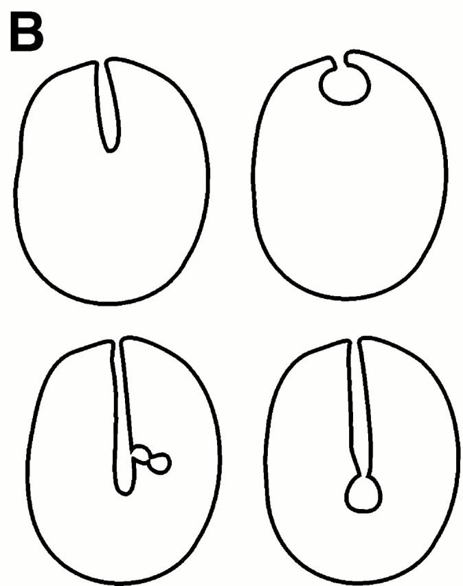
Many intracellular compartments of eukaryotic cells do not adopt a spherical shape, which would be expected in the absence of mechanisms organizing their structure. However, little is known about the principles determining the shape of organelles. We have observed very defined structural changes of vacuoles, the lysosome equivalents of yeast. The vacuolar membrane can form a large tubular invagination from which vesicles bud off into the lumen of the organelle. Formation of the tube is regulated via the Apg/Aut pathway. Its lumen is continuous with the cytosol, making this inverse budding reaction equivalent to microautophagocytosis. The tube is highly dynamic, often branched, and defined by a sharp kink of the vacuolar membrane at the site of invagination. The tube is formed by vacuoles in an autonomous fashion. It persists after vacuole isolation and, therefore, is independent of surrounding cytoskeleton. There is a striking lateral heterogeneity along the tube, with a high density of transmembrane particles at the base and a smooth zone devoid of transmembrane particles at the tip where budding occurs. We postulate a lateral sorting mechanism along the tube that mediates a depletion of large transmembrane proteins at the tip and results in the inverse budding of lipid-rich vesicles into the lumen of the organelle.


Figures

Figure 1
Figure 1
Tubular invaginations of vacuoles in living yeast cells. (A) CRY1 cells were grown logarithmically in YPD medium, stained with the vital dye FM4-64, and starved in SD(−N) for 1.5 h. The cells were viewed under a confocal microscope. The left images show the fluorescently stained vacuole; the right images show the same cell in transmitted light. Arrows indicate the position of the invagination. (B) Schematic summary of different shapes of tubes that can be observed. Bar, 3 μm.
Figure 1
Figure 1
Tubular invaginations of vacuoles in living yeast cells. (A) CRY1 cells were grown logarithmically in YPD medium, stained with the vital dye FM4-64, and starved in SD(−N) for 1.5 h. The cells were viewed under a confocal microscope. The left images show the fluorescently stained vacuole; the right images show the same cell in transmitted light. Arrows indicate the position of the invagination. (B) Schematic summary of different shapes of tubes that can be observed. Bar, 3 μm.
Figure 2
Figure 2
Budding of vesicles into the lumen of the vacuole. (A and B) CRY1 cells were grown logarithmically in YPD medium. Their vacuoles were stained with FM4-64 and the cells were starved in SD(−N) medium with 1 mM PMSF at an OD600 of 1 at 25°C. After 1 h of starvation they were viewed under a standard fluorescence microscope equipped with a video camera. The sequences of pictures shown were taken over a period of 45 s. The last picture shows the investigated cell under Nomarski optics at the end of the experiment. Arrows point to the nascent tube or vesicle. Bars, 5 μm.
Figure 3
Figure 3
Tubular invaginations are filled with cytosol. Yeast cells were grown logarithmically in YPD medium, transferred to SD(−N) medium, and starved. The cells were quick-frozen in liquid propane, freeze-substituted, and embedded. Thin sections were viewed in the electron microscope. (A and B) BJ3505 (a Pep4 mutant) starved for 1–2 h. The vacuole contains autophagic bodies (arrows). Note the high membrane curvature at the base of the tube (arrowheads). V, vacuole; N, nucleus. (C and D) DBY5734 starved for 4 h. Autophagic bodies did not accumulate because this strain is proteolytically competent. The intense dark staining of the vacuoles in C and D is a result of freeze-substitution in osmium tetroxide/acetone and embedding in Epon, whereas the cells in A and B were freeze-substituted in 0.5% uranyl acetate in ethanol and embedded in Lowicryl HM20. Bars, 1 μm.
Figure 4
Figure 4
Frequency of autophagic tubes is influenced by starvation and the autophagy pathway. (A) DBY5734 cells were grown logarithmically overnight and then transferred to rich medium (YPD) or starved for nitrogen in SD(−N) medium. After 4 h of incubation in these media, the vital dye FM4-64 (30 μM) was added to stain the vacuoles (1 h). Then, the cells were chased in the absence of the dye (40 min). The proportion of vacuoles with autophagic tubes was determined by fluorescence microscopy. A total of 200 cells were counted from at least 10 independent fields. (B) Same as in A, but the frequency of autophagic tubes was determined in starved wild-type (wt) (DBY5734) or its derivatives with deletions in aut1 (YTS1), aut7 (YTS3), apg5 (YTS5), or apg7 (YTS7). Values in both figures are the average of five to nine independent experiments. Error bars indicate SD.
Figure 5
Figure 5
Tubular invaginations are maintained on isolated vacuoles. Vacuoles were isolated from logarithmically growing cells by flotation through a Ficoll gradient (Sattler and Mayer 2000, this issue). The purified organelles were quick frozen in liquid propane, freeze–fractured, and replicated with platinum carbon for electron microscope analysis. Transmembrane particles can be seen on the limiting membrane of the vacuole and on the invaginated membrane of the tube (arrows). Note the high curvature of the membrane at the base of the tube (arrowhead). VL, vacuolar lumen. Bar, 1μm.
Figure 6
Figure 6
Distribution of transmembrane particles along autophagic tubes. (A, B, and C) Three vacuoles from DBY5734 cells processed for freeze–fracture analysis. The boxed areas are shown at higher magnifications in D, E, and F. Note the higher density of transmembrane particles at the more basal areas of the tube (arrows) and the absence of particles at the tips (arrowheads). VM, vacuolar membrane. Encircled arrowheads indicate the shadowing directions. Bars: (A–C, E, and F) 0.5 μm; (D) 0.25 μm.
Figure 7
Figure 7
Autophagic bodies are also devoid of transmembrane particles. Yeast cells with a deletion of the PEP4 gene (DBY5734-16) were starved on SD(−N) medium for 3 h and then processed for freeze–fracture analysis as described in the legend to Fig. 5. The picture shows a partial view of the vacuolar membrane (VM), which is particle rich, and of the vacuolar lumen (VL) containing autophagic bodies (arrows). Note the absence of transmembrane particles in the autophagic body membrane. CP, cytoplasm. The encircled arrowhead indicates the shadowing direction. Bar, 1 μm.
Figure 8
Figure 8
Smooth areas on the vacuolar membrane as precursors of tubular invaginations. Vacuoles from strain DBY5734 are shown after freeze–fracture analysis as described in the legend to Fig. 5. The arrows point to well-defined, particle-free areas. (A) Particle-free area on the vacuolar surface. (B) View of a particle-free zone on the surface in the process of invagination. (C) Same as in B, but the vacuole was fractured across its lumen. Only the invaginating smooth area was fractured along its surface. Encircled arrowheads indicate the shadowing directions. Bars, 0.5 μm.

References

    1. Abeliovich H., Darsow T., Emr S.D. Cytoplasm to vacuole trafficking of aminopeptidase I requires a t-SNARE-Sec1p complex composed of Tlg2p and Vps45. EMBO (Eur. Mol. Biol. Organ.) J. 1999;18:6005–6016. - PMC - PubMed
    1. Baba M., Takeshige K., Baba N., Ohsumi Y. Ultrastructural analysis of the autophagic process in yeastdetection of autophagosomes and their characterization. J. Cell Biol. 1994;124:903–913. - PMC - PubMed
    1. Baba M., Osumi M., Ohsumi Y. Analysis of the membrane structures involved in autophagy in yeast by freeze-replica method. Cell Struct. Funct. 1995;20:465–471. - PubMed
    1. Brown D.A., London E. Functions of lipid rafts in biological membranes. Annu. Rev. Cell Dev. Biol. 1998;14:111–136. - PubMed
    1. Bryant N.J., Piper R.C., Weisman L.S., Stevens T.H. Retrograde traffic out of the yeast vacuole to the TGN occurs via the prevacuolar/endosomal compartment. J. Cell Biol. 1998;142:651–663. - PMC - PubMed

Publication types

MeSH terms