. 2000 Oct 30;151(3):539-50.
doi: 10.1083/jcb.151.3.539.

Epidermal growth factor and membrane trafficking. EGF receptor activation of endocytosis requires Rab5a

Affiliations

Epidermal growth factor and membrane trafficking. EGF receptor activation of endocytosis requires Rab5a

M A Barbieri et al. J Cell Biol. .

Abstract

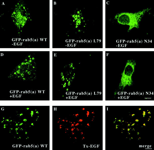
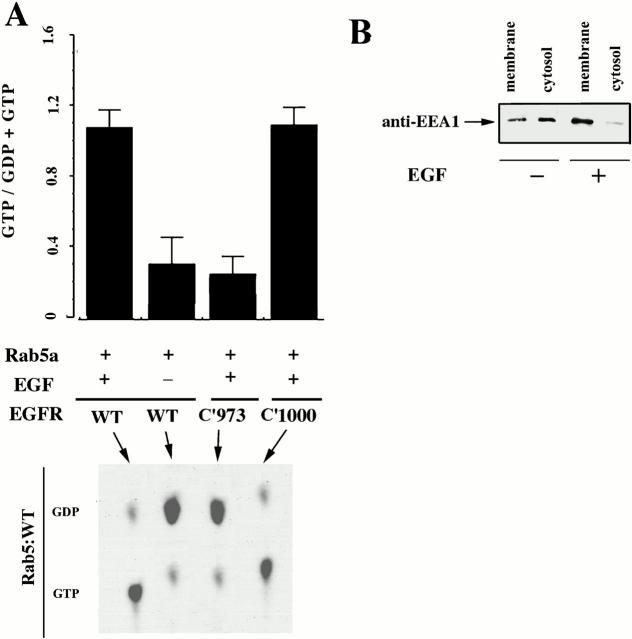
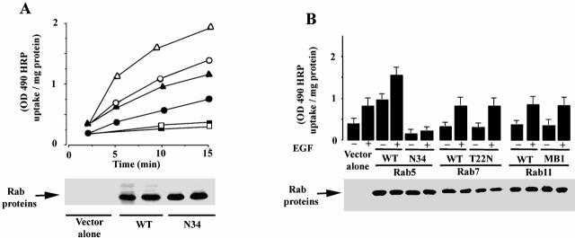
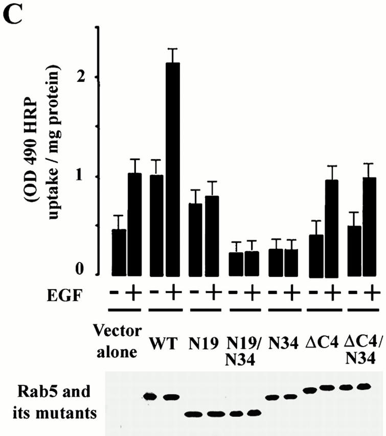
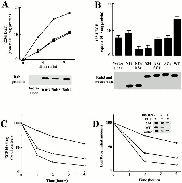
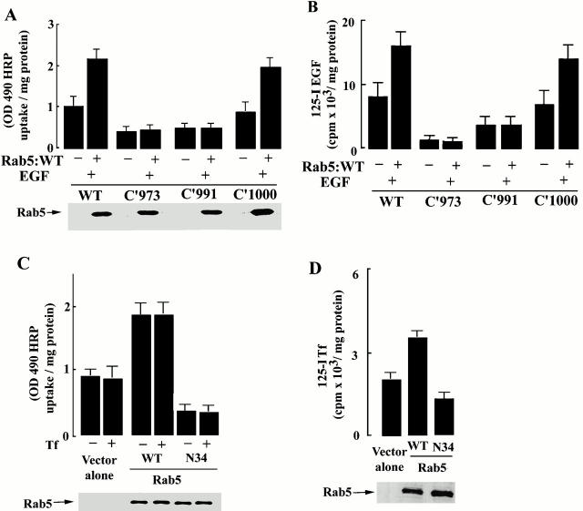
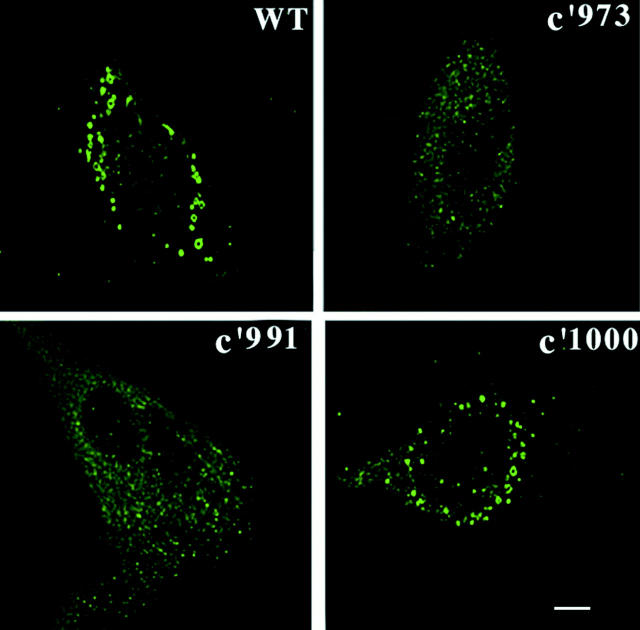
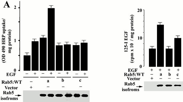
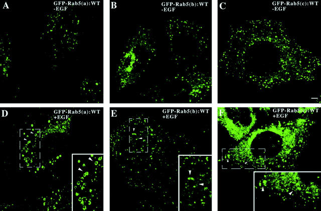
Activated epidermal growth factor receptors recruit various intracellular proteins leading to signal generation and endocytic trafficking. Although activated receptors are rapidly internalized into the endocytic compartment and subsequently degraded in lysosomes, the linkage between signaling and endocytosis is not well understood. Here we show that EGF stimulation of NR6 cells induces a specific, rapid and transient activation of Rab5a. EGF also enhanced translocation of the Rab5 effector, early endosomal autoantigen 1 (EEA1), from cytosol to membrane. The activation of endocytosis, fluid phase and receptor mediated, by EGF was enhanced by Rab5a expression, but not by Rab5b, Rab5c, or Rab5a truncated at the NH(2) and/or COOH terminus. Dominant negative Rab5a (Rab5:N34) blocked EGF-stimulated receptor-mediated and fluid-phase endocytosis. EGF activation of Rab5a function was dependent on tyrosine residues in the COOH-terminal domain of the EGF receptor (EGFR). Removal of the entire COOH terminus by truncation (c'973 and c'991) abrogated ligand-induced Rab5a activation of endocytosis. A "kinase-dead" EGFR failed to stimulate Rab5a function. However, another EGF receptor mutant (c'1000), with the kinase domain intact and a single autophosphorylation site effectively signaled Rab5 activation. These results indicate that EGFR and Rab5a are linked via a cascade that results in the activation of Rab5a and that appears essential for internalization. The results point to an interdependent relationship between receptor activation, signal generation and endocytosis.


Figures

Figure 1
Figure 1
EGF induces the formation of enlarged Rab5-positive endosomes. Cells expressing GFP-Rab5a (A and D), GFP-Rab5a:L79 (B and E), and GFP-Rab5a:N34 (C and F) were grown on coverslips and then were incubated in the presence or absence of 100 nM EGF for 15 min at 37°C. After stimulation, the cells were fixed with 2% paraformaldehyde, and then visualized by confocal microscopy. Cells expressing GFP-Rab5a (G–I) were incubated in the presence of 200 nM Texas red–EGF for 10 min at 37°C, fixed, and then visualized by confocal microscopy. Internalized Texas red–EGF (I) accumulated in the enlarged endosomes in cells expressing Rab5a (G). The yellow color indicates areas of colocalization of internalized Texas red–EGF and GFP-Rab5a (I). Optical sections viewed are 0.4 μM. Bars, 1 μM.
Figure 2
Figure 2
Membrane-cytosol redistribution of Rab proteins in the presence of EGF. (A) Cells expressing EGFR:WT (wild type) and mutants labeled with 32Pi, were transfected with GFP-Rab5a. Cells were incubated with 100 nM EGF for 15 min and the nucleotide status of Rab5 was analyzed as indicated in Materials and Methods (mean ± SD, n = 3.) (Bottom) The results from a typical experiment. (B) Cells expressing EGFR:WT were transfected with GFP-Rab5a. After transfection, the cells were incubated in the presence 100 nM EGF for 15 min and the membrane-cytosol distribution of EEA1 proteins was analyzed by Western blot. The experiment was repeated three times with similar results.
Figure 3
Figure 3
HRP-stimulated EGF-dependent endocytosis requires the small GTPase Rab5. (A) Cells expressing EGFR:WT were either transfected with virus alone (control: ○, •), virus encoding Rab5 (▴, ▵), or virus encoding Rab5:N34 (□, ▪). After transient transfection, the amount of HRP internalized at 37°C for the indicated times was quantified in the presence (▵, ○, □) or absence (•, ▴, ▪) of EGF. This experiment was repeated at least three times with similar results. (B) Cells expressing EGFR were transfected with virus encoding Rab5, Rab5:N34, Rab11, Rab11:mB1, Rab7, or Rab7:N22. After transfection, the internalization of HRP (37°C for 10 min) was quantified in the presence or absence of 100 nM EGF. Values are mean ± SD, n = 3. (C) Cells expressing EGFR:WT were either transfected with virus alone, virus encoding Rab5, virus encoding Rab5:N34, virus encoding Rab5:N34/ΔN19, virus encoding Rab5:ΔN19, virus encoding Rab5:ΔC4, or virus encoding Rab5:ΔC4/N34. After transient transfection, the amount of HRP internalized at 37°C for 15 min was quantified in the presence or absence of 100 nM EGF. Values are mean ± SD, n = 3.
Figure 3
Figure 3
HRP-stimulated EGF-dependent endocytosis requires the small GTPase Rab5. (A) Cells expressing EGFR:WT were either transfected with virus alone (control: ○, •), virus encoding Rab5 (▴, ▵), or virus encoding Rab5:N34 (□, ▪). After transient transfection, the amount of HRP internalized at 37°C for the indicated times was quantified in the presence (▵, ○, □) or absence (•, ▴, ▪) of EGF. This experiment was repeated at least three times with similar results. (B) Cells expressing EGFR were transfected with virus encoding Rab5, Rab5:N34, Rab11, Rab11:mB1, Rab7, or Rab7:N22. After transfection, the internalization of HRP (37°C for 10 min) was quantified in the presence or absence of 100 nM EGF. Values are mean ± SD, n = 3. (C) Cells expressing EGFR:WT were either transfected with virus alone, virus encoding Rab5, virus encoding Rab5:N34, virus encoding Rab5:N34/ΔN19, virus encoding Rab5:ΔN19, virus encoding Rab5:ΔC4, or virus encoding Rab5:ΔC4/N34. After transient transfection, the amount of HRP internalized at 37°C for 15 min was quantified in the presence or absence of 100 nM EGF. Values are mean ± SD, n = 3.
Figure 4
Figure 4
Endocytosis of EGF requires the small GTPase Rab5. (A) Cells expressing EGFR:WT were either transfected with virus alone (○), virus encoding Rab5a (•), virus encoding Rab7 (▴), or virus encoding Rab11 (▪). After transient transfection, the amount of 125I-EGF internalized at 37°C for the indicated times was quantified. This experiment was repeated at least four times, and the results were highly reproducible. (B) Cells expressing EGFR were transiently transfected with either virus alone, virus encoding Rab5, Rab5:N34, Rab5:N34/ΔN19, Rab5:ΔN19, Rab5:ΔC4, or Rab5:ΔC4N34. After transfection, uptake of 125I-EGF was carried out at 37°C for 10 min (mean ± SD, n = 3). (C) Cells transfected with virus alone or virus encoding Rab5:WT or Rab5:N34 were treated with EGF for different times. Surface EGFR was analyzed by 125I -EGF binding as described in Materials and Methods. This experiment was repeated at least three times, and the results were reproducible. (D) 35S-methionine–labeled NR6 cells transfected with virus alone, or virus encoding Rab5:WT or Rab5:N34, were treated with EGF. At the indicated times, aliquots were removed and EGFR was immunoprecipitated from cell lysates with anti–human EGFR antibody as described in Materials and Methods.
Figure 5
Figure 5
Endocytosis of HRP and EGF require an intact cytoplasmic domain of the EGFR. Cells expressing either EGFR:WT, EGFR:c'973, EGFR:c'991, or EGFR:c'1000 were transfected with virus alone or virus encoding Rab5. After transient transfection, the amount of HRP (A) or 125I-EGF (B) internalized at 37°C was quantified as described in Materials and Methods (mean ± SD, n = 4). (C and D) Cells were transfected with virus alone, or virus encoding Rab5, or Rab5:N34 and the internalization of either HRP (C) or 125I-Tf (D) was carried out for 10 min at 37°C in the presence or absence of 6 μg/ml transferrin. Mean ± SD, n = 3.
Figure 6
Figure 6
Formation of enlarged Rab5-positive endosomes requires an intact cytoplasmic domain of the EGFR. Cells expressing either EGFR:WT, EGFR:c'973, EGFR:c'991, or EGFR:c'1000 were transfected with virus encoding GFP-Rab5a. After transient transfection, the cells were incubated with EGF and the formation of enlarged endosomes were visualized by confocal microscopy as described in Materials and Methods. This experiment was repeated three times and the results were highly reproducible.
Figure 7
Figure 7
EGFR specifically regulates Rab5a function. (A) Cells expressing EGFR:WT were transfected with virus alone or virus encoding Rab5a, Rab5b, or Rab5c, and the internalization of HRP in the presence or absence of 100 nM EGF was carried out for 10 min at 37°C (mean ± SD, n = 3). (B) Cells expressing EGFR:WT were transfected as in A. After transfection, the uptake of 125I-EGF was carried out at 37°C for 10 min as described in Materials and Methods. Mean ± SD, n = 3.
Figure 8
Figure 8
The formation of enlarged endosomes by EGF is specific for the Rab5a isoform. Cells expressing EGFR:WT were transfected with virus encoding GFP-Rab5a (A and D), GFP-Rab5b (B and E), or GFP-Rab5c (C and F), and the visualization of the formation of enlarged endosomes were analyzed as described in Materials and Methods. This experiment was repeated three times and the results were highly reproducible.

References

    1. Alvarez-Dominguez C., Stahl P.D. Interferon-gamma selectively induces Rab5a synthesis and processing in mononuclear cells. J. Biol. Chem. 1998;273:33901–33904. - PubMed
    1. Alvarez-Dominguez C., Stahl P.D. Increased expression of Rab5a correlates directly with accelerated maturation of Listeria monocytogenes phagosomes. J. Biol. Chem. 1999;274:11459–11462. - PubMed
    1. Barbieri M.A., Hoffenberg S., Roberts R., Mukhopadhyay A., Pomrehn A., Dickey B.F., Stahl P.D. Evidence for a symmetrical requirement for Rab5:GTP in in vitro endosome–endosome fusion J. Biol. Chem 273 1998. 25850 25855a - PubMed
    1. Barbieri M.A., Kohn A.D., Roth R.A., Stahl P.D. Protein kinase B/akt and rab5 mediate Ras activation of endocytosis J. Biol. Chem 273 1998. 19367 19370b - PubMed
    1. Beguinot L., Lyall R.M., Willingham M.C., Pastan I. Down-regulation of the epidermal growth factor receptor in KB cells is due to receptor internalization and subsequent degradation in lysosomes. Proc. Natl. Acad. Sci. USA. 1984;81:2384–2388. - PMC - PubMed

Publication types

MeSH terms