. 2000 Oct 30;151(3):587-600.
doi: 10.1083/jcb.151.3.587.

A selective transport route from Golgi to late endosomes that requires the yeast GGA proteins

Affiliations

A selective transport route from Golgi to late endosomes that requires the yeast GGA proteins

M W Black et al. J Cell Biol. .

Abstract

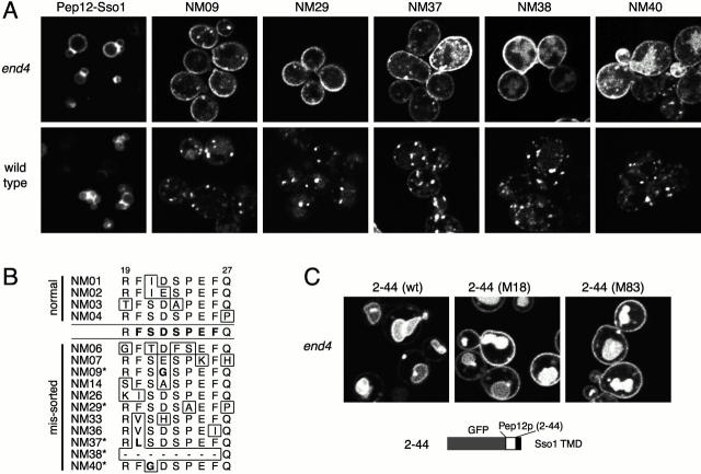
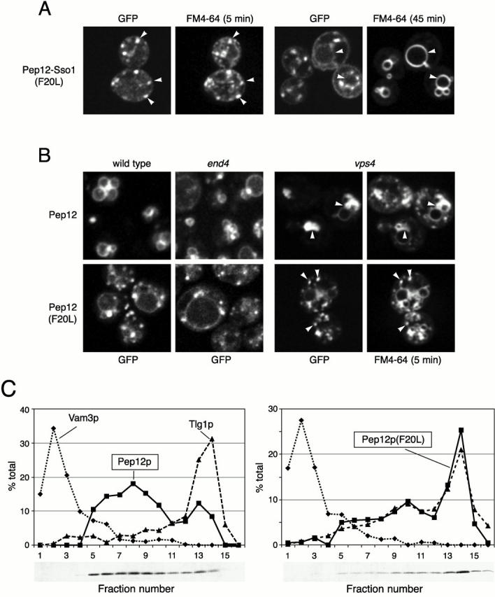
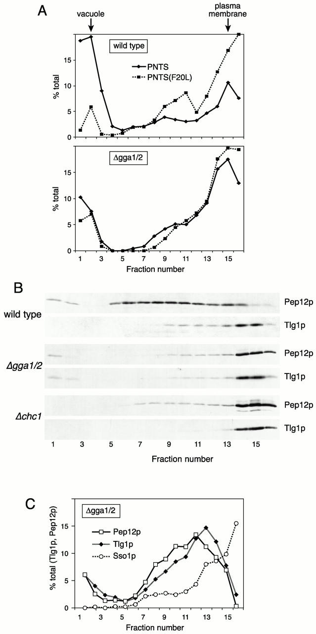
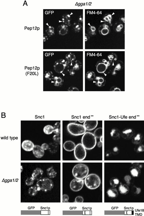
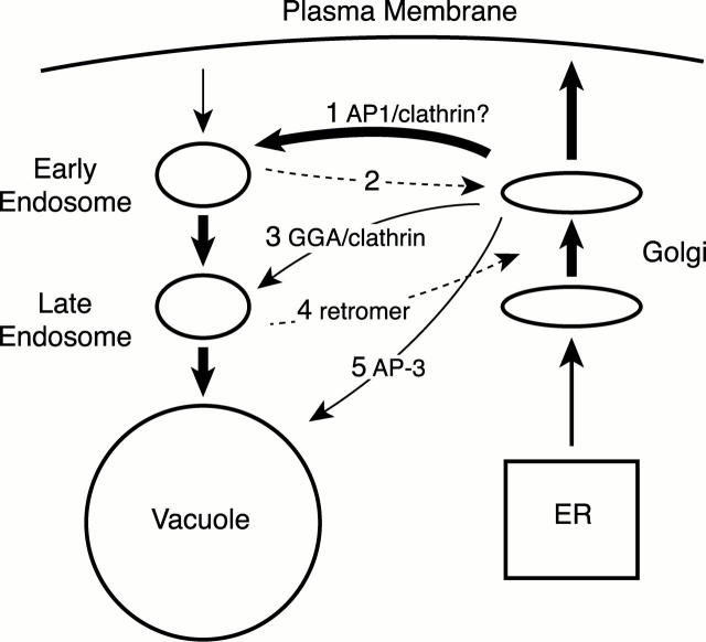
Pep12p is a yeast syntaxin located primarily in late endosomes. Using mutagenesis of a green fluorescent protein chimera we have identified a sorting signal FSDSPEF, which is required for transport of Pep12p from the exocytic pathway to late endosomes, from which it can, when overexpressed, reach the vacuole. When this signal is mutated, Pep12p instead passes to early endosomes, a step that is determined by its transmembrane domain. Surprisingly, Pep12p is then specifically retained in early endosomes and does not go on to late endosomes. By testing appropriate chimeras in mutant strains, we found that FSDSPEF-dependent sorting was abolished in strains lacking Gga1p and Gga2p, Golgi-associated coat proteins with homology to gamma adaptin. In the gga1 gga2 double mutant endogenous Pep12p cofractionated with the early endosome marker Tlg1p, and recycling of Snc1p through early endosomes was defective. Pep12p sorting was also defective in cells lacking the clathrin heavy or light chain. We suggest that specific and direct delivery of proteins to early and late endosomes is required to maintain the functional heterogeneity of the endocytic pathway and that the GGA proteins, probably in association with clathrin, help create vesicles destined for late endosomes.


Figures

Figure 2
Figure 2
Targeted mutagenesis of the sorting signal. (A) Images of the parental construct (GFP-tagged Pep12-Sso1) and missorted mutants in wild type and end4 strains. (B) Sequence changes in constructs that were missorted or showed a normal location. Dashes in the NM38 sequence indicate a deletion. *Mutants shown in A. (C) Distribution in end4 cells of a construct containing only residues 2–44 of Pep12p (structure as diagrammed), or the M18 and M83 mutant versions (see Fig. 1).
Figure 1
Figure 1
Mutations that affect targeting of Pep12p. (A) GFP-tagged Pep12p is on vacuolar membranes, identified by labeling with FM4-64. (B) GFP-tagged Pep12p bearing the Sso1p TMD (Pep12-Sso1), and examples of PCR-generated mutants, expressed in wild-type and end4 cells. (C) Sequence changes in mutants. Changes within the region shown (residues 2–102 of Pep12p) were sufficient for mistargeting to the cell surface in end4 cells. Black dots indicate the heptad repeat of the first predicted helix; the black bar indicates the smallest region that contains a change in each of the mutants sequenced.
Figure 3
Figure 3
Missorted mutants retain function. (A) Serial 10-fold dilutions of wild-type, Δpep12, and the Δpep12 strain expressing untagged versions of Pep12-Sso1 or mutant derivatives (see Fig. 2) from the PEP12 promoter were grown at 25° or 37°C. (B) CPY secreted by patches of the same cells was detected by antibody staining.
Figure 4
Figure 4
The F20L mutation allows Pep12p to accumulate in early endosomes. (A) Wild-type cells expressing GFP-tagged Pep12-Sso1 (F20L) were incubated with FM4-64 for 15 min, chased for 5 or 45 min as indicated, and imaged. FM4-64 labels the dots containing the GFP-labeled protein at early times, and then passes to the vacuole. (B) GFP-labeled Pep12p (F20L) with its own TMD is punctate in wild-type, end4, and vps4 cells, whereas unmutated Pep12p is in vacuoles or, in vps4, the PVC. The vps4 cells were incubated with FM4-64 and chased for 5 min to reveal labeling of punctate early endosomes containing the mutant protein. (C) Extracts of Δpep12 cells expressing untagged wild-type, Pep12p, or the F20L mutant at normal levels (from the PEP12 promoter) were fractionated on sucrose density gradients. Fractions were assayed for Pep12p, Vam3p (vacuolar marker), and Tlg1p (early endosomal marker) by immunoblotting. The Pep12p blots are shown beneath the graphs.
Figure 5
Figure 5
Properties of the NH2-terminal domain of Pep12p. (A) GFP-tagged PNTP and the F20L mutant version both reach vacuoles in wild-type cells. PNTS, the equivalent construct with the Sso1 TMD, goes to vacuoles in wild-type and end4 cells, whereas the F20L mutant goes to the plasma membrane. (B) GFP-labeled derivatives of Ste18p (as diagrammed) were expressed in a Δste18 strain and imaged. Equivalent constructs without GFP, or no construct (−), were expressed in Δste18 cells carrying a FUS1-lacZ reporter and the cells assayed for β-galactosidase activity. Note the log scale of the graph.
Figure 6
Figure 6
Sorting of the NH2-terminal domain of Pep12p in various strains. GFP-tagged PNTS (see Fig. 5) was expressed in the indicated mutants, and in wild-type cells (wt) that coexpressed from a multicopy vector non–GFP-tagged versions (competitor) of either PNTS itself or the F20L mutant version.
Figure 7
Figure 7
Missorting of PNTS by clathrin and gga mutants. GFP-tagged PNTS or the F20L mutant version were expressed in the indicated mutants. A marker for the AP-3 pathway to the vacuole, GNS (structure as diagrammed) was also expressed in the indicated mutants.
Figure 8
Figure 8
Subcellular fractionation of PNTS and Pep12p in mutants. (A) Wild-type and gga1/2 mutant cells expressing GFP-tagged PNTS or its F20L mutant variant were fractionated on sucrose gradients as in Fig. 4 C and the expressed proteins detected by immunoblotting. The peak positions of markers for the vacuole (Vam3p) and plasma membrane (Sso1p) are indicated at the top. (B) Extracts of wild-type, gga1/2, and chc1 cells were fractionated as in A and analyzed by immunoblotting for endogenous Pep12p and Tlg1p. (C) The gga1/2 cells were fractionated on a gradient of higher density (see Materials and Methods) to resolve the early endosome region, and the distributions of Tlg1p, Pep12p, and Sso1p were measured by immunoblotting. The Sso1p profile has been scaled by a factor of 0.6 for clarity.
Figure 9
Figure 9
Sorting properties of the gga 1/2 mutant. (A) Distribution of GFP-tagged Pep12p and the F20L mutant in gga1/2 cells. Cells were double labeled with FM4-64 (chased for 30 min) to reveal vacuoles. (B) GFP-tagged versions of Snc1p, a double point mutant Snc1p that is endocytosis-defective (end) and a derivative of this bearing a modified Ufe1p TMD that targets it from the Golgi to the vacuole (ufe18; Lewis et al. 2000) were expressed in wild-type and gga1/2 double mutant cells.
Figure 10
Figure 10
Summary of the proposed connections between the exocytic and endocytic pathways in yeast. The known or postulated coat proteins involved in some of the steps are indicated. See text for details.

Similar articles

Cited by

References

    1. Abeliovich H., Darsow T., Emr S.D. Cytoplasm to vacuole trafficking of aminopeptidase I requires a t-SNARE-Sec1p complex composed of Tlg2p and Vps45p. EMBO (Eur. Mol. Biol. Organ.) J. 1999;18:6005–6016. - PMC - PubMed
    1. Ausubel, F.M., R. Brent, R.E. Kingston, D.D. Moore, J.G. Seidman, J.A. Smith, and K. Struhl. 1995. Current Protocols in Molecular Biology. Vol. 1. J. Wiley and Sons, Inc., Boston, MA. 13.6.2–13.6.3.
    1. Babst M., Sato T.K., Banta L.M., Emr S.D. Endosomal transport function in yeast requires a novel AAA-type ATPase, Vps4p. EMBO (Eur. Mol. Biol. Organ.) J. 1997;16:1820–1831. - PMC - PubMed
    1. Becherer K.A., Rieder S.E., Emr S.D., Jones E.W. Novel syntaxin homologue, Pep12p, required for the sorting of lumenal hydrolases to the lysosome-like vacuole in yeast. Mol. Biol. Cell. 1996;7:579–594. - PMC - PubMed
    1. Boman A.L., Zhang C., Zhu X., Kahn R.A. A family of ADP-ribosylation factor effectors that can alter membrane transport through the trans-Golgi. Mol. Biol. Cell. 2000;11:1241–1255. - PMC - PubMed

Publication types

MeSH terms