. 2000 Oct 30;151(3):601-12.
doi: 10.1083/jcb.151.3.601.

Rabenosyn-5, a novel Rab5 effector, is complexed with hVPS45 and recruited to endosomes through a FYVE finger domain

Affiliations

Rabenosyn-5, a novel Rab5 effector, is complexed with hVPS45 and recruited to endosomes through a FYVE finger domain

E Nielsen et al. J Cell Biol. .

Abstract

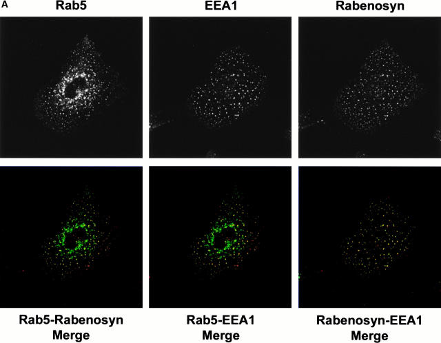
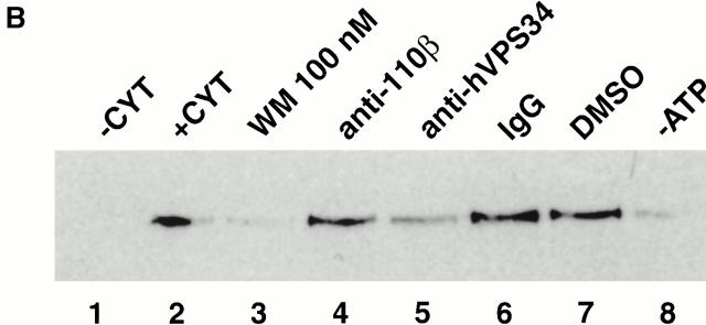
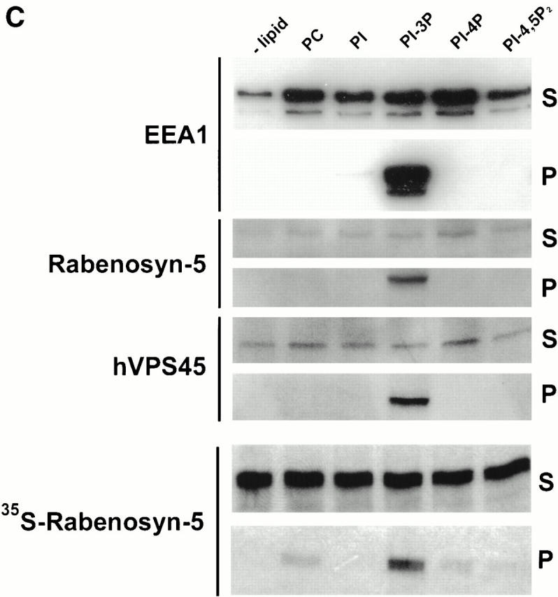
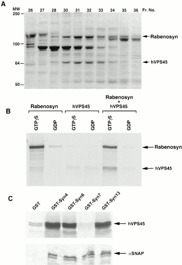
Rab5 regulates endocytic membrane traffic by specifically recruiting cytosolic effector proteins to their site of action on early endosomal membranes. We have characterized a new Rab5 effector complex involved in endosomal fusion events. This complex includes a novel protein, Rabenosyn-5, which, like the previously characterized Rab5 effector early endosome antigen 1 (EEA1), contains an FYVE finger domain and is recruited in a phosphatidylinositol-3-kinase-dependent fashion to early endosomes. Rabenosyn-5 is complexed to the Sec1-like protein hVPS45. hVPS45 does not interact directly with Rab5, therefore Rabenosyn-5 serves as a molecular link between hVPS45 and the Rab5 GTPase. This property suggests that Rabenosyn-5 is a closer mammalian functional homologue of yeast Vac1p than EEA1. Furthermore, although both EEA1 and Rabenosyn-5 are required for early endosomal fusion, only overexpression of Rabenosyn-5 inhibits cathepsin D processing, suggesting that the two proteins play distinct roles in endosomal trafficking. We propose that Rab5-dependent formation of membrane domains enriched in phosphatidylinositol-3-phosphate has evolved as a mechanism for the recruitment of multiple effector proteins to mammalian early endosomes, and that these domains are multifunctional, depending on the differing activities of the effector proteins recruited.


Figures

Figure 1
Figure 1
Rabenosyn-5 is a novel Rab5 effector protein with FYVE finger and C2H2 zinc finger domains. (A) Specific binding of Rab5 effector proteins to GST-Rab5-GTPγS. Rab5-interacting proteins purified by GST-Rab5 affinity chromatography were subjected to two-dimensional SDS-PAGE followed by silver staining (left), or transferred to nitrocellulose and gel-overlay assay with GST-Rab5-GTPγS (middle), or GST-Rab5-GDP (right). Positions of the known Rab5 effector proteins, EEA1 (arrowhead), and Rabaptin-5α (arrow) were determined by immunoblotting. The position of a novel 110-kD Rab5 effector, Rabenosyn-5, is marked with an asterisk. (B) Alignment of the FYVE finger domain of Rabenosyn-5 with other Rab effector proteins. The FYVE finger domains of human Rabenosyn-5 (sequence data available from EMBL/GenBank/DDBJ under accession no. AY009133), S. cerevisiae Vac1p (accession no. P32609), S. pombe Vac1p homologous protein (accession no. Z99162), and human EEA1 (accession no. S44243) were aligned using CLUSTALW. (C) Domain organization of Rabenosyn-5, Vac1p, and EEA1. Each protein is represented as a line; the relative lengths are proportional to the length of the coding sequence. Positions of C2H2, RING, and FYVE zinc fingers, and the NPF motif–containing domains are indicated. (D) The five NPF-containing motifs of Rabenosyn-5, and their consensus sequence. (E) Schematic diagram of the truncation mutations of Rabenosyn-5.
Figure 1
Figure 1
Rabenosyn-5 is a novel Rab5 effector protein with FYVE finger and C2H2 zinc finger domains. (A) Specific binding of Rab5 effector proteins to GST-Rab5-GTPγS. Rab5-interacting proteins purified by GST-Rab5 affinity chromatography were subjected to two-dimensional SDS-PAGE followed by silver staining (left), or transferred to nitrocellulose and gel-overlay assay with GST-Rab5-GTPγS (middle), or GST-Rab5-GDP (right). Positions of the known Rab5 effector proteins, EEA1 (arrowhead), and Rabaptin-5α (arrow) were determined by immunoblotting. The position of a novel 110-kD Rab5 effector, Rabenosyn-5, is marked with an asterisk. (B) Alignment of the FYVE finger domain of Rabenosyn-5 with other Rab effector proteins. The FYVE finger domains of human Rabenosyn-5 (sequence data available from EMBL/GenBank/DDBJ under accession no. AY009133), S. cerevisiae Vac1p (accession no. P32609), S. pombe Vac1p homologous protein (accession no. Z99162), and human EEA1 (accession no. S44243) were aligned using CLUSTALW. (C) Domain organization of Rabenosyn-5, Vac1p, and EEA1. Each protein is represented as a line; the relative lengths are proportional to the length of the coding sequence. Positions of C2H2, RING, and FYVE zinc fingers, and the NPF motif–containing domains are indicated. (D) The five NPF-containing motifs of Rabenosyn-5, and their consensus sequence. (E) Schematic diagram of the truncation mutations of Rabenosyn-5.
Figure 3
Figure 3
The Rabenosyn-5 FYVE domain is sufficient to target Rabenosyn-5 to early endosomes. HeLa cells coexpressing Rab5Q79L and myc-tagged truncation mutants of Rabenosyn-5 were processed for immunofluorescence and analyzed by laser scanning confocal microscopy for the extent of colocalization of myc-tagged truncations of Rabenosyn-5 (red, Merge) with Rab5Q79L-positive endosomal structures (green, Merge).
Figure 2
Figure 2
Localization of Rabenosyn-5 with early endosomes is PI-3-kinase dependent. (A) Rabenosyn-5 colocalizes with EEA1 on early endosomes in A431 cells. A431 cells expressing EGFP-Rab5 were processed for immunofluorescence and analyzed by laser scanning confocal microscopy to detect the extent of colocalization of EGFP-Rab5 fluorescence (top left), EEA1 was detected with human antiserum (top middle), and Rabenosyn-5 was detected with affinity-purified rabbit antibodies (top right). EGFP-Rab5 (green) colocalized significantly with Rabenosyn-5 (red; bottom left) and EEA1 (red; bottom middle); EEA1 (green) and Rabenosyn-5 (red) displayed complete colocalization (bottom right). (B) Recruitment of Rabenosyn-5 on early endosomes. Reactions containing early endosomes, cytosol (3 mg/ml), and an ATP-regenerating system were incubated for 30 min at 37°C (+CYT, lane 2), membranes were recovered by centrifugation, resuspended in SDS-PAGE buffer, and analyzed by immunoblotting with antibodies against Rabenosyn-5. Reactions were carried out in the absence of cytosol (−CYT, lane 1) or an ATP-regenerating system (−ATP, lane 8). Other reactions were carried with both cytosol and an ATP-regenerating system (lanes 2–7): alone (+CYT, lane 2); in the presence of 100 nM wortmannin (WM 100 nM, lane 3); function blocking antibodies against p110β (anti-110β, lane 4); hVPS34 (anti-hVPS34, lane 5); control nonspecific IgG (IgG, lane 6); or control concentration of DMSO (DMSO, lane 7). (C) Recruitment of Rabenosyn-5 on artificial liposomes. Reactions containing cytosol (5 mg/ml), or in vitro–translated, [35S]methionine-labeled Rabenosyn-5 were incubated for 15 min at room temperature with liposomes (100 μg total lipid) consisting of PC alone (100% total lipid), or PC mixed with PI (2% total lipid), PI-3P (2%), PI-4P (2%), or PI-4,5P2 (2%). Supernatants (S) and membrane pellets (P) were separated by centrifugation, resuspended in SDS-PAGE buffer (10% of total supernatants), and analyzed by immunoblotting with antibodies specific to Rabenosyn-5, EEA1, and hVPS45, or by fluorography to detect [35S]methionine-labeled Rabenosyn-5.
Figure 2
Figure 2
Localization of Rabenosyn-5 with early endosomes is PI-3-kinase dependent. (A) Rabenosyn-5 colocalizes with EEA1 on early endosomes in A431 cells. A431 cells expressing EGFP-Rab5 were processed for immunofluorescence and analyzed by laser scanning confocal microscopy to detect the extent of colocalization of EGFP-Rab5 fluorescence (top left), EEA1 was detected with human antiserum (top middle), and Rabenosyn-5 was detected with affinity-purified rabbit antibodies (top right). EGFP-Rab5 (green) colocalized significantly with Rabenosyn-5 (red; bottom left) and EEA1 (red; bottom middle); EEA1 (green) and Rabenosyn-5 (red) displayed complete colocalization (bottom right). (B) Recruitment of Rabenosyn-5 on early endosomes. Reactions containing early endosomes, cytosol (3 mg/ml), and an ATP-regenerating system were incubated for 30 min at 37°C (+CYT, lane 2), membranes were recovered by centrifugation, resuspended in SDS-PAGE buffer, and analyzed by immunoblotting with antibodies against Rabenosyn-5. Reactions were carried out in the absence of cytosol (−CYT, lane 1) or an ATP-regenerating system (−ATP, lane 8). Other reactions were carried with both cytosol and an ATP-regenerating system (lanes 2–7): alone (+CYT, lane 2); in the presence of 100 nM wortmannin (WM 100 nM, lane 3); function blocking antibodies against p110β (anti-110β, lane 4); hVPS34 (anti-hVPS34, lane 5); control nonspecific IgG (IgG, lane 6); or control concentration of DMSO (DMSO, lane 7). (C) Recruitment of Rabenosyn-5 on artificial liposomes. Reactions containing cytosol (5 mg/ml), or in vitro–translated, [35S]methionine-labeled Rabenosyn-5 were incubated for 15 min at room temperature with liposomes (100 μg total lipid) consisting of PC alone (100% total lipid), or PC mixed with PI (2% total lipid), PI-3P (2%), PI-4P (2%), or PI-4,5P2 (2%). Supernatants (S) and membrane pellets (P) were separated by centrifugation, resuspended in SDS-PAGE buffer (10% of total supernatants), and analyzed by immunoblotting with antibodies specific to Rabenosyn-5, EEA1, and hVPS45, or by fluorography to detect [35S]methionine-labeled Rabenosyn-5.
Figure 4
Figure 4
Rabenosyn-5 recruits the Sec1-like protein hVPS45 to Rab5. (A) SDS-PAGE analysis and Coomassie blue staining of Rab5-interacting proteins separated by Superose-6 size-exclusion chromatography. Fraction numbers are indicated at the top of each lane. MS/MS tandem mass spectroscopy sequencing identified Rabenosyn-5 and hVPS45 proteins. (B) Rabenosyn-5 recruits hVPS45 to GST-Rab5. Glutathione-sepharose beads loaded with GST-Rab5-GTPγS (GTPγS) or GST-Rab5-GDP (GDP) were incubated with [35S]methionine-labeled in vitro–translated Rabenosyn-5 alone (Rabenosyn), hVPS45 alone (hVPS45), or both Rabenosyn-5 and hVPS45 cotranslated together (Rabenosyn + hVPS45). Bound proteins were eluted and analyzed by SDS-PAGE followed by fluorography. (C) hVPS45 interacts with multiple syntaxin isoforms. Glutathione-sepharose beads loaded with GST-syntaxin fusion proteins (GST-syntaxin 4, GST-Syn4; GST-syntaxin 6, GST-Syn6; GST-syntaxin 7, GST-Syn7; and GST-syntaxin 13, GST-Syn13), or GST alone (GST), and incubated with [35S]methionine-labeled in vitro–translated hVPS45 (top), or α-SNAP (bottom). GST fusions and associated proteins were eluted and analyzed by SDS-PAGE followed by fluorography.
Figure 5
Figure 5
Requirement of Rabenosyn-5 for homotypic early endosome–early endosome, and heterotypic CCV–early endosome fusion. (A) Immunodepletion of Rabenosyn-5 from the cytosol. 100 μg of cytosol (cytosol), cytosol immunodepleted of Rabenosyn-5 (anti-Rabenosyn), or cytosol immunodepleted with nonspecific IgG (IgG) were suspended in SDS-PAGE buffer, and analyzed by immunoblotting with antibodies specific to EEA1, Rabenosyn-5, and hVPS45. (B) Fusion of CCVs loaded with biotinylated transferrin (donor) and early endosomes loaded with antitransferrin antibody (acceptor), or donor and acceptor loaded early endosomes was performed under standard conditions (see Materials and Methods). Reactions were carried out either in the absence of cytosol (−Cytosol), in the presence of untreated cytosol (Basal), in the presence of untreated cytosol but with no ATP-regenerating system (−Energy), in the presence of cytosol immunodepleted of Rabenosyn-5 (−Rabenosyn), or in the presence of cytosol treated with nonspecific IgG (IgG). Inhibition of fusion observed upon immunodepletion of Rabenosyn-5 could be rescued with Rab5 effector fractions containing Rabenosyn-5 (Fraction 33), but not with fractions containing EEA1 (Fraction 19). If Rabenosyn-5 was immunodepleted from fraction 33, the ability of this fraction to rescue fusion was abolished (anti-Rabenosyn depleted Fraction 33).
Figure 5
Figure 5
Requirement of Rabenosyn-5 for homotypic early endosome–early endosome, and heterotypic CCV–early endosome fusion. (A) Immunodepletion of Rabenosyn-5 from the cytosol. 100 μg of cytosol (cytosol), cytosol immunodepleted of Rabenosyn-5 (anti-Rabenosyn), or cytosol immunodepleted with nonspecific IgG (IgG) were suspended in SDS-PAGE buffer, and analyzed by immunoblotting with antibodies specific to EEA1, Rabenosyn-5, and hVPS45. (B) Fusion of CCVs loaded with biotinylated transferrin (donor) and early endosomes loaded with antitransferrin antibody (acceptor), or donor and acceptor loaded early endosomes was performed under standard conditions (see Materials and Methods). Reactions were carried out either in the absence of cytosol (−Cytosol), in the presence of untreated cytosol (Basal), in the presence of untreated cytosol but with no ATP-regenerating system (−Energy), in the presence of cytosol immunodepleted of Rabenosyn-5 (−Rabenosyn), or in the presence of cytosol treated with nonspecific IgG (IgG). Inhibition of fusion observed upon immunodepletion of Rabenosyn-5 could be rescued with Rab5 effector fractions containing Rabenosyn-5 (Fraction 33), but not with fractions containing EEA1 (Fraction 19). If Rabenosyn-5 was immunodepleted from fraction 33, the ability of this fraction to rescue fusion was abolished (anti-Rabenosyn depleted Fraction 33).
Figure 6
Figure 6
Rabenosyn-5 overexpression impairs cathepsin D trafficking. (A) Time course analysis of cathepsin D trafficking. HeLa cells overexpressing Rabenosyn-5, EEA1, or mock transfected were metabolically labeled with [35S]methionine for 30 min, chased with cold methionine for the indicated times, and then cellular cathepsin D was immunoprecipitated and relative quantities of precursor (left) or processed intermediate (right) cathepsin D were analyzed by SDS-PAGE followed by autoradiography. The signal was quantified by densitometric analysis of the autoradiograms. (B) Effect of overexpression of Rabenosyn-5 truncation mutants upon cathepsin D trafficking. Experiments were performed as described in A, except quantification of relative percentages of precursor cathepsin D (black bars) and processed intermediate cathepsin D (white bars) were performed after 4 h of chase time.
Figure 6
Figure 6
Rabenosyn-5 overexpression impairs cathepsin D trafficking. (A) Time course analysis of cathepsin D trafficking. HeLa cells overexpressing Rabenosyn-5, EEA1, or mock transfected were metabolically labeled with [35S]methionine for 30 min, chased with cold methionine for the indicated times, and then cellular cathepsin D was immunoprecipitated and relative quantities of precursor (left) or processed intermediate (right) cathepsin D were analyzed by SDS-PAGE followed by autoradiography. The signal was quantified by densitometric analysis of the autoradiograms. (B) Effect of overexpression of Rabenosyn-5 truncation mutants upon cathepsin D trafficking. Experiments were performed as described in A, except quantification of relative percentages of precursor cathepsin D (black bars) and processed intermediate cathepsin D (white bars) were performed after 4 h of chase time.

Similar articles

Cited by

References

    1. Abeliovich H., Darsow T., Emr S.D. Cytoplasm to vacuole trafficking of aminopeptidase I requires a t-SNARE–Sec1p complex composed of Tlg2p and Vps45p. EMBO (Eur. Mol. Biol. Organ.) J. 1999;18:6005–6016. - PMC - PubMed
    1. Burd C.G., Peterson M., Cowles C.R., Emr S.D. A novel Sec18p/NSF-dependent complex required for Golgi-to-endosome transport in yeast. Mol. Biol. Cell. 1997;8:1089–1104. - PMC - PubMed
    1. Carr C.M., Grote E., Munson M., Hughson F.M., Novick P.J. Sec1p binds to SNARE complexes and concentrates at sites of secretion. J. Cell Biol. 1999;146:333–344. - PMC - PubMed
    1. Christoforidis S., Zerial M. Purification and identification of novel Rab effectors using affinity chromatography. Methods. 2000;20:403–410. - PubMed
    1. Christoforidis S., McBride H.M., Burgoyne R.D., Zerial M. The Rab5 effector EEA1 is a core component of endosome docking Nature 397 1999. 621 625a - PubMed

Publication types

MeSH terms

Associated data