. 2000 Oct 30;151(3):673-84.
doi: 10.1083/jcb.151.3.673.

The dendritic cell receptor for endocytosis, DEC-205, can recycle and enhance antigen presentation via major histocompatibility complex class II-positive lysosomal compartments

Affiliations

The dendritic cell receptor for endocytosis, DEC-205, can recycle and enhance antigen presentation via major histocompatibility complex class II-positive lysosomal compartments

K Mahnke et al. J Cell Biol. .

Abstract

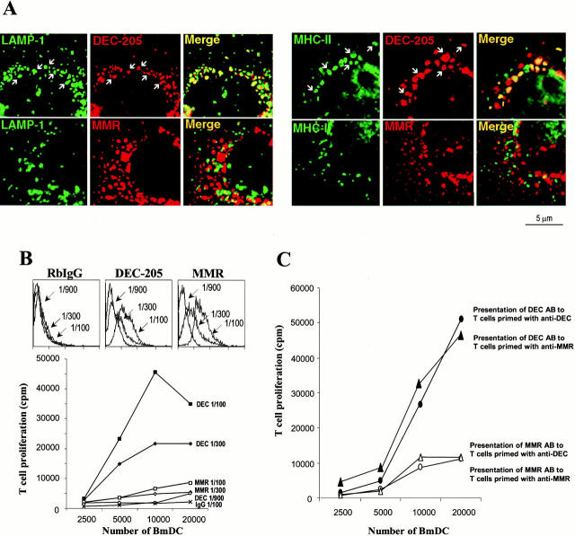
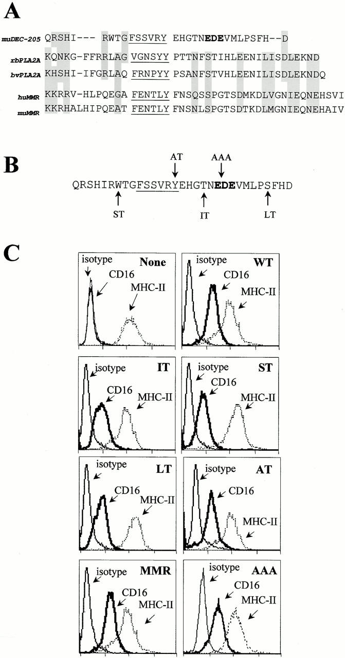
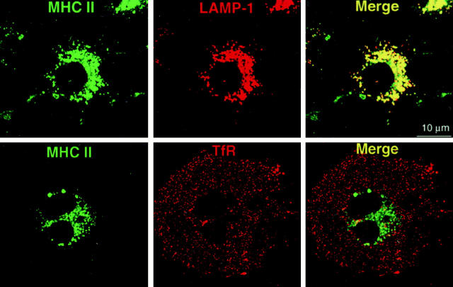
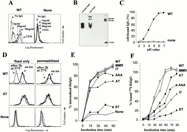
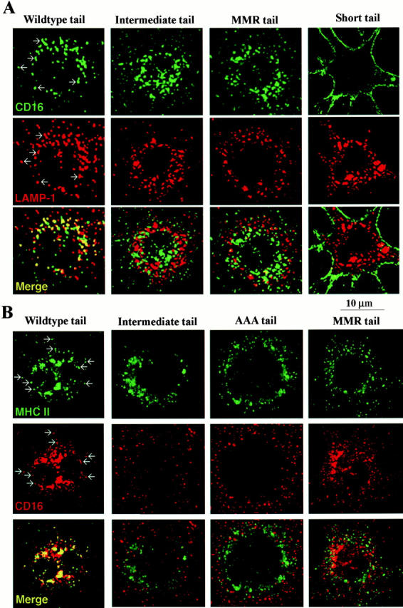
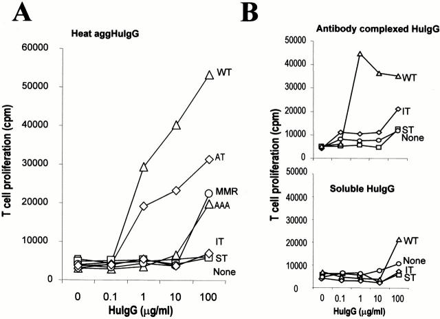
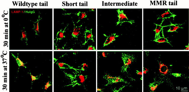
Many receptors for endocytosis recycle into and out of cells through early endosomes. We now find in dendritic cells that the DEC-205 multilectin receptor targets late endosomes or lysosomes rich in major histocompatibility complex class II (MHC II) products, whereas the homologous macrophage mannose receptor (MMR), as expected, is found in more peripheral endosomes. To analyze this finding, the cytosolic tails of DEC-205 and MMR were fused to the external domain of the CD16 Fcgamma receptor and studied in stable L cell transfectants. The two cytosolic domains each mediated rapid uptake of human immunoglobulin (Ig)G followed by recycling of intact CD16 to the cell surface. However, the DEC-205 tail recycled the CD16 through MHC II-positive late endosomal/lysosomal vacuoles and also mediated a 100-fold increase in antigen presentation. The mechanism of late endosomal targeting, which occurred in the absence of human IgG, involved two functional regions: a membrane-proximal region with a coated pit sequence for uptake, and a distal region with an EDE triad for the unusual deeper targeting. Therefore, the DEC-205 cytosolic domain mediates a new pathway of receptor-mediated endocytosis that entails efficient recycling through late endosomes and a greatly enhanced efficiency of antigen presentation to CD4(+) T cells.


Figures

Figure 1
Figure 1
Distinct intracellular localization and function of DEC-205 and MMR in DCs. (A) Localization. Immature day 6 bone marrow DCs (BmDC) (Inaba et al. 1992) were seeded on coverslips, fixed, and double stained with rabbit anti–DEC-205 and MMR (red), and the late endosomal/lysosomal components, LAMP-1 or MHC II (green). Examples of individual granules that contain red and green label are marked by arrows, and colocalization is further shown in yellow in the merged images. (B) Surface binding and presentation of anti–DEC-205 and MMR antibodies. Day 6, immature marrow-derived DCs were incubated with different dilutions (as indicated) of anti–DEC-205, anti-MMR, or preimmune rabbit serum on ice for 1 h. Unbound antibody was washed away. The cells were incubated with FITC-labeled anti-Ig, to assess amounts of surface bound antibody by FACS® (top), or cocultured in graded doses with rabbit IgG–primed T cells, to measure presentation of peptides from rabbit antibodies (bottom). (C) DCs were incubated with 1:100 dilution of anti–DEC-205 or anti-MMR rabbit serum on ice for 1 h. Unbound antibody was washed away, and presentation of peptides from rabbit antibodies was assayed by coculturing the DCs with graded doses of T cells primed with anti–DEC-205 or anti-MMR antiserum, respectively.
Figure 2
Figure 2
Cytosolic domains used to study the targeting of endocytosis receptors. (A) Sequences of the cytoplasmic domain of three multilectin receptors for adsorptive endocytosis: DEC-205, PLA2R, and MMR. Underlining indicates a consensus sequence for endocytosis via coated pits; boldface indicates a cluster of acidic residues (EDE), postulated to guide DEC-205 beyond early endosomes; gray shading shows similar amino acids in the tails. (B) Mutations were introduced into the DEC tail (arrows), leading to truncations at the sites denoted (ST, short tail; IT, intermediate tail; LT, long tail), or to amino acid substitutions: Y to A in alternate tail (AT), and EDE to AAA in the AAA tail. (C) Surface expression of CD16 and MHC II in untransfected DCEK.ICAM.Hi7 cells (none) or cells transfected with CD16 fusion receptors (WT, LT, IT, ST, AT, AAA, and MMR). Staining was with anti-CD16 (clone 3G8), anti–I-Ek (clone HB32), or isotype control anti–I-Ak (clone 10.216), followed by FITC goat anti–mouse F(ab′)2 antibodies.
Figure 4
Figure 4
Presence of MHC II+ lysosomal compartments in CD16 transfectants (WT-DEC:CD16 shown here). Stably transfected DCEK.ICAM.Hi7 cells on tissue culture chamber slides were fixed and double labeled for MHC II and lysosomal (LAMP-1) proteins (top) or TfR (bottom). Primary antibodies were visualized by FITC- and Texas red–labeled secondary antibodies. By confocal laser scan microscopy, MHC II colocalizes with LAMP-1 in all cells (yellow) but not with the TfR.
Figure 3
Figure 3
Ligand binding, uptake, and recycling via CD16 DEC-205 chimeric receptors. (A) Binding of aggHuIgG to transfected cells. WT-DEC/CD16 transfected cells (WT) and untransfected cells (None) were incubated on ice for 1 h with graded doses of aggHuIgG. After washing, surface-bound IgG was detected with FITC goat anti-HuIgG F(ab′)2 and FACS®. Cells were also stained for surface CD16 using 3G8 and FITC goat anti–mouse F(ab′)2. Binding to WT cells and all other transfectants (see Fig. 1) was similar. (B) To detect the small amount of aggHuIgG in the heated HuIgG preparations, samples were analyzed under reducing conditions by SDS-PAGE analysis. (C) Elution of HuIgG to CD16 at acidic pH. Elution of aggHuIgG to CD16 was performed at different pH's, predicting a release of ligand in mildly acidic compartments. (D) Endocytosis of aggHuIgG. Untransfected cells (none) and transfectants with different DEC:CD16 chimeric receptors (WT, ST) were incubated on ice for 1 h with aggHuIgG (10 μg/ml). Aliquots were fixed and stained for surface-bound HuIgG immediately (bold line) or incubated at 37°C for 4 h (thin line) before fixation and staining with FITC-labeled anti-HuIgG (first column, fixed only). Cells were also permeabilized before staining with FITC-labeled anti-HuIgG (second column) to reveal endocytosed HuIgG. (E) Uptake of HuIgG. Cell lines were incubated with aggHuIgG on ice, transferred to 37°C for different times, and stained with FITC-labeled anti-HuIgG F(ab′)2 without or with prior permeabilization. The mean fluorescence was determined by FACS® analysis, and the amount of internalized IgG was plotted as a percentage of total surface-bound IgG. (F) Recycling of CD16 chimeric receptors. Cells were treated for 2 h with CHX and then incubated with 100 μg/ml aggHuIgG, and transferred on ice, to saturate binding of all CD16 chimeric receptors (see Fig. 2 A). After washing, the cells were incubated at 37°C in the continued presence of CHX for different times, and recycling CD16 chimeric receptors were detected by binding of 125I-HuIgG. The amount of recycled receptors is plotted as a percentage of the amount of surface-bound 125I-HuIgG obtained with untreated cells.
Figure 5
Figure 5
Typical distribution of CD16 chimeric receptors in DCEK.ICAM.Hi7 cells in the absence (shown here) or presence of ligand, 10 μg/ml aggHuIgG. (A) CD16 and LAMP-1 double labeling. Cells on tissue culture chamber slides were fixed and stained for CD16 (green, FITC-labeled anti–mouse Ig) and LAMP-1 (red, Texas red–labeled anti–rat Ig). Colocalization in discrete vesicles in WT transfected cells is indicated by arrows, and for all the transfectants is displayed in yellow (bottom row). (B) CD16 and MHC II double labeling. Cells grown on tissue chamber slides were fixed and stained for CD16 (red) or MHC II (green) with species-specific secondary reagents. Slides were analyzed by confocal laser scan microscopy. Examples of colocalization in single vesicles in WT transfected cells are indicated by arrows and displayed in yellow (bottom row).
Figure 7
Figure 7
Antigen presentation by DEC-CD16 transfected cells. (A) Presentation of aggHuIgG. Transfected and untransfected (none) DCEK.ICAM.Hi7 cell lines were incubated for 6 h with aggHuIgG, washed, and used to present antigen to 250,000 purified lymph node T cells from mice primed to HuIgG. At 72 h, the cells were pulsed with [3H]thymidine for 12 h, and incorporated radioactivity was measured. (B) Presentation of immune complexes of HuIgG. As in A, transfected and untransfected (none) DCEK.ICAM.Hi7 cell lines were incubated with either soluble or antibody aggregated HuIgG. Antigen presenting assays were then performed.
Figure 6
Figure 6
Intracellular localization of endocytosed HuIgG in CD16 transfectants. (A) Double labeling for endocytosed HuIgG and LAMP-1. Cells, grown on tissue chamber slides, were incubated with 10 μg/ml HuIgG for 1 h on ice. Unbound HuIgG was washed away and cells were either fixed immediately or further incubated in medium for 30 min at 37°C. Cells were double stained with FITC-labeled anti-HuIgG (green) and with anti–LAMP-1 antibodies (red; Texas red–labeled secondary antibodies).

Similar articles

Cited by

References

    1. Amigorena S., Bonnerot C., Drake J.R., Choquet D., Hunziker W., Guillet J.-G., Webster P., Sautes C., Mellman I., Fridman W.H. Cytoplasmic domain heterogeneity and functions of IgG Fc receptors in B lymphocytes Science. 256 1992. 1808 1812a - PubMed
    1. Amigorena S., Salamero J., Davoust J., Fridman W.H., Bonnerot C. Tyrosine-containing motif that transduces cell activation signals also determines internalization and antigen presentation via type III receptors for IgG Nature. 358 1992. 337 341b - PubMed
    1. Bonifacino J.S., Dell'Angelica E.C. Molecular bases for the recognition of tyrosine-based sorting signals. J. Cell Biol. 1999;145:923–926. - PMC - PubMed
    1. Bonnerot C., Lankar D. Role of B cell receptor Igα and Igβ subunits in MHC class II-restricted antigen presentation. Immunity. 1995;3:335–347. - PubMed
    1. Bonnerot C., Amigorena S., Choquet D., Pavlovich R., Choukroun V., Fridman W.H. Role of associated gamma-chain in tyrosine kinase activation via murine Fc gamma RIII. EMBO (Eur. Mol. Biol. Organ.) J. 1992;11:2747–2757. - PMC - PubMed

Publication types

MeSH terms