. 2000 Oct 30;151(3):685-96.
doi: 10.1083/jcb.151.3.685.

Evidence that beta3 integrin-induced Rac activation involves the calpain-dependent formation of integrin clusters that are distinct from the focal complexes and focal adhesions that form as Rac and RhoA become active

Affiliations

Evidence that beta3 integrin-induced Rac activation involves the calpain-dependent formation of integrin clusters that are distinct from the focal complexes and focal adhesions that form as Rac and RhoA become active

K Bialkowska et al. J Cell Biol. .

Abstract

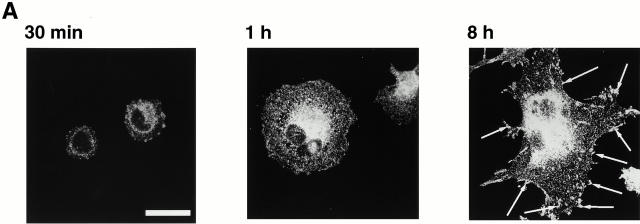
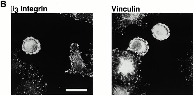
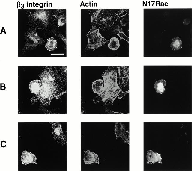
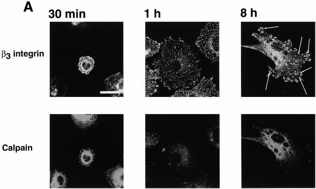
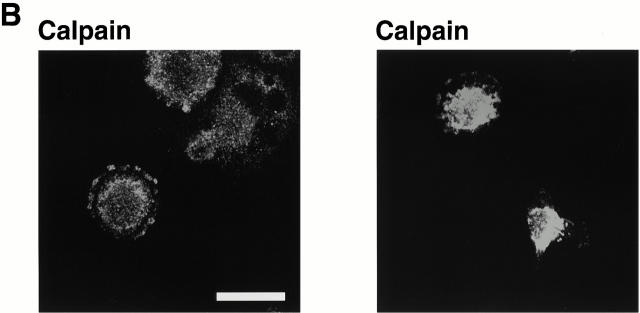
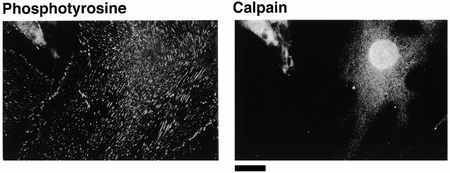
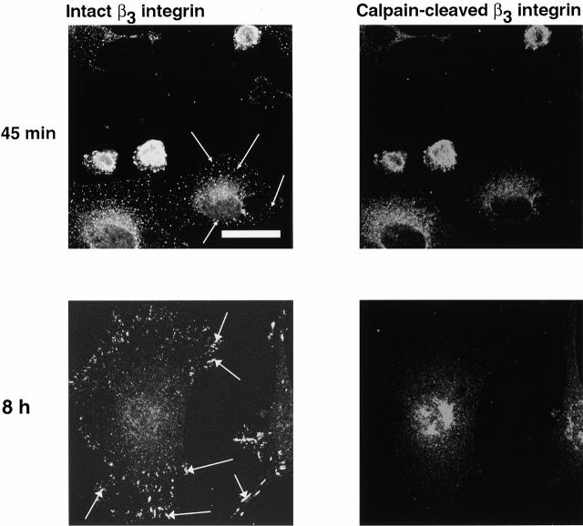
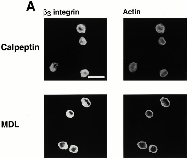
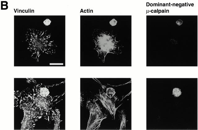
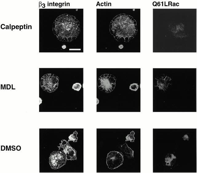
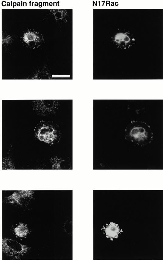
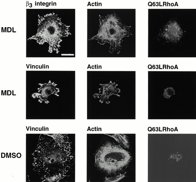
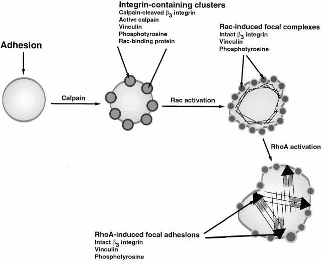
Interaction of integrins with the extracellular matrix leads to transmission of signals, cytoskeletal reorganizations, and changes in cell behavior. While many signaling molecules are known to be activated within Rac-induced focal complexes or Rho-induced focal adhesions, the way in which integrin-mediated adhesion leads to activation of Rac and Rho is not known. In the present study, we identified clusters of integrin that formed upstream of Rac activation. These clusters contained a Rac-binding protein(s) and appeared to be involved in Rac activation. The integrin clusters contained calpain and calpain-cleaved beta3 integrin, while the focal complexes and focal adhesions that formed once Rac and Rho were activated did not. Moreover, the integrin clusters were dependent on calpain for their formation. In contrast, while Rac- and Rho-GTPases were dependent on calpain for their activation, formation of focal complexes and focal adhesions by constitutively active Rac or Rho, respectively, occurred even when calpain inhibitors were present. Taken together, these data are consistent with a model in which integrin-induced Rac activation requires the formation of integrin clusters. The clusters form in a calpain-dependent manner, contain calpain, calpain-cleaved integrin, and a Rac binding protein(s). Once Rac is activated, other integrin signaling complexes are formed by a calpain-independent mechanism(s).


Figures

Figure 1
Figure 1
Distribution of β3 integrin and vinculin in spreading BAE cells. BAE cells were allowed to spread on fibronectin-coated coverslips for 30 min, 1 h, or 8 h (A) or on vitronectin-coated coverslips for 30 min (B). Cells were fixed and permeabilized. Cells were stained with antibodies against β3 integrin or vinculin. Arrows in A indicate focal adhesions. Bars, 20 μm.
Figure 1
Figure 1
Distribution of β3 integrin and vinculin in spreading BAE cells. BAE cells were allowed to spread on fibronectin-coated coverslips for 30 min, 1 h, or 8 h (A) or on vitronectin-coated coverslips for 30 min (B). Cells were fixed and permeabilized. Cells were stained with antibodies against β3 integrin or vinculin. Arrows in A indicate focal adhesions. Bars, 20 μm.
Figure 2
Figure 2
β3 integrin-containing clusters form upstream of Rac activation. BAE cells were grown to 80% confluency and transiently transfected with plasmid encoding myc-tagged dominant-negative N17Rac. Cells were detached and replated on fibronectin-coated coverslips for 1 h (A and B) or on vitronectin-coated coverslips for 1 h (C), fixed, and permeabilized. Cells were stained with antibodies against β3 integrin. Transfected cells were detected with anti–myc antibody and actin filaments were detected with TRITC-labeled phalloidin. Bar, 20 μm.
Figure 3
Figure 3
Calpain is present in the integrin-containing clusters. BAE cells were allowed to spread on fibronectin-coated coverslips for 30 min, 1 h, or 8 h, fixed, and permeabilized. (A) Cells were stained with antibody against β3 integrin and antibody against calpain. (Right) Arrows indicate focal adhesions. (B) Two additional fields were stained only for calpain. Bars, 20 μm.
Figure 3
Figure 3
Calpain is present in the integrin-containing clusters. BAE cells were allowed to spread on fibronectin-coated coverslips for 30 min, 1 h, or 8 h, fixed, and permeabilized. (A) Cells were stained with antibody against β3 integrin and antibody against calpain. (Right) Arrows indicate focal adhesions. (B) Two additional fields were stained only for calpain. Bars, 20 μm.
Figure 4
Figure 4
Calpain is absent from the focal adhesions of BAE cells overexpressing μ-calpain. BAE cells overexpressing μ-calpain were allowed to spread on fibronectin-coated coverslips for 24 h, fixed, and permeabilized. Cells were stained with antibodies against phosphotyrosine and calpain. Bar, 20 μm.
Figure 6
Figure 6
Calpain-cleaved β3 integrin is present in the integrin-containing clusters. BAE cells were allowed to spread on fibronectin-coated coverslips for 45 min or 8 h, fixed, and permeabilized. Cells were stained with antibody against αvβ3 integrin and antibodies against calpain-cleaved β3 integrin. Small arrowheads indicate focal complexes and large arrowheads indicate focal adhesions. Bar, 20 μm.
Figure 7
Figure 7
Calpain inhibitors and dominant-negative μ-calpain prevent formation of integrin-containing clusters. (A) BAE cells were detached and replated on fibronectin-coated coverslips in the presence of calpeptin (100 μg/ml) or MDL (250 μM) for 1 h, fixed, and permeabilized. Cells were stained with antibodies against β3 integrin, actin filaments were detected with TRITC-labeled phalloidin. (B) BAE cells were grown to 80% confluency and transiently transfected with plasmid encoding HA-tagged dominant-negative μ-calpain. Cells were detached and replated on fibronectin-coated coverslips for 1–3 h, fixed, and permeabilized. Cells were stained with antibodies against vinculin. Transfected cells were detected with HA-myc antibody and actin filaments were detected with TRITC-labeled phalloidin. Two independent fields are shown. Bar, 20 μm.
Figure 7
Figure 7
Calpain inhibitors and dominant-negative μ-calpain prevent formation of integrin-containing clusters. (A) BAE cells were detached and replated on fibronectin-coated coverslips in the presence of calpeptin (100 μg/ml) or MDL (250 μM) for 1 h, fixed, and permeabilized. Cells were stained with antibodies against β3 integrin, actin filaments were detected with TRITC-labeled phalloidin. (B) BAE cells were grown to 80% confluency and transiently transfected with plasmid encoding HA-tagged dominant-negative μ-calpain. Cells were detached and replated on fibronectin-coated coverslips for 1–3 h, fixed, and permeabilized. Cells were stained with antibodies against vinculin. Transfected cells were detected with HA-myc antibody and actin filaments were detected with TRITC-labeled phalloidin. Two independent fields are shown. Bar, 20 μm.
Figure 8
Figure 8
Rac-induced focal complexes contain β3 integrin and vinculin and form by a mechanism that does not involve calpain. BAE cells were grown to 80% confluency and transiently transfected with plasmid encoding HA-tagged constitutively active Q61LRac. Cells were detached and replated on fibronectin-coated coverslips in the presence of calpeptin (100 μg/ml), MDL (250 μM), or corresponding volume of DMSO for 1 h, fixed, and permeabilized. Cells were stained with antibodies against β3 integrin. Transfected cells were detected with anti–HA epitope antibody, actin filaments were detected with TRITC-labeled phalloidin. Bar, 20 μm.
Figure 5
Figure 5
Autolytic fragment of calpain is present in the integrin-containing clusters, which form upstream of Rac activation. BAE cells were transiently transfected with cDNA encoding myc-tagged dominant-negative N17Rac. Cells were detached and replated on fibronectin-coated coverslips for 1 h, fixed, and permeabilized. Transfected cells were detected with anti–myc antibody and autolysed calpain with an antibody that only recognizes the autolysed fragment. Three different transfected cells are shown. Bar, 20 μm.
Figure 9
Figure 9
RhoA-induced focal adhesions contain β3 integrin and vinculin and form by a mechanism that does not require calpain. BAE cells were grown to 80% confluency and transiently transfected with plasmid encoding constitutively active Q63LRhoA. Cells were detached and replated on fibronectin-coated coverslips in the presence of MDL (250 μM) or DMSO for 1 h, fixed, and permeabilized. Cells were stained with antibodies against β3 integrin or vinculin. Transfected cells were detected with anti–HA epitope antibody, actin filaments were detected with TRITC-labeled phalloidin. Bar, 20 μm.
Figure 10
Figure 10
Schematic representation of the formation of integrin-containing clusters, focal complexes, and focal adhesions. In this model, integrin engagement leads to calpain activation and formation of transient integrin-containing clusters. The clusters contain calpain-cleaved β3 integrin, active calpain, vinculin, and phosphotyrosine. Recruitment of Rac to the clusters leads to its activation and subsequent formation of Rac-induced focal complexes. These complexes do not require calpain activation for their formation, and they appear to have a different composition than the initial integrin clusters. Rac activation is followed by RhoA activation and formation of arrowhead-shaped focal adhesions, which also form by a mechanism that is independent of calpain activation.

References

    1. Banno Y., Nakashima S., Hachiya T., Nozawa Y. Endogenous cleavage of phospholipase C-b3 by agonist-induced activation of calpain in human platelets. J. Biol. Chem. 1995;270:4318–4324. - PubMed
    1. Beckerle M.C., Burridge K., DeMartino G.N., Croall D.E. Colocalization of calcium-dependent protease II and one of its substrates at sites of cell adhesion. Cell. 1987;51:569–577. - PubMed
    1. Boguski M.S., McCormick F. Proteins regulating Ras and its relatives. Nature. 1993;366:643–654. - PubMed
    1. Bokoch G.M., Der C.J. Emerging concepts in the Ras superfamily of GTP-binding proteins. FASEB J. 1993;7:750–759. - PubMed
    1. Bollag G., McCormick F. Regulators and effectors of ras proteins. Annu. Rev. Cell Biol. 1991;7:601–632. - PubMed

Publication types

MeSH terms