. 2000 Oct 30;151(3):709-18.
doi: 10.1083/jcb.151.3.709.

Chlamydomonas IFT88 and its mouse homologue, polycystic kidney disease gene tg737, are required for assembly of cilia and flagella

Affiliations

Chlamydomonas IFT88 and its mouse homologue, polycystic kidney disease gene tg737, are required for assembly of cilia and flagella

G J Pazour et al. J Cell Biol. .

Abstract

Intraflagellar transport (IFT) is a rapid movement of multi-subunit protein particles along flagellar microtubules and is required for assembly and maintenance of eukaryotic flagella. We cloned and sequenced a Chlamydomonas cDNA encoding the IFT88 subunit of the IFT particle and identified a Chlamydomonas insertional mutant that is missing this gene. The phenotype of this mutant is normal except for the complete absence of flagella. IFT88 is homologous to mouse and human genes called Tg737. Mice with defects in Tg737 die shortly after birth from polycystic kidney disease. We show that the primary cilia in the kidney of Tg737 mutant mice are shorter than normal. This indicates that IFT is important for primary cilia assembly in mammals. It is likely that primary cilia have an important function in the kidney and that defects in their assembly can lead to polycystic kidney disease.


Figures
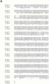
Figure 1
Figure 1
Sequence and structure of the Chlamydomonas IFT88 protein. (a) Chlamydomonas IFT88 is homologous to the mouse and human Tg737 proteins. The sequences of the Chlamydomonas (Cr_IFT88, accession number AF298884), mouse (Mm_Tg737, accession number AAB59705), and human (Hs_Tg737, accession number AAA86720) proteins were aligned using ClustalW (Thompson et al. 1994). (*) Complete conservation; :, highly conserved substitutions; and (.) semiconservative substitutions (below the alignment). (b) IFT88 contains 10 TPR repeats. Residues matching the TPR consensus sequence (bottom) are indicated by bold font. (c) The 10 TPR repeats (shaded boxes) are organized in a group of three in the NH2-terminal half of the protein and a group of seven in the COOH-terminal half of the protein.
Figure 1
Figure 1
Sequence and structure of the Chlamydomonas IFT88 protein. (a) Chlamydomonas IFT88 is homologous to the mouse and human Tg737 proteins. The sequences of the Chlamydomonas (Cr_IFT88, accession number AF298884), mouse (Mm_Tg737, accession number AAB59705), and human (Hs_Tg737, accession number AAA86720) proteins were aligned using ClustalW (Thompson et al. 1994). (*) Complete conservation; :, highly conserved substitutions; and (.) semiconservative substitutions (below the alignment). (b) IFT88 contains 10 TPR repeats. Residues matching the TPR consensus sequence (bottom) are indicated by bold font. (c) The 10 TPR repeats (shaded boxes) are organized in a group of three in the NH2-terminal half of the protein and a group of seven in the COOH-terminal half of the protein.
Figure 2
Figure 2
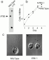
Phenotype of the ift88-1 mutant cells. (a) Southern blot showing that the V79 cell line has an insertion in the IFT88 gene. DNA was isolated from wild-type and V79 cells, digested with PstI, and analyzed by Southern blotting with a 365-bp fragment of IFT88 genomic DNA as a probe. The single band in wild-type cells is split into two bands in the mutant. (b) Disruption of IFT88 does not affect growth rate. Wild-type and ift88-1 mutant cells were grown in liquid culture and monitored as described in Pazour et al. 1998. (c) In contrast to wild-type cells that have two ∼10-μm-long flagella, flagella are not formed on ift88-1 cells. Cells were recorded by differential interference contrast microscopy as described in Pazour et al. 1998.
Figure 3
Figure 3
Ultrastructure of the ift88-1 flagella. The flagella on ift88-1 mutant cells are very short and the microtubules do not extend beyond the transition zone (arrows). The microtubules in wild-type cells start at the basal body, extend through the transition zone, and continue on to the flagellar tip. The wild-type flagellum shown here leaves the plane of section shortly after passing through the cell wall.
Figure 4
Figure 4
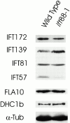
Western blot showing the effect of the ift88-1 mutation on the levels of IFT motor and particle proteins. Equal amounts of whole-cell extracts of ift88-1 (3276.2) and wild-type cells (CC124) were separated by SDS-PAGE, transferred to membrane and probed with antibodies to IFT particle proteins (IFT172, IFT139, IFT81, IFT57) and IFT motor proteins (FLA10, DHC1b). An antibody to α-tubulin (α-Tub) was used to confirm that equivalent amounts of wild-type and ift88-1 protein extracts were loaded on the gel.
Figure 5
Figure 5
Presence of the IFT88 gene correlates with the wild-type phenotype in meiotic products. Strain V79 was transformed with a BAC clone containing the IFT88 gene. Transformed cells recovered the ability to swim and were enriched by taking inoculi from the top part of an unmixed culture. After enrichment, a pure culture of one of the transformants (V79/40-B3#2.5) was isolated and mated to a wild-type cell line (CC124) of the opposite mating type. Tetrads were dissected and the offspring scored for motility by light microscopy. DNA was isolated from the parents, the four products of one tetrad, and random single products of 10 additional tetrads. The DNA was analyzed by Southern blotting using a 386-bp fragment of IFT88 genomic DNA as a probe. Cells that swam normally carried at least one copy of the wild-type gene, whereas the nonmotile cells did not carry a copy of the wild-type gene.
Figure 6
Figure 6
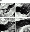
Primary cilia in the kidney of Tg737 mutant mice are shorter than normal. (a) Kidneys from 4-d-old pups were fixed with glutaraldehyde, freeze fractured, metal impregnated, and examined by scanning electron microscopy. Numerous cilia were found on the epithelial cells in the tubules and collecting ducts of the wild-type mice (+/+). Cilia were also found in the homozygous mutant (−/−) pups, but they were usually <2-μm long and most were only short stubs. (b) Cilia length in kidneys of 4-d-old mice. Cilia were measured in scanning electron micrographs of tubules located distal to the proximal tubule. Wild-type cilia averaged 3.1 ± 1.4 μm (n = 50), whereas the mutant cilia averaged 1.0 ± 0.6 μm (n = 50). (c) Cilia length in kidneys of 7-d-old mice. The cilia lengths were measured in scanning electron micrographs of tubules located distal to the proximal tubule. Wild-type cilia averaged 3.5 ± 1.7 μm (n = 50), whereas the mutant cilia averaged 1.3 ± 0.6 μm (n = 50).
Figure 6
Figure 6
Primary cilia in the kidney of Tg737 mutant mice are shorter than normal. (a) Kidneys from 4-d-old pups were fixed with glutaraldehyde, freeze fractured, metal impregnated, and examined by scanning electron microscopy. Numerous cilia were found on the epithelial cells in the tubules and collecting ducts of the wild-type mice (+/+). Cilia were also found in the homozygous mutant (−/−) pups, but they were usually <2-μm long and most were only short stubs. (b) Cilia length in kidneys of 4-d-old mice. Cilia were measured in scanning electron micrographs of tubules located distal to the proximal tubule. Wild-type cilia averaged 3.1 ± 1.4 μm (n = 50), whereas the mutant cilia averaged 1.0 ± 0.6 μm (n = 50). (c) Cilia length in kidneys of 7-d-old mice. The cilia lengths were measured in scanning electron micrographs of tubules located distal to the proximal tubule. Wild-type cilia averaged 3.5 ± 1.7 μm (n = 50), whereas the mutant cilia averaged 1.3 ± 0.6 μm (n = 50).

Similar articles

Cited by

References

    1. Afzelius B.A. The immotile-cilia syndrome and other ciliary diseases. Int. Rev. Exp. Pathol. 1979;19:1–43. - PubMed
    1. Andrews P.M., Porter K.R. A scanning electron microscopic study of the nephron. Am. J. Anat. 1974;140:81–116. - PubMed
    1. Baccetti B., Burrini A.G., Capitani S., Collodel G., Moretti E., Piomboni P., Renieri T. Notulae seminologicae. 2. The ‘short tail’ and ‘stump’ defect in human spermatozoa. Andrologia. 1993;25:331–335. - PubMed
    1. Barr M.M., Sternberg P.W. A polycistic kidney-disease gene homologue required for male mating behaviour in C. elegans . Nature. 1999;401:386–389. - PubMed
    1. Beech P.L., Pagh-Roehl K., Noda Y., Hirokawa N., Burnside B., Rosenbaum J.L. Localization of kinesin superfamily proteins to the connecting cilium of fish photoreceptors. J. Cell Sci. 1996;109:889–897. - PubMed

Publication types

MeSH terms