. 2000 Oct;33(5):317-29.
doi: 10.1046/j.1365-2184.2000.00177.x.

A quantitative model for differential motility of gliomas in grey and white matter

Affiliations

A quantitative model for differential motility of gliomas in grey and white matter

K R Swanson et al. Cell Prolif. 2000 Oct.

Abstract

We have extended a mathematical model of gliomas based on proliferation and diffusion rates to incorporate the effects of augmented cell motility in white matter as compared to grey matter. Using a detailed mapping of the white and grey matter in the brain developed for a MRI simulator, we have been able to simulate model tumours on an anatomically accurate brain domain. Our simulations show good agreement with clinically observed tumour geometries and suggest paths of submicroscopic tumour invasion not detectable on CT or MRI images. We expect this model to give insight into microscopic and submicroscopic invasion of the human brain by glioma cells. This method gives insight in microscopic and submicroscopic invasion of the human brain by glioma cells. Additionally, the model can be useful in defining expected pathways of invasion by glioma cells and thereby identify regions of the brain on which to focus treatments.


Figures
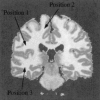
Figure 1
Figure 1
Computational domain with tumour locations. Grey matter appears grey and white matter appears white. Position 1 is the location of an inferior fronto‐parietal tumour, position 2 is the location for a superior fronto‐parietal tumour, position 3 is the location for a temporal lobe tumour.
Figure 2
Figure 2
Simulation of tumour invasion of a high‐grade glioma in position 2 in the superior cerebral hemisphere: (a) (b) at diagnosis; (c) (d) at death; (a) (c) as seen by our standard threshold of detection; (b) (d) as calculated out to 1.25% of the threshold (boundary) cell concentration defining the sensitive threshold of detection.
Figure 3
Figure 3
Simulation of tumour invasion of a high‐grade glioma in position 1 in the inferior cerebral hemisphere: (a) (b) at diagnosis; (c) (d) at death; (a) (c) as seen by our standard threshold of detection; (b) (d) as calculated out to 1.25% of the threshold (boundary) cell concentration defining the sensitive threshold of detection.
Figure 4
Figure 4
Simulation of tumour invasion of a high‐grade glioma in position 3 in the temporal lobe: (a) (b) at diagnosis; (c) (d) at death; (a) (c) as seen by our standard threshold of detection; (b) (d) as calculated out to 1.25% of the threshold (boundary) cell concentration defining the sensitive threshold of detection.)
Figure 5
Figure 5
Percentage of position 1 tumour detectable by our standard threshold of detection at diagnosis vs. the growth rate ρ and the diffusion coefficient in grey matter Dg. Note that the greatest proportion of detectable tumour occurs with high growth rate and low diffusion coefficient; conversely, the least proportion is seen with gliomas having low growth rate and high diffusion coefficient.
Figure 6
Figure 6
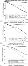
Percentage of detectable volume of tumour vs. the threshold of detection at various positions. The 40 000 cells/cm2 represents our standard, the threshold of detection for an enhanced CT scan.

References

    1. Alvord EC Jr, Shaw CM (1991) Neoplasms affecting the nervous system in the elderly In: Duckett S. ed. The Pathology of the Aging Human Nervous System,210–281. Philadelphia : Lea & Febiger.
    1. Blankenberg FG, Teplitz RL, Ellis W et al. (1995) The influence of volume tric tumour doubling time, DNA ploidy, and histologic grade on the survival of patients with intrcranial astrocytomas. Am. J. Neuro Rad 16,1001–1012. - PMC - PubMed
    1. Burgess PK, Kulesa PM, Murray JD, Alvord EC Jr (1997) The interaction of growth rates and diffusion coefficients in a three‐dimensional mathematical model of gliomas. J. Neuropath Exp Neuro 56,704–713. - PubMed
    1. Chicoine MR, Silbergeld DL (1995) Assessment of brain tumour cell motility in vivo and in vitro. J. Neurosurg 82,615–622. - PubMed
    1. Cocosco CA, Kollokian V, Kwan RK‐S, Evans AC (1997) Brainweb: Online interface to a 3D MRI simulated brain database. Neuroimage 5,S425.

Publication types