. 2000 Nov;106(9):1115-26.
doi: 10.1172/JCI9594.

Nitric oxide and atrial natriuretic factor stimulate cGMP-dependent membrane insertion of aquaporin 2 in renal epithelial cells

Affiliations

Nitric oxide and atrial natriuretic factor stimulate cGMP-dependent membrane insertion of aquaporin 2 in renal epithelial cells

R Bouley et al. J Clin Invest. 2000 Nov.

Abstract

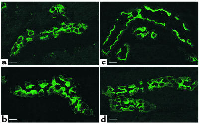
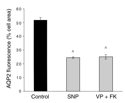
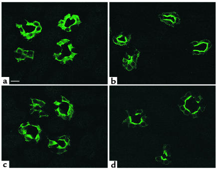
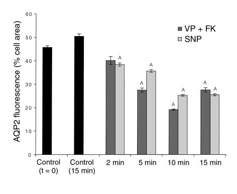
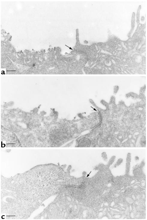
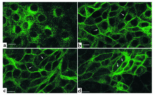
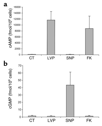
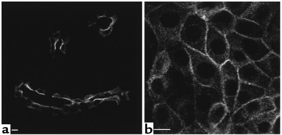
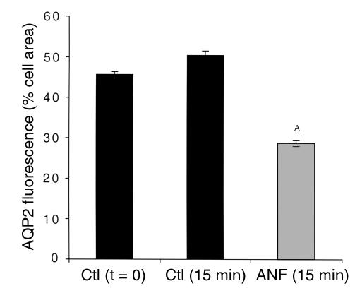
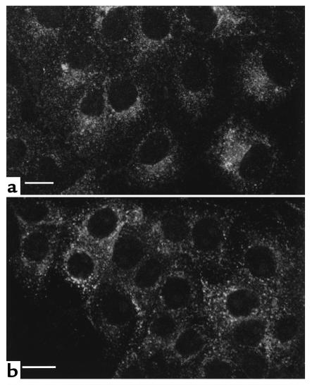
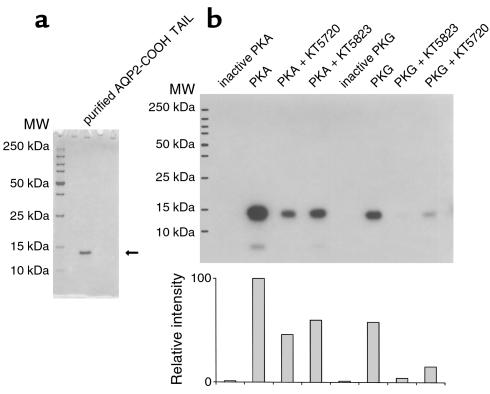
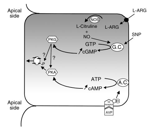
In collecting duct principal cells, aquaporin 2 (AQP2) is shuttled from intracellular vesicles to the plasma membrane upon vasopressin (VP) stimulation. VP activates adenylyl cyclase, increases intracellular cAMP, activating protein kinase A (PKA) to phosphorylate AQP2 on the COOH-terminal residue, serine 256. Using rat kidney slices and LLC-PK1 cells stably expressing AQP2 (LLC-AQP2 cells), we now show that AQP2 trafficking can be stimulated by cAMP-independent pathways. In these systems, the nitric oxide (NO) donors sodium nitroprusside (SNP) and NONOate and the NO synthase substrate L-arginine mimicked the effect of VP, stimulating relocation of AQP2 from cytoplasmic vesicles to the plasma membrane. Unlike VP, these other agents did not increase intracellular cAMP. However, SNP increased intracellular cGMP, and exogenous cGMP stimulated AQP2-membrane insertion. Atrial natriuretic factor, which signals via cGMP, also stimulated AQP2 translocation. The VP and SNP effects were blocked by the kinase inhibitor H89. SNP did not stimulate membrane insertion of AQP2 in LLC-PK1 cells expressing the phosphorylation-deficient mutant 256SerAla-AQP2, indicating that phosphorylation of Ser256 is required for signaling. Both PKA and cGMP-dependent protein kinase G phosphorylated AQP2 on this COOH-terminal residue in vitro. These results demonstrate a novel, cAMP-independent and cGMP-dependent pathway for AQP2 membrane insertion in renal epithelial cells.


Figures

Figure 1
Figure 1
Confocal microscope images of tissue slices showing AQP2 in principal cells from inner stripe (IS) collecting ducts. A kidney from one rat was cut into thin slices and incubated in vitro with or without agonists for 15 minutes before fixation by immersion, sectioning, and immunostaining to detect AQP2. (a) Control, buffer alone; (b) 10 nM VP plus 10 μM forskolin; (c) 1 mM SNP; (d) 10 mM L-arginine. Under control conditions (a), AQP2 is located in numerous intracellular vesicles and shows no apparent membrane staining. AVP plus forskolin, SNP, and L-arginine all induce a marked redistribution of AQP2 to the apical plasma membrane. Intercalated cells in the collecting duct epithelium are unstained. This result is representative of three sets of experiments using a different animal each time. Bar, 10 μm.
Figure 2
Figure 2
Quantification of the effect of VP plus forskolin and SNP on AQP2 redistribution in collecting duct (inner stripe) principal cells. Total principal cell area (excluding the nucleus) and the area occupied by AQP2 fluorescence were determined using IP Lab Spectrum software on confocal microscope images as in Figure 1. Results are expressed as the percentage of total cell area occupied by AQP2 fluorescence. VP plus forskolin and SNP both induce a similar and marked reduction in the area of AQP2 fluorescence, indicating that these agonists cause the AQP2 staining to become concentrated in a smaller region of the cell, i. e., the apical pole. AP < 0.05 compared with the control value. FK, forskolin.
Figure 3
Figure 3
Confocal images showing the time course of VP/forskolin–induced and SNP-induced (10 μM) translocation of AQP2 to the apical plasma membrane in collecting duct (inner stripe) principal cells. (a) Collecting duct stained 2 minutes after addition of VP/forskolin; (b) stained 10 minutes after addition of VP/forskolin; (c) stained 2 minutes after addition of SNP; (d) stained 10 minutes after addition of SNP. AQP2 is distributed intracellularly before agonist addition, but shows a progressive translocation to the apical cell surface during incubation with VP/forskolin and SNP (see Figure 4 for a quantification of the time course of agonist-induced AQP2 translocation). Some apical staining is already detectable 2 minutes after agonist addition, but a significant amount of intracellular staining is still present at this early time point (a, c). The maximum effect is seen after 10 minutes of treatment, when most of the staining is located on the apical membrane (b, d). Bar, 10 μm.
Figure 4
Figure 4
Quantification of the time course of AQP2 translocation to the apical plasma membrane of principal cells after VP/forskolin and SNP stimulation. The quantification of AQP2-membrane insertion was performed in the same way as described in Figure 2. AQP2 was distributed throughout the cytoplasm before addition of agonists, and this distribution was slightly (but not significantly) more pronounced after a further 15 minutes of in vitro incubation in the absence of agonists. A redistribution of AQP2 was already detectable 2 minutes after VP/forskolin or SNP addition and became progressively more marked after 5 and 10 minutes of treatment. After 15 minutes, the SNP effect was similar to the 10-minute response, while the effect of VP/forskolin was somewhat less pronounced after 15 minutes of treatment, although the difference was not statistically significant compared with the 10-minute time point. AP < 0.05 compared with control values (controls, both t = 0 and t = 15 minutes).
Figure 5
Figure 5
Immunogold electron microscopy showing apical plasma membrane insertion of AQP2 induced by VP/forskolin and SNP treatment of kidney slices. AQP2 was localized in nonpermeabilized tissues with an Ab against an external epitope of AQP2. (a) SNP treatment for 15 minutes. Abundant gold particles, representing AQP2 antigenic sites, are located on the apical plasma membrane of a principal cell (left). An adjacent intercalated cell (right) is unlabeled. (b) Apical plasma membrane localization of gold labeling for AQP2 in a principal cell after VP/forskolin treatment. The adjacent intercalated cell is unlabeled. (c) Control tissue incubated for 15 minutes in the absence of agonist. Very few gold particles are seen on the apical membrane, indicating that most of the AQP2 is inside the cell in this condition, supporting the confocal data shown in Figures 1 and 3. In all figures, most of the gold particles are on the external surface of the apical membrane, consistent with the use of an Ab raised against an external epitope of AQP2. The position of the cell junction between the principal cell (left) and the intercalated cell (right) is indicated with an arrow in each figure. Bar, 1 μm.
Figure 6
Figure 6
Immunofluorescence localization of AQP2 in LLC-AQP2 cells showing that SNP, forskolin, and dibutyryl cGMP stimulate AQP2 translocation to the plasma membrane. In nonstimulated cells (a), the c-myc Ab stains many cytoplasmic vesicles, but the plasma membrane is virtually unstained. In contrast, AQP2 shows a plasma membrane localization (arrows) after incubation for 10 minutes with 1mM SNP (b), 10 μM forskolin (c), and cGMP (d). Bar, 5 μm.
Figure 7
Figure 7
Effect of VP and SNP treatment on intracellular levels of cAMP and cGMP in LLC-AQP2 cells. Cells were incubated for 10 minutes with 10 nM VP, 1 mM SNP, or 10 μM forskolin (FK). After incubation, the cells were solubilized and supernatants were used to measure the intracellular cAMP (a) and cGMP (b) using an ELISA assay. Each point represents the mean ± SEM of triplicate determinations. Similar data were obtained in two more separate experiments.
Figure 8
Figure 8
ANF stimulates plasma membrane insertion of AQP2 in kidney epithelial cells. Rat kidney slices and LLC-AQP2 cells were incubated with 10 μM ANF for 10 minutes, followed by localization of AQP2 by indirect immunofluorescence. Under these conditions, AQP2 was located in the apical plasma membrane of principal cells in inner-stripe collecting ducts (a) and in the basolateral plasma membrane of LLC-AQP2 cells in culture (b). Bar, 5 μm.
Figure 9
Figure 9
Quantification of the effect of ANF on AQP2 redistribution in collecting duct (inner stripe) principal cells. Total principal cell area (excluding the nucleus) and the area occupied by AQP2 fluorescence were determined using IP Lab Spectrum software on confocal microscope images such as those shown in Figure 8a. Results are expressed as the percentage of total cell area occupied by AQP2 fluorescence. ANF treatment (10 μM ANF for 10 minutes) induces a marked reduction in the area of AQP2 fluorescence, indicating it causes the AQP2 staining to become concentrated in a smaller region of the cell, i.e., the apical pole. AP < 0.05 compared with the control values. This ANF experiment was performed in parallel with the time course study shown in Figure 4, and the controls (t = 0 and t = 15 minutes) are, therefore, the same.
Figure 10
Figure 10
SNP does not induce AQP2 relocalization in LLC-AQP2 cells pretreated for 30 minutes with the PKA-inhibitor H89 (30 μM) or in cells expressing the S256A-mutant form of AQP2. Using Ab’s against the c-myc epitope tag on the AQP2 COOH-terminus, AQP2 was detected on perinuclear intracellular vesicles under basal conditions. This location did not change with SNP in H89 treated cells (a) or in cells expressing the S256A mutated form of AQP2 (b), in contrast to the effect of SNP in cells expressing the wild-type AQP2 (see Figure 6b). Bar, 5 μm.
Figure 11
Figure 11
In vitro phosphorylation of the AQP2 COOH tail by PKA and PKG. Purified AQP2 COOH tail was used as a substrate for the phosphorylation assay. The AQP2 COOH tail was purified and runs as a single band at 13 kDa on SDS-PAGE (a). Phosphorylation of this recombinant protein (b). It is phosphorylated by both PKA and PKG, and the phosphorylation is partially inhibited by 10 μM KT5720 and KT5823, respectively. However, these “specific” reagents also inhibit phosphorylation in crossover assays, although somewhat less effectively than when they are used to inhibit their “specific” target (PKA for KT5720 and PKG for KT5823). No AQP2 was phosphorylated when the PKA and PKG catalytic subunits were heat inactivated before inclusion in the assays (inactive PKA and inactive PKG). Neither PKA nor PKG phosphorylated the fusion protein in the absence of the AQP2 COOH tail (not shown). A quantification (by NIH Image software) of the degree of phosphorylation is shown in the lower panel of b.
Figure 12
Figure 12
In current models of VP action, adenylate cyclase is stimulated by the interaction of VP with its receptor (V2R), and intracellular cAMP increases. PKA is activated, resulting in AQP2 phosphorylation on serine 256, followed by AQP2 translocation from vesicles to the cell surface. Our present data suggest an alternative pathway of AQP2 translocation. SNP (and ANF) activates guanylate cyclase resulting in increased cytosolic cGMP and PKG activation. Whether PKG directly phosphorylates AQP2 in intact cells (as it can in vitro — see Figure 11), or whether it activates PKA (either directly or indirectly), which in turn phosphorylates AQP2, remains unknown.

References

    1. Gross SS, Wolin MS. Nitric oxide: pathophysiological mechanisms. Annu Rev Physiol. 1995;57:737–769. - PubMed
    1. Bredt DS, Snyder SH. Nitric oxide: a physiologic messenger molecule. Annu Rev Biochem. 1994;63:175–195. - PubMed
    1. Ahn KY, Mohaupt MG, Madsen KM, Kone BC. In situ hybridization localization of mRNA encoding inducible nitric oxide synthase in rat kidney. Am J Physiol. 1994;267:F748–F757. - PubMed
    1. Mohaupt MG, et al. Differential expression and induction of mRNAs encoding two inducible nitric oxide synthases in rat kidney. Kidney Int. 1994;46:653–665. - PubMed
    1. Terada Y, Tomita K, Nonoguchi H, Marumo F. Polymerase chain reaction localization of constitutive nitric oxide synthase and soluble guanylate cyclase messenger RNAs in microdissected rat nephron segments. J Clin Invest. 1992;90:659–665. - PMC - PubMed

Publication types

MeSH terms