. 2000 Nov;106(9):1175-84.
doi: 10.1172/JCI10902.

The bile acid synthetic gene 3beta-hydroxy-Delta(5)-C(27)-steroid oxidoreductase is mutated in progressive intrahepatic cholestasis

Affiliations

The bile acid synthetic gene 3beta-hydroxy-Delta(5)-C(27)-steroid oxidoreductase is mutated in progressive intrahepatic cholestasis

M Schwarz et al. J Clin Invest. 2000 Nov.

Abstract

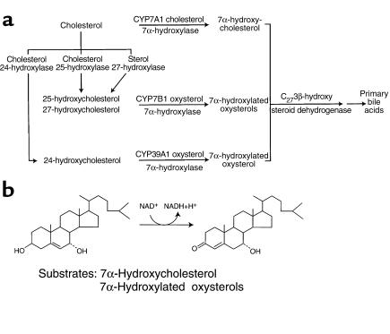
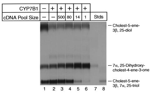
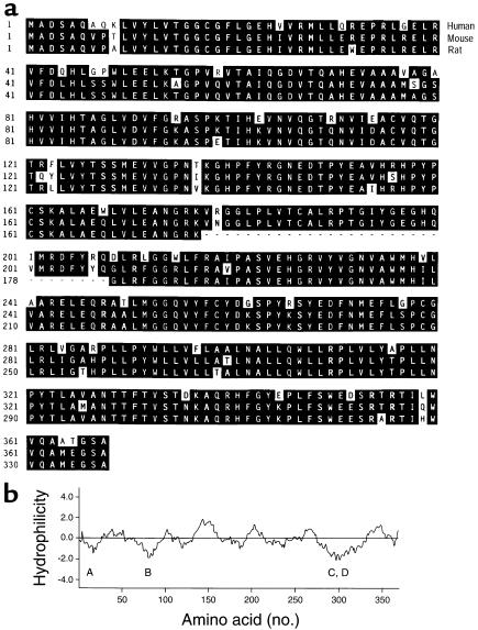
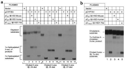
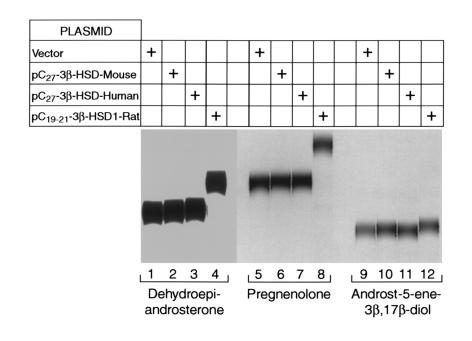
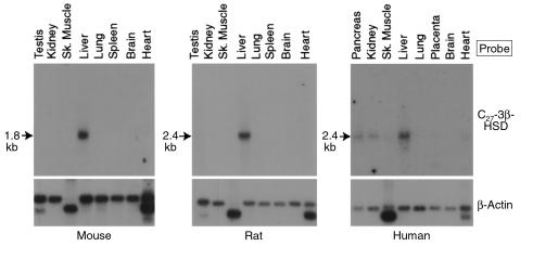
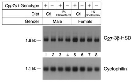
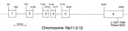
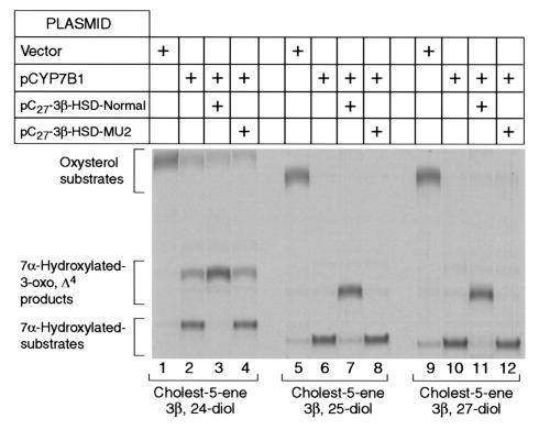
We used expression cloning to isolate cDNAs encoding a microsomal 3beta-hydroxy-Delta(5)-C(27)-steroid oxidoreductase (C(27) 3beta-HSD) that is expressed predominantly in the liver. The predicted product shares 34% sequence identity with the C(19) and C(21) 3beta-HSD enzymes, which participate in steroid hormone metabolism. When transfected into cultured cells, the cloned C(27) 3beta-HSD cDNA encodes an enzyme that is active against four 7alpha-hydroxylated sterols, indicating that a single C(27) 3beta-HSD enzyme can participate in all known pathways of bile acid synthesis. The expressed enzyme did not metabolize several different C(19/21) steroids as substrates. The levels of hepatic C(27) 3beta-HSD mRNA in the mouse are not sexually dimorphic and do not change in response to dietary cholesterol or to changes in bile acid pool size. The corresponding human gene on chromosome 16p11.2-12 contains six exons and spans 3 kb of DNA, and we identified a 2-bp deletion in the C27 3beta-HSD gene of a patient with neonatal progressive intrahepatic cholestasis. This mutation eliminates the activity of the enzyme in transfected cells. These findings establish the central role of C(27) 3beta-HSD in the biosynthesis of bile acids and provide molecular tools for the diagnosis of a third type of neonatal progressive intrahepatic cholestasis associated with impaired bile acid synthesis.


Figures

Figure 1
Figure 1
(a) Schematic showing branching in the early steps of the bile acid synthesis pathways. Individual enzymes and their catalyzed reactions are indicated. (b) The reaction catalyzed by the C27 3β-hydroxysteroid dehydrogenase isolated in this report is shown for the bile acid synthesis pathway involving cholesterol 7α-hydroxylase.
Figure 2
Figure 2
Expression cloning of mouse cDNAs encoding C27 3β-HSD enzyme activity. Cultured HEK 293 cells were cotransfected with a cDNA encoding the CYP7B1 oxysterol 7α-hydroxylase and pools of cDNAs derived from a mouse liver library. After 24 hours, the transfected cells were assayed for enzyme activity by thin-layer chromatography using [3H]cholest-5-ene-3β,25-diol (25-hydroxycholesterol, 1.2 μM) as substrate (see Methods). In lane 1, cells transfected with a control plasmid lacking a cDNA insert exhibited basal oxysterol 7α-hydroxylase activity, which converts 25-hydroxycholesterol into cholest-5-ene-3β,7α,25-triol, and low levels of C27 3β-HSD enzyme activity, which converts the product of the CYP7B1 enzyme into 7α,25-dihydroxy-cholest-4-ene-3-one. In lane 2, transfection with the CYP7B1 oxysterol 7α-hydroxylase cDNA alone increased the synthesis of cholest-5-ene-3β,7α,25-triol. In lane 3, transfection of a pool of 500 cDNAs containing a cDNA encoding C27 3β-HSD enzyme activity increased the synthesis of 7α,25-dihydroxy-cholest-4-ene-3-one. In lanes 4–6, transfection of progressively smaller (more enriched) pools containing the cDNA encoding the C27 3β-HSD activity resulted in a gradual increase in the synthesis of 7α,25-dihydroxy-cholest-4-ene-3-one and a decrease in the 7α-hydroxylated intermediate. In lanes 7 and 8 are radiolabeled sterol standards whose identities were confirmed previously by gas chromatography-mass spectrometry (12).
Figure 3
Figure 3
(a) Alignment of C27 3β-HSD enzyme sequences deduced from the human, mouse, and rat cDNAs. Identities between enzymes are shaded in black. Amino acids are numbered on the left. A sequence of 31 amino acids present in the human and mouse enzymes but absent in that of the rat is indicated by dashes. The GenBank/EBI Data Bank accession numbers for the human, mouse, and rat sequences are AF277719, AF277718, and BAA22931, respectively. (b) A hydropathy plot was generated for the amino acid sequence of the human C27 3β-HSD using the Kyte-Doolittle algorithm. The window size was 17 amino acids. Hydrophobic sequences fall below the central dividing line, and hydrophilic sequences rise above this line. Letters below the plot indicate four putative transmembrane domains.
Figure 4
Figure 4
Substrate specificities of the human and mouse C27 3β-HSD enzymes. Cultured HEK 293 cells were transfected with the indicated cDNA-expression plasmids and assayed for 3β-HSD enzyme activity using different radiolabeled sterol intermediates of the bile acid synthesis pathways. In (a), intermediates arising in the oxysterol 7α-hydroxylase pathways were added to cells at a final concentration of 3 μM, and the incubations continued for 24 hours: lanes 1–5, results obtained with [3H]cholest-5-ene-3β,24-diol (24-hydroxycholesterol); lanes 6–10, [3H]cholest-5-ene-3β,25-diol (25-hydroxycholesterol); and lanes 11–15, [3H]cholest-5-ene-3β,27-diol (27-hydroxycholesterol). In (b), [14C]cholesterol was added to cyclodextrin-treated cells at a final concentration of 3 μM, and the incubations continued for 24 hours. The positions to which sterols of known structure migrated on the thin-layer chromatograms are shown on the left.
Figure 5
Figure 5
Activities of different 3β-HSD enzymes against C19/21 steroid substrates. Cultured HEK 293 cells were transfected with the indicated cDNA expression plasmids and assayed for 3β-HSD enzyme activity using different radiolabeled intermediates of steroid hormone metabolism. In lanes 1–4, [14C]dehydroepiandrosterone (C19) was added to cells at a concentration of 3 μM and the incubation continued for 24 hours. In lanes 5–8, [3H]pregnenolone (C21) was added, and in lanes 9–12, [3H]androst-5-ene-3β, 17β-diol (C19) was added.
Figure 6
Figure 6
Tissue distribution of C27 3β-HSD mRNAs in the mouse, rat, and human. Aliquots (2 μg) of poly(A)+-enriched mRNA from the indicated tissues and species were size fractionated by agarose-gel electrophoresis, transferred to nylon membranes, and subjected to blot hybridization using radiolabeled probes derived from the mouse (mouse and rat membranes) or human (human membrane) C27 3β-HSD cDNAs. After washing, the mouse, rat, and human filters were exposed to x-ray film for 72, 16, and 168 hours, respectively. The size of the C27 3β-HSD mRNA detected in each experiment is shown to the left of the autoradiograms. To ensure that mRNA was present in each lane of the individual membranes, they were stripped of radioactivity after the initial hybridization and rehybridized with a [32P]-labeled probe derived from a human β-actin cDNA. The filters were then washed and exposed to x-ray film. The resulting autoradiograms, with signals in each lane, are shown below those derived with the C27 3β-HSD cDNA probes.
Figure 7
Figure 7
Expression of C27 3β-HSD mRNA in wild-type and cholesterol 7α-hydroxylase (Cyp7a1) knockout mice fed low- and high-cholesterol diets. Poly(A)+-enriched mRNA was isolated from the livers (n = 5) of male and female wild-type mice (+) or cholesterol 7α-hydroxylase (Cyp7a1) knockout mice () fed normal chow containing 0.02% (wt/wt) cholesterol (Ctl) or chow supplemented with 1% (wt/wt) cholesterol for 21 days. Aliquots (5 μg) of this RNA were size fractionated with agarose-gel electrophoresis, transferred to nylon membranes, and subjected to blot hybridization using a radiolabeled probe derived from the mouse C27 3β-HSD cDNA. After washing, the filter was exposed to x-ray film for 72 hours. The location to which the C27 3β-HSD mRNA migrated in the experiment and its interpolated size (1.8 kb) are shown on the left of the autoradiogram. To ensure that equal amounts of mRNA were present in each lane of the membrane, it was stripped of radioactivity after the initial hybridization and rehybridized with a [32P]-labeled probe derived from a rat cyclophilin cDNA. The filter was washed and exposed to x-ray film. The resulting autoradiogram, with a signal of equal intensity in each lane, is shown below that derived with the C27 3β-HSD cDNA probe. The calculated size of the cyclophilin mRNA (1.1 kb) is shown to the left of the autoradiogram.
Figure 8
Figure 8
Structure of the human C27 3β-HSD gene. A schematic of the gene is shown drawn to scale with six exons indicated by numbered boxes and five introns indicated by connecting lines. Individual amino acids occurring at the exon-intron junctions are shown in single letter code above the gene. The location of a 2-bp deletion (Δ1057-1058) found in homozygous form in the DNA of a patient (MU2) with progressive intrahepatic cholestasis is shown in exon 6 below the gene. Radiation hybrid panel–mapping experiments indicated that the human C27 3β-HSD gene is located on chromosome 16p11.2-12 (see Methods).
Figure 9
Figure 9
Expression of normal and mutant C27 3β-HSD genes in cultured HEK 293 cells. The C27 3β-HSD gene was amplified using PCR from the genomic DNA of either a normal subject (Normal) or an individual with intrahepatic cholestasis (MU2) and inserted into the plasmid pCMV6 as described in Methods. The resulting expression vectors were introduced together with a plasmid expressing the mouse CYP7B1 oxysterol 7β-hydroxylase cDNA (pCYP7B1) into HEK 293 cells by cotransfection. After 24 hours, the ability of the transfected cells to convert [3H]cholest-5-ene-3β,24-diol (24-hydroxycholesterol, lanes 1–4), [3H]cholest-5-ene-3β,25-diol (25-hydroxycholesterol, lanes 5–8), and [3H]cholest-5-ene-3β,27-diol (27-hydroxycholesterol, lanes 9–12) was determined by thin-layer chromatography. The chromatogram was exposed to x-ray film for 6 days.

References

    1. Russell DW, Setchell KDR. Bile acid biosynthesis. Biochemistry. 1992;31:4737–4749. - PubMed
    1. Gourley, G.R. 1994. Bilirubin metabolism and neonatal jaundice. In Liver disease in children. F.J. Suchy, editor. Mosby. St. Louis, Missouri, USA. 105–125.
    1. Clayton PT, et al. Familial giant cell hepatitis associated with synthesis of 3β,7α-dihydroxy- and 3β,7α,12α-trihydroxy-5-cholenoic acids. J Clin Invest. 1987;79:1031–1038. - PMC - PubMed
    1. Setchell KD, et al. Δ4-3-oxosteroid 5β-reductase deficiency described in identical twins with neontal hepatitis. A new inborn error in bile acid synthesis. J Clin Invest. 1988;82:2148–2157. - PMC - PubMed
    1. Setchell KDR, et al. Identification of a new inborn error in bile acid synthesis: mutation of the oxysterol 7α-hydroxylase gene causes severe neonatal liver disease. J Clin Invest. 1998;102:1690–1703. - PMC - PubMed

Publication types

MeSH terms