. 2000 Nov 6;192(9):1213-22.
doi: 10.1084/jem.192.9.1213.

Induction of interleukin 10-producing, nonproliferating CD4(+) T cells with regulatory properties by repetitive stimulation with allogeneic immature human dendritic cells

Affiliations

Induction of interleukin 10-producing, nonproliferating CD4(+) T cells with regulatory properties by repetitive stimulation with allogeneic immature human dendritic cells

H Jonuleit et al. J Exp Med. .

Abstract

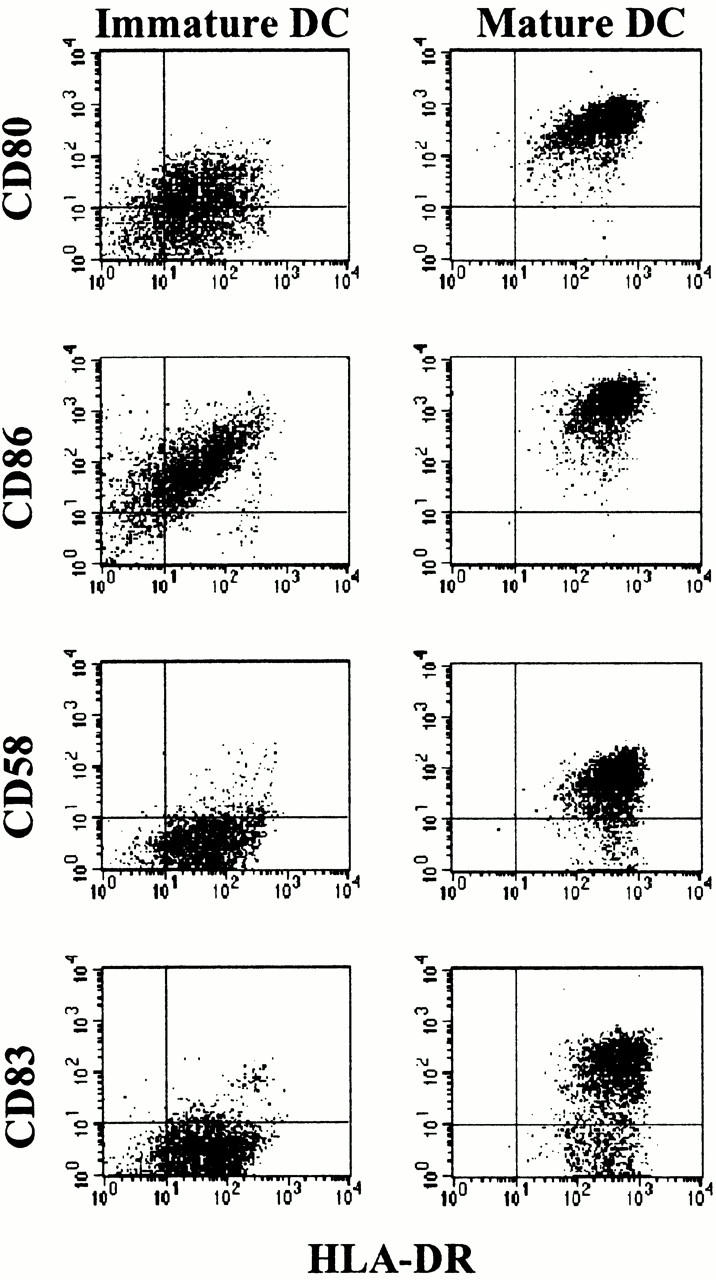
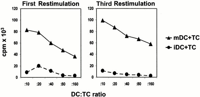
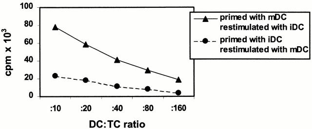
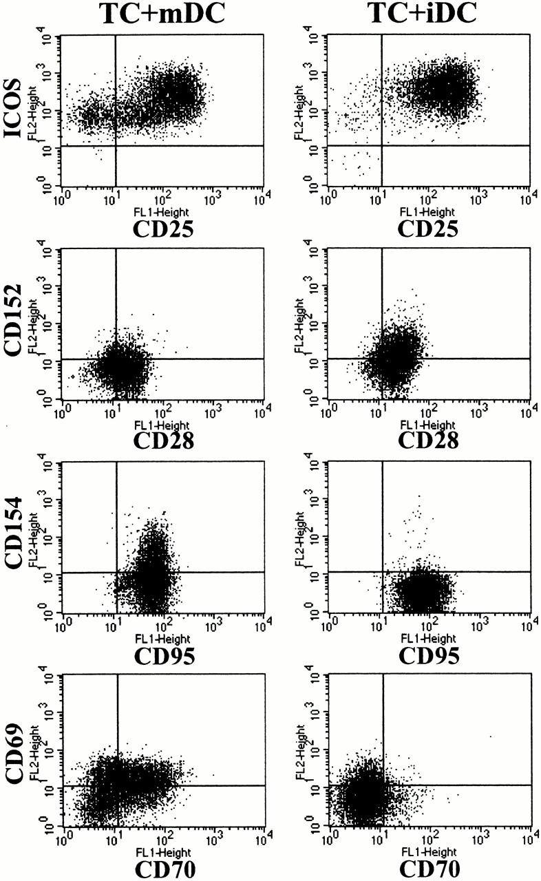
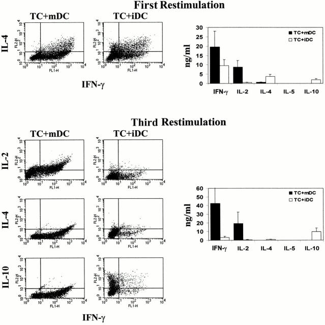
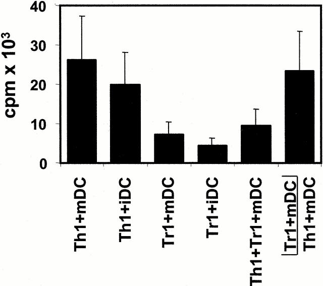
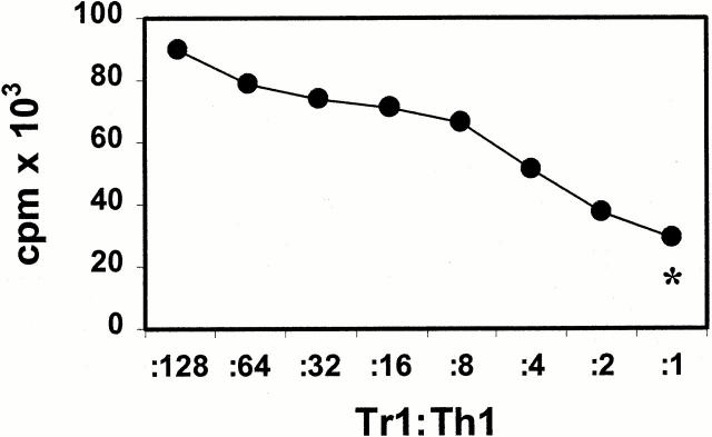
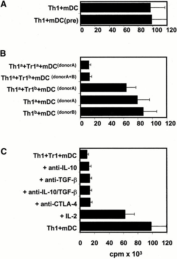
The functional properties of dendritic cells (DCs) are strictly dependent on their maturational state. To analyze the influence of the maturational state of DCs on priming and differentiation of T cells, immature CD83(-) and mature CD83(+) human DCs were used for stimulation of naive, allogeneic CD4(+) T cells. Repetitive stimulation with mature DCs resulted in a strong expansion of alloreactive T cells and the exclusive development of T helper type 1 (Th1) cells. In contrast, after repetitive stimulation with immature DCs the alloreactive T cells showed an irreversibly inhibited proliferation that could not be restored by restimulation with mature DCs or peripheral blood mononuclear cells, or by the addition of interleukin (IL)-2. Only stimulation of T cells with mature DCs resulted in an upregulation of CD154, CD69, and CD70, whereas T cells activated with immature DCs showed an early upregulation of the negative regulator cytotoxic T lymphocyte-associated molecule 4 (CTLA-4). These T cells lost their ability to produce interferon gamma, IL-2, or IL-4 after several stimulations with immature DCs and differentiated into nonproliferating, IL-10-producing T cells. Furthermore, in coculture experiments these T cells inhibited the antigen-driven proliferation of Th1 cells in a contact- and dose-dependent, but antigen-nonspecific manner. These data show that immature and mature DCs induce different types of T cell responses: inflammatory Th1 cells are induced by mature DCs, and IL-10-producing T cell regulatory 1-like cells by immature DCs.


Figures

Figure 1
Figure 1
Phenotype of iDCs and mDCs. iDCs were generated from leukapheresis products by culture of adherent monocytes for 6 d in X-VIVO-15 plus 1% autologous plasma in the presence of GM-CSF and IL-4 (left panels); maturation of DCs was induced by stimulation with a cocktail of proinflammatory cytokines and PGE2 for 24 h (right panels). Surface expression of both populations was analyzed at day 7. The results were similar in 10 independent experiments.
Figure 2
Figure 2
Proliferation of alloreactive CD4+ T cells induced by iDCs or mDCs. Naive CD4+ T cells purified by MACS sorting from cord blood were primed and restimulated (every week) with allogeneic iDCs or mDCs from the same donor (DC/T cell ratio of 1:10) in serum-free X-VIVO-20. Proliferation of alloreactive T cells (5 × 10⁴/well, triplicate cultures) was determined by [3H]TdR incorporation. A representative result of four independent experiments is shown.
Figure 3
Figure 3
Cross-stimulation of alloreactive T cells induced with iDCs or mDCs. Naive CD4+ T cells were primed with allogeneic iDCs or mDCs from the same donor at a DC/T cell ratio of 1:10. T cells were restimulated two times under the same conditions as in the primary culture. 7 d after the second restimulation, alloreactive T cells primed with iDCs were cultured with mDCs, and alloreactive T cells primed with mDCs were cultured with iDCs (5 × 10⁴ T cells/well, triplicate cultures). Proliferation of T cells was determined by the addition of [3H]TdR after 4 d of coculture. Similar results were obtained in three independent experiments.
Figure 4
Figure 4
Phenotype of alloreactive T cells after stimulation with allogeneic DCs. Naive CD4+ T cells were primed and restimulated with allogeneic iDCs or mDCs from the same donor. The phenotype of CD4+ T cells 42 h after coculture with allogeneic DCs (first restimulation) is shown (representative result of seven independent experiments).
Figure 5
Figure 5
Induction of IL-10–producing CD4+ T cells by allogeneic iDCs. 106 naive cord blood CD4+ T cells were cultured with allogeneic iDCs or mDCs at a DC/T cell ratio of 1:10 in serum-free medium. Repetitive stimulations under the same conditions were repeated weekly. Cytokines in the supernatants (right) were detected by ELISA 48 h after restimulation. The induced cytokine profiles (left) were also analyzed by intracellular staining of activated T cells. Cells were collected and washed 6 d after coculture with allogeneic DCs and activated with 2.4 μg/ml PHA plus 1 ng/ml PMA for 6 h. Monensin (1.33 μM/ml) was added for the last 4 h of culture. The cells were fixed and stained for detection of intracellular cytokines using FITC- or PE-conjugated specific mAbs. Cytokine profiles and production after the first and after the third restimulation are shown. One of six experiments with similar results is shown.
Figure 7
Figure 7
The inhibitory effect of Tr1-like cells on the proliferation of Th1 cells requires direct cell contact. T cells with a Th1 or Tr1 cytokine profile were induced by repetitive stimulations of naive cord blood–derived CD4+ T cells with allogeneic mDCs or iDCs from the same donor. 7 d after the third restimulation, 106 Th1/Tr1 cells were stimulated with 105 allogeneic iDCs or mDCs in 24-well Transwell plates. Additionally, 106 Tr1 cells were added directly to the coculture of Th1 plus mDCs or were placed in Transwell chambers in the same well with 105 mDCs. After 4 d of culture, 105 activated T cells/well were transferred in 96-well plates to measure incorporation of [3H]TdR for the final 16 h. Results representative of three independent experiments are presented as mean cpm of triplicate determinations.
Figure 6
Figure 6
Inhibition of antigen-specific proliferation of Th1 cells after coculture with Tr1-like cells. Th1 and Tr1 cell lines were induced by repetitive stimulation of naive CD4+ T cells with allogeneic mDCs (Th1 cytokine profile) or iDCs (Tr1 cytokine profile) at DC/T cell ratios of 1:10. 7 d after the third restimulation, the Th1 cells (5 × 10⁴ cells/well) were restimulated with mDCs (5 × 10³ cells/well) in the presence of different numbers of Tr1-like cells from the same cord blood fraction induced with iDCs from the same DC donor. Proliferation of T cells was determined by [3H]TdR incorporation after 4 d of culture. *Background proliferation of Tr1-like cells plus mDCs. One of three experiments is shown.
Figure 8
Figure 8
The inhibitory effect of Tr1-like cells was antigen nonspecific and partially reversible by the addition of IL-2. (A) Day 7 mDCs were cultured for 48 h alone or in the presence of Tr1-like cells. Thereafter, T cells were depleted using CD2 beads and the resulting mDCs (mDC[pre]; 104/well) were used for stimulation of alloreactive Th1 cells (105 cells/well) compared with untreated mDCs. (B) Polarized Th1 cells and Tr1-like cells from the same donor were induced by repetitive stimulation of naive T cells with either allogeneic iDCs/mDCs from donor A (Tr1a/Th1a) or donor B (Tr1b/Th1b). Both T cell populations (105 cells/well) were cocultured after the second restimulation in the presence of mDCs (104 cells/well) from donor A and/or donor B as indicated. (C) 10 μg/ml anti–IL-10, 2 μg/ml anti–TGF-β, and 10 μg/ml anti–CTLA-4 were added at the onset of the cultures that were prepared as described under B. After 4 d of culture, [3H]TdR was added for an additional 16 h. Results representative of three independent experiments are presented as mean cpm of triplicate determinations.

References

    1. Young J.W., Steinman R.M. Dendritic cells stimulate primary human cytolytic lymphocyte responses in the absence of CD4+ helper T cells. J. Exp. Med. 1990;171:1315–1332. - PMC - PubMed
    1. Banchereau J., Steinman R.M. Dendritic cells and the control of immunity. Nature. 1998;392:245–252. - PubMed
    1. Cella M., Sallusto F., Lanzavecchia A. Origin, maturation and antigen presenting function of dendritic cells. Curr. Opin. Immunol. 1997;9:10–16. - PubMed
    1. Cella M., Engering A., Pinet V., Pieters J., Lanzavecchia A. Inflammatory stimuli induce accumulation of MHC class II complexes on dendritic cells. Nature. 1997;388:782–787. - PubMed
    1. Pierre P., Turley S.J., Gatti E., Hull M., Meltzer J., Mirza A., Inaba K., Steinman R.M., Mellman I. Developmental regulation of MHC class II transport in mouse dendritic cells. Nature. 1997;388:787–792. - PubMed

Publication types

MeSH terms