. 2000 Nov 6;192(9):1289-300.
doi: 10.1084/jem.192.9.1289.

Host cell invasion by Trypanosoma cruzi is potentiated by activation of bradykinin B(2) receptors

Affiliations

Host cell invasion by Trypanosoma cruzi is potentiated by activation of bradykinin B(2) receptors

J Scharfstein et al. J Exp Med. .

Abstract

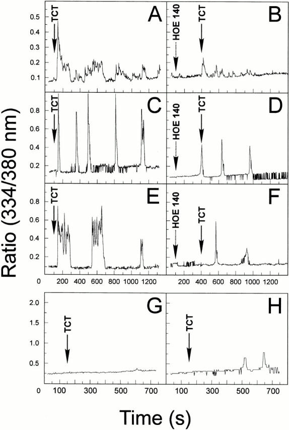
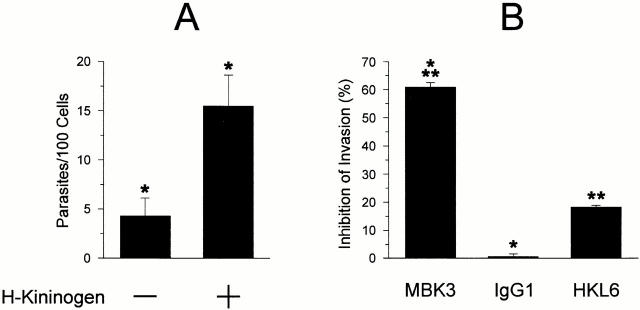
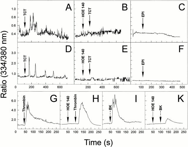
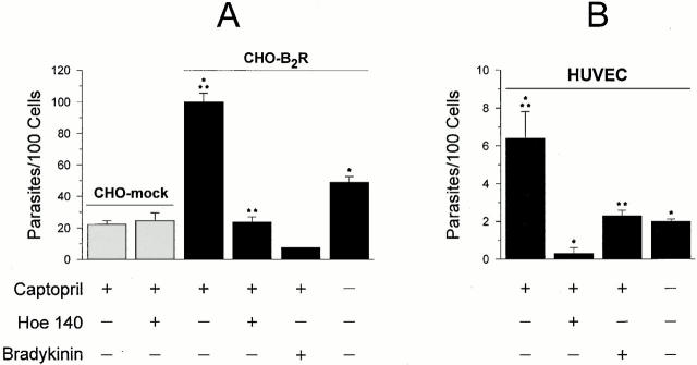
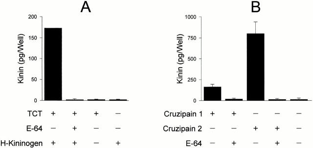
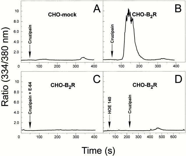
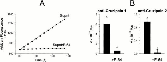
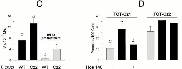
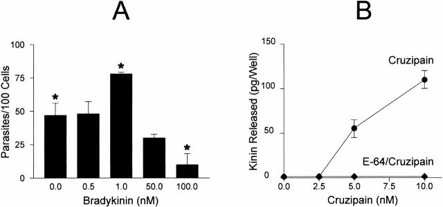
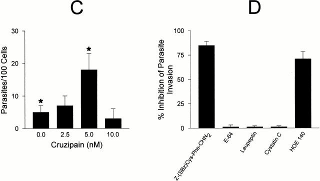
The parasitic protozoan Trypanosoma cruzi employs multiple molecular strategies to invade a broad range of nonphagocytic cells. Here we demonstrate that the invasion of human primary umbilical vein endothelial cells (HUVECs) or Chinese hamster ovary (CHO) cells overexpressing the B(2) type of bradykinin receptor (CHO-B(2)R) by tissue culture trypomastigotes is subtly modulated by the combined activities of kininogens, kininogenases, and kinin-degrading peptidases. The presence of captopril, an inhibitor of bradykinin degradation by kininase II, drastically potentiated parasitic invasion of HUVECs and CHO-B(2)R, but not of mock-transfected CHO cells, whereas the B(2)R antagonist HOE 140 or monoclonal antibody MBK3 to bradykinin blocked these effects. Invasion competence correlated with the parasites' ability to liberate the short-lived kinins from cell-bound kininogen and to elicit vigorous intracellular free calcium ([Ca(2+)](i)) transients through B(2)R. Invasion was impaired by membrane-permeable cysteine proteinase inhibitors such as Z-(SBz)Cys-Phe-CHN(2) but not by the hydrophilic inhibitor 1-trans-epoxysuccinyl-l-leucyl-amido-(4-guanidino) butane or cystatin C, suggesting that kinin release is confined to secluded spaces formed by juxtaposition of host cell and parasite plasma membranes. Analysis of trypomastigote transfectants expressing various cysteine proteinase isoforms showed that invasion competence is linked to the kinin releasing activity of cruzipain, herein proposed as a factor of virulence in Chagas' disease.


Figures

Figure 1
Figure 1
Trypomastigotes induce [Ca2+]i transients in CHO cells through the B2R. The changes of [Ca2+]i were determined on CHO-mock or CHO-B2R preloaded with Fura 2-AM in Ham's F12 medium supplemented with 1 mg/ml of BSA, 12.5 mM Hepes, and 25 μM captopril. The tracings represent cytosolic [Ca2+]i transients that addition of TCTs (parasite/host cell ratio of 10:1) induces in either a single cell or average responses of 20–30 cells. (A) TCTs (arrow) added to CHO-B2R, average responses. (B) TCTs added to CHO-B2R that were pretreated for 200 s with 100 nM HOE 140 (left arrow), average responses. (C and E) TCTs added to CHO-B2R, individual cell response. (D and F) TCTs added to CHO-B2R pretreated with HOE 140, individual cell responses. (G) TCTs added to CHO-mock, individual cell responses. (H) TCTs added to CHO-mock, average responses. CHO-mock failed to induce significant [Ca2+]i elevations upon BK application (up to 50 nM).
Figure 4
Figure 4
H-kininogen potentiates parasite invasion of HUVECs. (A) The effects of addition of H-kininogen on parasite invasion. Interaction was carried out in CAP medium at a parasite/host cell ratio of 2:1 for 3 h in the presence or absence of 9 nM H-kininogen. Data are expressed as means ± SD of three independent experiments. The statistical significance between pairs of data is indicated by asterisks (P < 0.05). (B) The effects of mAbs directed against different kininogen epitopes. CAP medium was supplemented with 200 nM MBK3, IgG1 myeloma protein (MOPC 31c), or HKL6, and the interaction was carried out at a ratio of 5:1 (parasites/host cells) for 3 h. Results are expressed as percentage of inhibition of invasion, in relation to values obtained in cultures containing CAP medium alone.
Figure 2
Figure 2
TCTs induce cytosolic [Ca2+]i transients in HUVECs through the B2R. The changes in [Ca2+]i were determined by adding TCTs (parasite/host cell ratio of 10:1) to monolayers of HUVECs preloaded with Fura 2-AM, loaded, plated on gelatin-coated glass coverslips, maintained in M199 medium with 12.5 mM Hepes, 1 mg/ml BSA, and 25 μM captopril. (A) TCTs added to HUVECs, average responses; (D) individual cell response; (B) TCTs added to HUVECs that were previously treated with 100 nM HOE 140, average cell responses; (E) individual cell response; (C) epimastigotes (EPI) added to HUVECs, average responses; (F) epimastigotes added to HUVECs, individual cell responses. (G–I) Internal controls showing the sensitivity of HUVECs to BK and selectivity of the B2R antagonist HOE 140. Tracings represent average cell responses. (G) Cytosolic [Ca2+]i changes induced by 14 nM α-thrombin; (H) effect of 100 nM HOE 140 on thrombin-induced responses; (I) responses induced by 5 nM BK; (K) effect of 100 nM HOE 140 on responses induced by BK.
Figure 3
Figure 3
T. cruzi invasion is enhanced in mammalian cells overexpressing B2R. (A) Invasion assays were performed with CHO-B2R or CHO-mock in HAM's F12 medium containing 1 mg/ml of BSA in the presence or absence of 25 μM captopril. Dm28c TCTs were added at a ratio of 2:1 (parasite vs. CHO) and the interaction proceeded for 3 h at 37°C in the presence or absence of 100 nM HOE 140 or BK, as indicated. (B) TCTs were added to HUVECs (ratio of 2:1) in M199 medium containing 1 mg/ml BSA. The interaction proceeded for 3 h in the presence or absence of 25 μM captopril. HOE 140 or BK, 100 nM each, was added to the CAP medium immediately before addition of the parasites. Values represent the number of intracellular parasites per 100 cells as means ± SD of three independent experiments. Significant differences (P < 0.05) between paired bars are marked by asterisks (*/*; **/**).
Figure 5
Figure 5
Kinin-releasing activity of TCTs and recombinant cruzipain isoforms. (A) Freshly released parasites, harvested from supernatants of Vero cultures maintained in DMEM-FCS, were washed and resuspended in Ham's F12 medium containing 12.5 mM Hepes, 1 mg/ml of BSA, and 25 μM captopril at pH 6.5. Purified H-kininogen (10 nM, final concentration) was added to 250 μl of the parasites (3 × 10⁶ cells), and the suspension was incubated at 37°C for 30 min. Involvement of cysteine proteases was examined by preincubating the TCTs in medium supplemented with E-64 (75 μM). After removing the parasites by centrifugation (3,000 rpm for 15 min at 4°C), the supernatants were filtered, deproteinized with ice-cold TCA, and the kinin concentrations were determined by a competitive ELISA. (B) Kinin-releasing activity of recombinant cruzipain-1 or cruzipain-2. The reaction was carried out by incubating each of the recombinant proteases (20 nM) with H-kininogen (10 nM) in 200 μl of 50 mM Na2HPO4, pH 6.5, 200 mM NaCl, 5 mM EDTA, and 0.25 mM DTT for 2 h at 37°C. Controls were carried out by preinactivating the proteinases with E-64. Values represent the mean ± SD of three independent assays.
Figure 7
Figure 7
Cruzipain induces [Ca2+]i transients in CHO cells through the B2R. Activated cruzipain-1 or E-64–treated cruzipain-1 were added to monolayers of CHO cells in Ham's F12 medium supplemented with 1 mg/ml of BSA, 25 μM captopril. The protease was diluted 10-fold to 5 nM. (A) CHO-mock; (B) CHO-B2R exposed to cruzipain; (C) CHO-B2R exposed to E-64–treated cruzipain; (D) CHO-B2R pretreated with 100 nM HOE 140 for 120 s and then exposed to cruzipain-1 (indicated by arrows). Addition of the proteinase buffer alone did not elicit any [Ca2+]i transients in CHO cells. The y-axis represents the ratio of fluorescent absorbances at 334 and 380 nm. The tracings represent average cell responses.
Figure 6
Figure 6
Differential infectivity of TCTs overexpressing cruzipain isoforms. (A) Analysis of the cysteine protease activity present in supernatants from wild-type Dm28c TCTs. Parasite suspensions (2 × 10⁷ cells/ml) were incubated at 37°C for 2 h in DMEM without FCS. The filtered supernatants were tested for peptidase activity at 37°C using 20 μM ε-NH2-Cap-Leu-(SBz)Cys-MCA in 50 mM Na2HPO4, pH 6.5, 200 mM NaCl, 5 mM EDTA, and 5 mM DTT. The graph depicts enzyme activity found in trypomastigote supernatants (Supnt) activated with DTT (▪) or pretreated with 30 μM E-64 (•). (B) Protein A–agarose beads were loaded with anti–cruzipain-1 or anti–cruzipain-2 and the immunobeads were added to supernatants obtained from wild-type TCT suspensions. After 2 h of incubation at room temperature, the immune complexes associated with the beads were washed with PBS and the initial rates of hydrolysis by enzymes associated with the solid phase were monitored in the presence or absence of 30 μM E-64, as specified above. Results (initial rates) are represented by the mean of three independent experiments. (C) The alkaline pH stability of the cysteine proteases from wild-type (WT) Dm28c TCTs or TCT-cz2 was assessed by mixing 2 μl of the Triton X-100 lysates (1 mg/ml of protein) with 100 μl of 0.1 M glycine buffer, pH 12. After 5 s, the lysates were diluted in the reaction buffer and the residual E-64–sensitive peptidase activity was measured for TCT-cz2 transfectants or wild-type parasites with ε-l-NH2-Cap-l-(SBz)C-MCA. Gray bars depict rates obtained after alkaline treatment of lysates. The values for initial rates of hydrolysis are expressed as means ± SD of three independent experiments. (D) Invasion assays performed by adding the TCT-cz1 or TCT-cz2 transfectants to monolayers of CHO-B2R (black bars) or CHO-mock (gray bars) in Ham's F12 medium containing 1 mg/ml of BSA in the presence of 25 μM captopril for 3 h at a parasite/host cell ratio of 2:1. Assays were carried out in the presence or absence of 100 nM HOE 140 as indicated, and the number of intracellular parasites per 100 cells was calculated. Results are given as means ± SD of three independent experiments. Statistical significance (P < 0.05) is indicated by asterisks.
Figure 6
Figure 6
Differential infectivity of TCTs overexpressing cruzipain isoforms. (A) Analysis of the cysteine protease activity present in supernatants from wild-type Dm28c TCTs. Parasite suspensions (2 × 10⁷ cells/ml) were incubated at 37°C for 2 h in DMEM without FCS. The filtered supernatants were tested for peptidase activity at 37°C using 20 μM ε-NH2-Cap-Leu-(SBz)Cys-MCA in 50 mM Na2HPO4, pH 6.5, 200 mM NaCl, 5 mM EDTA, and 5 mM DTT. The graph depicts enzyme activity found in trypomastigote supernatants (Supnt) activated with DTT (▪) or pretreated with 30 μM E-64 (•). (B) Protein A–agarose beads were loaded with anti–cruzipain-1 or anti–cruzipain-2 and the immunobeads were added to supernatants obtained from wild-type TCT suspensions. After 2 h of incubation at room temperature, the immune complexes associated with the beads were washed with PBS and the initial rates of hydrolysis by enzymes associated with the solid phase were monitored in the presence or absence of 30 μM E-64, as specified above. Results (initial rates) are represented by the mean of three independent experiments. (C) The alkaline pH stability of the cysteine proteases from wild-type (WT) Dm28c TCTs or TCT-cz2 was assessed by mixing 2 μl of the Triton X-100 lysates (1 mg/ml of protein) with 100 μl of 0.1 M glycine buffer, pH 12. After 5 s, the lysates were diluted in the reaction buffer and the residual E-64–sensitive peptidase activity was measured for TCT-cz2 transfectants or wild-type parasites with ε-l-NH2-Cap-l-(SBz)C-MCA. Gray bars depict rates obtained after alkaline treatment of lysates. The values for initial rates of hydrolysis are expressed as means ± SD of three independent experiments. (D) Invasion assays performed by adding the TCT-cz1 or TCT-cz2 transfectants to monolayers of CHO-B2R (black bars) or CHO-mock (gray bars) in Ham's F12 medium containing 1 mg/ml of BSA in the presence of 25 μM captopril for 3 h at a parasite/host cell ratio of 2:1. Assays were carried out in the presence or absence of 100 nM HOE 140 as indicated, and the number of intracellular parasites per 100 cells was calculated. Results are given as means ± SD of three independent experiments. Statistical significance (P < 0.05) is indicated by asterisks.
Figure 8
Figure 8
The involvement of cruzipain in parasite invasion. (A) TCTs were added to HUVECs (parasite/host cell ratio of 5:1) in M199 CAP medium containing BK (0, 10, 50, and 100 nM) and incubated at 37°C for 3 h. (B) The graph illustrates the dose dependence of kinin-releasing activity of cruzipain-1. Enzyme stocks (50 nM) preactivated with DTT were subsequently diluted to the appropriate concentration in 50 mM Na2HPO4, pH 6.5, 5 mM EDTA, and 0.25 mM DTT supplemented with 10 nM H-kininogen. After a 30-min incubation at 37°C, the reaction was stopped by the addition of 75 μM E-64 and 25 μM captopril. Kinin release by activated cruzipain-1 (•) or E-64–treated cruzipain-1 (♦) was determined by competitive ELISA. Controls carried out with buffer in the absence of cruzipain did not lead to kinin release. (C) Invasion assays were carried out by adding aliquots of preactivated cruzipain-1 (50 nM stocks) to CAP medium before addition of TCTs to HUVECs (parasite/host cell ratio of 5:1, 1 h). (D) Invasion assays carried out at a parasite/host cell ratio of 5:1 for 3 h in CAP medium supplemented with Z-(SBz)Cys-Phe-CHN2 (10 μM), E-64 (75 μM), leupeptin (10 μM), cystatin C (1 μM), and HOE 140 (100 nM). The addition of DMSO vehicle (1%) did not interfere with invasion. The activity of each compound was expressed as the percentage of inhibition of parasite invasion with reference to assays performed in CAP medium alone. Data are expressed as means ± SD of at least three independent experiments. The statistical analysis was done by one-way analysis of variance; significance was considered at *P < 0.05.
Figure 8
Figure 8
The involvement of cruzipain in parasite invasion. (A) TCTs were added to HUVECs (parasite/host cell ratio of 5:1) in M199 CAP medium containing BK (0, 10, 50, and 100 nM) and incubated at 37°C for 3 h. (B) The graph illustrates the dose dependence of kinin-releasing activity of cruzipain-1. Enzyme stocks (50 nM) preactivated with DTT were subsequently diluted to the appropriate concentration in 50 mM Na2HPO4, pH 6.5, 5 mM EDTA, and 0.25 mM DTT supplemented with 10 nM H-kininogen. After a 30-min incubation at 37°C, the reaction was stopped by the addition of 75 μM E-64 and 25 μM captopril. Kinin release by activated cruzipain-1 (•) or E-64–treated cruzipain-1 (♦) was determined by competitive ELISA. Controls carried out with buffer in the absence of cruzipain did not lead to kinin release. (C) Invasion assays were carried out by adding aliquots of preactivated cruzipain-1 (50 nM stocks) to CAP medium before addition of TCTs to HUVECs (parasite/host cell ratio of 5:1, 1 h). (D) Invasion assays carried out at a parasite/host cell ratio of 5:1 for 3 h in CAP medium supplemented with Z-(SBz)Cys-Phe-CHN2 (10 μM), E-64 (75 μM), leupeptin (10 μM), cystatin C (1 μM), and HOE 140 (100 nM). The addition of DMSO vehicle (1%) did not interfere with invasion. The activity of each compound was expressed as the percentage of inhibition of parasite invasion with reference to assays performed in CAP medium alone. Data are expressed as means ± SD of at least three independent experiments. The statistical analysis was done by one-way analysis of variance; significance was considered at *P < 0.05.

References

    1. Andrade Z.A. Immunopathology of Chagas' disease Mem. Inst. Oswaldo Cruz. 94Suppl. I1999. 71 80 - PubMed
    1. Rossi M. Microvascular changes as a cause of chronic cardiomyopathy in Chagas' disease. Am. Heart J. 1990;120:233–236. - PubMed
    1. Morris S.A., Tanowitz H., Wittner M., Bilezikjan J.P. Pathophysiological insights into the cardiomyopathy of Chagas' disease. Circulation. 1990;82:1900–1909. - PubMed
    1. Tardieux I., Webster P., Ravesloot J., Boron W., Lunn J.A., Andrews N.W. Lysosome recruitment and fusion are early events required for Trypanosoma invasion of mammalian cells. Cell. 1992;71:1117–1130. - PubMed
    1. Burleigh B.A, Andrews N.W. The mechanisms of Trypanosoma cruzi invasion of mammalian cells. Annu. Rev. Microbiol. 1995;49:175–200. - PubMed

Publication types

MeSH terms