. 2000 Nov 6;192(9):1317-26.
doi: 10.1084/jem.192.9.1317.

Influenza virus lung infection protects from respiratory syncytial virus-induced immunopathology

Affiliations

Influenza virus lung infection protects from respiratory syncytial virus-induced immunopathology

G Walzl et al. J Exp Med. .

Abstract

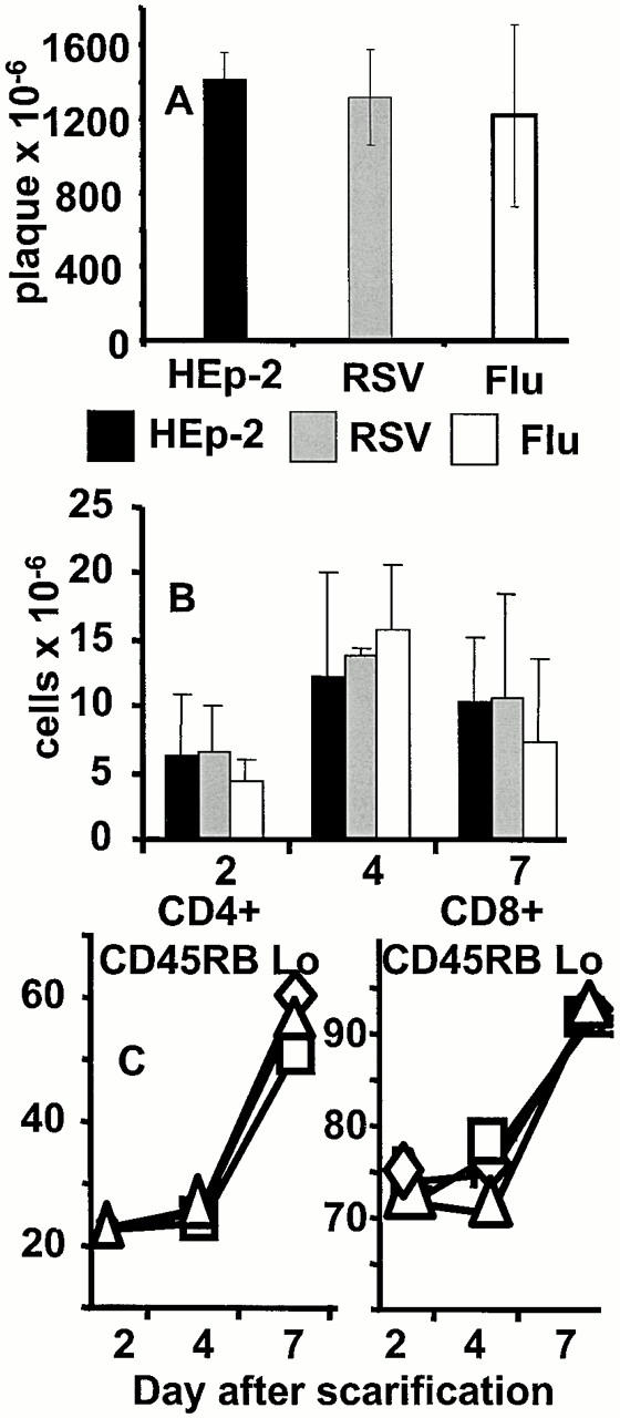
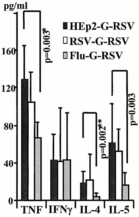
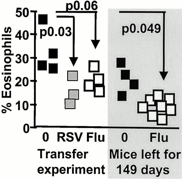
The effect of infection history is ignored in most animal models of infectious disease. The attachment protein of respiratory syncytial virus (RSV) induces T helper cell type 2-driven pulmonary eosinophilia in mice similar to that seen in the failed infant vaccinations in the 1960s. We show that previous influenza virus infection of mice: (a) protects against weight loss, illness, and lung eosinophilia; (b) attenuates recruitment of inflammatory cells; and (c) reduces cytokine secretion caused by RSV attachment protein without affecting RSV clearance. This protective effect can be transferred via influenza-immune splenocytes to naive mice and is long lived. Previous immunity to lung infection clearly plays an important and underestimated role in subsequent vaccination and infection. The data have important implications for the timing of vaccinations in certain patient groups, and may contribute to variability in disease susceptibility observed in humans.


Figures

Figure 1
Figure 1
Previous infection prevents illness during secondary responses to the attachment protein of RSV. (A) Weight loss was monitored on the day of, and during, the final RSV infection. The mean and standard deviation from five to six mice per group are shown. (B) Illness was monitored by a blinded observer using a standard grading system based on the degree of cachexia, ruffled fur, and mobility. The results from five to six mice per group per day were added (cumulative disease severity score). Mice experiencing secondary responses to the G protein are represented by the black bars; those preinfected with RSV and influenza are represented by the gray and white bars, respectively. (C) Eosinophils were enumerated in hematoxylin and eosin–stained cytocentrifuge preparations of cells recovered by BAL. The results represent individual mice from two independent experiments. The groups are designated as follows: A, HEp-2–G-RSV; B, HEp-2–β-gal–RSV; C, RSV–β-gal–RSV; D, Flu–β-gal–RSV; E, RSV–G-RSV; and F, Flu–G-RSV.
Figure 2
Figure 2
Previous lung infection changes the nature of the illness induced in rVV-G–primed, RSV-challenged mice. Lungs were inflated with 2% formalin in buffered saline, removed, and embedded in paraffin. Representative sections from HEp-2–G-RSV– (right), RSV–G-RSV– (middle), and influenza–G-RSV– (left) infected animals were stained with hematoxylin and eosin. The top panels show the extent of inflammatory infiltrate in representative sections of lung (original magnification: ×100); the bottom panels show the presence of eosinophils (indicated by arrows; original magnification: ×1,000, oil immersion).
Figure 3
Figure 3
Previous lung infection does not alter the replication of rVV-G or the recruitment of cells to the inguinal lymph node. (A) Mice were intranasally infected with HEp-2, RSV, or influenza as shown and scarified with rVV-G after 3 wk. The number of plaques in the scarified area of skin was assessed after 24 h in HEp-2 monolayers. (B) Inguinal lymph nodes were also recovered and mechanically disrupted. Total viable cell recovery on days 2, 4, and 7 is shown. (C) The proportion of CD45RBlow CD4 (left), and CD8 (right) T cells was determined by flow cytometry from HEp-2- (□), RSV- (▵), and influenza- (⋄) preprimed mice. The results represent the mean and standard deviation of three to four mice per group.
Figure 4
Figure 4
RSV- and influenza-specific CD8+ T cells are recruited back to the lung during the final RSV infection. (A) BAL cells from mice sequentially primed with RSV–G-RSV do not bind the influenza NP–specific MHC tetramer. This sample was used to set the position of the quadrant. (B) M2-specific cell lines bind the RSV M2–specific MHC tetramer. The quadrant position was determined by staining the same sample with the influenza NP–specific tetramer. This cell line was generated from the spleen of rVV-M2–primed and RSV-challenged mice that had been grown in vitro for 3 wk. (C and D) The presence of NP- and M2-specific CD8+ T cells in mice sequentially infected with Flu–G-RSV and RSV–G-RSV, respectively. BAL was collected 7 d after the final RSV infection and samples were stained with MHC tetramers and antibodies to CD8. 40,000 lymphocytes were analyzed by flow cytometry. The quadrant positions were determined from staining cells in the RSV–G-RSV group with the NP-specific tetramer. (E and F) Anti-CD8– and tetramer-stained cells were then permeabilized with saponin and stained with FITC-conjugated antibody to IFN-γ. The proportion of CD8+, NP (E), and M2 (F) tetramer–stained cells expressing IFN-γ is shown. These results are representative of four to five mice per group.
Figure 5
Figure 5
Previous infection reduces Th2 cytokines during secondary responses to the G protein. Mice were infected with HEp-2, RSV, or influenza, scarified with rVV-G 21 d later, and finally infected with RSV after 14 d. 7 d later, cytokine levels in lavage fluid were examined by ELISA and concentrations were calculated using a standard curve and linear regression analysis. The mean and standard deviation from individual mice in two independent experiments are shown.
Figure 6
Figure 6
Transfer of splenocytes from RSV- and influenza-immune mice partially protects G-primed, RSV-challenged mice from illness. Mice were intranasally infected with RSV and influenza and left for 21–35 d. 3 × 10⁷ spleen cells were then transferred intravenously into naive animals. 1 d later, these mice were scarified with rVV-G and intranasally infected with RSV after an additional 14 d. Eosinophils were assessed in lung lavage samples in Giemsa-stained cytocentrifuge preparations. The shaded part represents naive mice (▪) or those infected with influenza (□), and left for 149 d before G priming and RSV infection.
Figure 7
Figure 7
Splenocytes from influenza-primed mice do not kill RSV-infected targets. Spleens from RSV- or influenza-infected mice were removed after 14 d and disrupted to a single cell suspension. After 5 d in the presence of 2 PFU/cell of RSV or 1 HA unit of influenza virus, remaining cells were assessed for cytotoxic activity. RSV-immune mice effectively killed RSV-infected targets (♦). Influenza-immune splenocytes similarly killed influenza (▴) but not RSV-infected (▪) targets. (B) Influenza tetramer–positive cells were purified from the lung and mediastinal lymph nodes of 10 influenza-infected and RSV-challenged mice using avidin-coated magnetic beads and MACS. A cytotoxicity assay with influenza- (•) or RSV- (⋄) infected P815 cells was then performed. Spontaneous release has been subtracted from all results and never exceeded 10% of maximal chromium release.

References

    1. Prescott S.L., Macaubas C., Smallacombe T., Holt B.J., Sly P.D., Holt P.G. Development of allergen-specific T-cell memory in atopic and normal children. Lancet. 1999;353:196–200. - PubMed
    1. Romagnani S. Induction of TH1 and TH2 responsesa key role for the ‘natural’ immune response? Immunol. Today. 1992;13:379–381. - PubMed
    1. Martinez F.D., Wright A.L., Taussig L.M., Holberg C.J., Halonen M., Morgan W.J. Asthma and wheezing in the first six years of life. N. Engl. J. Med. 1995;332:133–138. - PubMed
    1. Holt P.G., Macaubas C. Development of long-term tolerance versus sensitization to environmental allergens during the perinatal period. Curr. Opin. Immunol. 1997;9:782–787. - PubMed
    1. Shirakawa T., Enomoto T., Shimazu S., Hopkin J.M. The inverse association between tuberculin responses and atopic disorder. Science. 1997;275:77–79. - PubMed

Publication types

MeSH terms