. 2000 Nov 6;192(9):1327-38.
doi: 10.1084/jem.192.9.1327.

Identification of a novel prostaglandin f(2alpha) synthase in Trypanosoma brucei

Affiliations

Identification of a novel prostaglandin f(2alpha) synthase in Trypanosoma brucei

B K Kubata et al. J Exp Med. .

Abstract

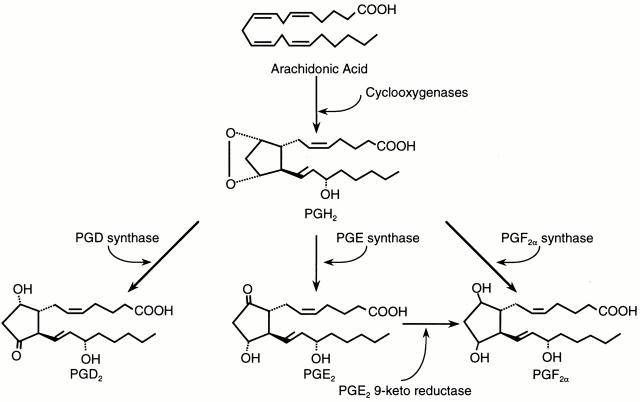
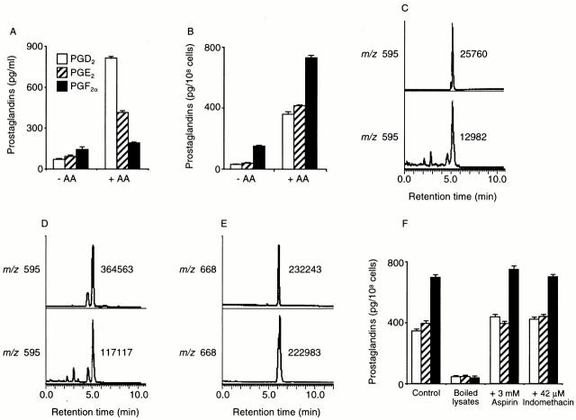
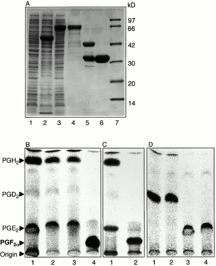
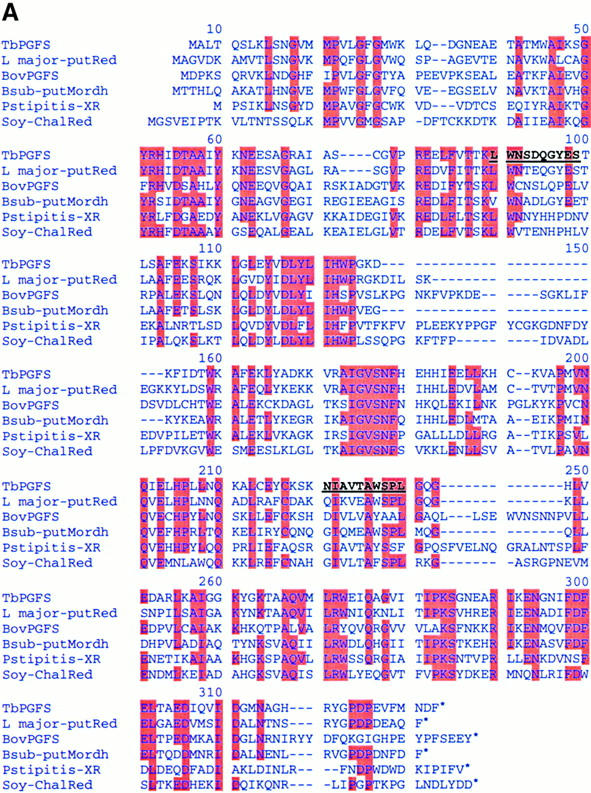
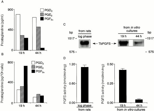
Members of the genus Trypanosoma cause African trypanosomiasis in humans and animals in Africa. Infection of mammals by African trypanosomes is characterized by an upregulation of prostaglandin (PG) production in the plasma and cerebrospinal fluid. These metabolites of arachidonic acid (AA) may, in part, be responsible for symptoms such as fever, headache, immunosuppression, deep muscle hyperaesthesia, miscarriage, ovarian dysfunction, sleepiness, and other symptoms observed in patients with chronic African trypanosomiasis. Here, we show that the protozoan parasite T. brucei is involved in PG production and that it produces PGs enzymatically from AA and its metabolite, PGH(2). Among all PGs synthesized, PGF(2alpha) was the major prostanoid produced by trypanosome lysates. We have purified a novel T. brucei PGF(2alpha) synthase (TbPGFS) and cloned its cDNA. Phylogenetic analysis and molecular properties revealed that TbPGFS is completely distinct from mammalian PGF synthases. We also found that TbPGFS mRNA expression and TbPGFS activity were high in the early logarithmic growth phase and low during the stationary phase. The characterization of TbPGFS and its gene in T. brucei provides a basis for the molecular analysis of the role of parasite-derived PGF(2alpha) in the physiology of the parasite and the pathogenesis of African trypanosomiasis.


Figures

Figure 1
Figure 1
Biosynthetic pathways of PGs from AA.
Figure 2
Figure 2
PG production by T. brucei and mass spectra of trypanosomal PGs. (A) The PGs secreted into the culture media by live trypanosomes when the organisms were cultured with or without AA were quantified. − AA and + AA indicate growth in the absence and presence of 66 μM AA, respectively. PG detection limits were <25, 11, and 10 pg/assay for PGD2, PGE2, and PGF, respectively. The values shown are the means from three independent experiments along with SE. (B) PG production by T. brucei lysates. PGs were produced during the incubation of 1 mM AA with lysates of bloodstream-form trypanosomes. The values shown are the means from three independent experiments along with SE. (C–E) To identify and quantify the prostanoids by GC-MS, PGs from the incubation of trypanosome lysates with AA were extracted and resolved into PGD2, PGE2, and PGF as described in Materials and Methods. After treatment (see Materials and Methods), the prostanoids were subjected to GC-MS. (C) Selected ion recordings monitoring characteristic ions for PGD2 at m/z 595. (D) Those for PGE2 at m/z 595. (E) Those for PGF at m/z 668. The number indicates the ion intensity for each peak. Upper mass spectra indicate traces of the selected ion recordings of the ME-MO-DMiPS ether derivatives of authentic PGD2 (C), PGE2 (D), and PGF (E); and lower ones, traces of the selected ion recordings of the ME-MO-DMiPS ether derivatives of PGD2 (C), PGE2 (D), and PGF (E) produced by trypanosome lysates. (F) Effects of heat treatment and of NSAIDs on PG production by trypanosome lysates. Nonboiled (control) and boiled T. brucei lysates were incubated with 1 mM AA at 37°C for 30 min in the absence of NSAIDs, and PGs produced were measured by EIA. To investigate the effects of NSAIDs, we incubated nonboiled T. brucei lysates with 1 mM AA in the absence (control) or presence of the indicated concentrations of aspirin or indomethacin. The values shown are the means from three independent experiments along with SE.
Figure 3
Figure 3
De novo synthesis of PGD2 and PGF from PGH2 by T. brucei lysates. Membrane or cytosolic fractions from long slender bloodstream-form parasites (250 μg protein) were incubated with 5 μM 1-[14C] PGH2 at 37°C for 2 min. Arrows indicate the positions of PGH2, PGD2, PGE2, and PGF. (A) T. brucei PGD synthase activity. Lane 1, control reaction without membrane fraction; lane 2, reaction with membrane fraction. (B) TbPGFS synthase activity was assayed as described in Materials and Methods. Lane 1, control; lane 2, addition of cytosolic proteins; lane 3, addition of heat-treated cytosolic proteins. (C) SDS-PAGE of purified TbPGFS. 5 μg of protein was subjected to electrophoresis on a 14% SDS–polyacrylamide gel, and the protein bands were detected by silver staining. Lane 1, molecular mass marker proteins; lane 2, purified active fraction from Hiload 16/60 Superdex 200 pg.
Figure 5
Figure 5
Expression of TbPGFS in E. coli and TbPGFS activity of the recombinant protein. (A) TbPGFS was expressed as a fusion protein with MBP in E. coli and purified as described in Materials and Methods. Lane 1, noninduced lysate of E. coli DH5α; lane 2, IPTG-induced lysate of E. coli DH5α transformed with empty pMAL-c2 vector after 4-h induction; lane 3, IPTG-induced lysate of E. coli DH5α transformed with pMAL-c2-TbPGFS after 4 h; lane 4, purified MBP–TbPGFS fusion protein; lane 5, enzyme after purification by gel filtration on Superdex 75, after cleavage with factor Xa to separate TbPGFS from MBP; lane 6, purified TbPGF synthase after Mono S column chromatography; lane 7, molecular mass marker proteins. (B) Reduction of PGH2 by recombinant TbPGFS. Lane 1, substrate incubated in the absence of enzyme; lane 2, substrate incubated with 100 μg total protein from lysate of the host E. coli DH5α; lane 3, substrate incubated with 100 μg protein from lysate of induced E. coli cells containing empty pMAL-c2 vector; lane 4, incubation with 25 μg of lysate from induced E. coli that contained pMAL-c2-TbPGFS. (C) Effect of phenanthrenequinone on TbPGFS activity. The enzymatic activity was determined as described in Materials and Methods. 1-[14C] PGH2 (5 μM) was incubated in the presence of 40 μM of phenanthrenequinone without (lane 1) or with 14 μg of purified TbPGFS (lane 2). (D) Lack of reduction of PGD2 and PGE2 by purified TbPGFS. 1-[14C]PGD2 and 1-[14C]PGE2 (10 μM), produced as described in Materials and Methods, were incubated without (lanes 1 and 3) or with (lanes 2 and 4) 14 μg of purified TbPGFS.
Figure 4
Figure 4
Sequence homology of TbPGFS with members of the AKR superfamily. (A) Multiple sequence alignment of deduced TbPGFS amino acid sequence with representative members of the AKR superfamily. The amino acid sequences were taken from the sequence databases SwissProt (SP), Protein Identification Resource (PIR), and GenBank (GB). TbPGF synthase is aligned with: L major-putRed, L. major putative reductase (SP P22045) 57; BovPGFS, bovine lung PGFS (GB J03570) 58; Bsub-putMordh, B. subtilis putative morphine dehydrogenase (GB AF008220) 59; Pstipitis-XR, Pichia stipitis xylose reductase (SP P31867) 60; and Soy-ChalRed, soybean chalcone reductase (SP P26690) 61. Dashes show the gap used to maximize the similarity. Conserved residues with TbPGF synthase are highlighted in red. Underlined black bold letters indicate the peptide sequences identified from purified native TbPGFS and used for primer design. (B) Neighbor-joining tree of TbPGFS and some members of the AKR superfamily. Amino acid sequences of oxidoreductases from pig, cow, human, mouse, L. major, apple, soybean, yeast, and bacteria were selected as representatives for mammals, protozoa, plants, and prokaryotes to simplify the phylogenetic analysis. Numbers at the nodes represent bootstrap proportions based on 1,000 replicates. Por-ALR, porcine aldose reductase (SP P80276); Bov-ADR, bovine aldehyde reductase (SP P16116); Hum-3a-HSDdh, human 3α-hydroxysteroid dehydrogenase (GB NM003739); Mou-17b-HSDdh, mouse estradiol 17β-(hydroxysteroid) dehydrogenase (PIR A56424); App-S6Pdh, apple NADP-dependent sorbitol-6-phosphate dehydrogenase (SP P28475); Spombe-OxidoRed, Schizosaccharomyces pombe probable oxidoreductase (SP Z99165); Cb-25dkg, Corynebacterium sp. 2,5-diketo-gulonate reductase (SP P15339); Ecoli-OxidoRed, E. coli hypothetical oxidoreductase (SP Q46857); Mtub-Rv2971, Mycobacterium tuberculosis hypothetical protein Rv2971 (GB Z83018); Strepm-putRed, Streptomyces coelicolor putative oxidoreductase (GB AL034443); Ps put-Mordh, Pseudomonas putida morphine dehydrogenase (GB M94775); and Therm-OxidoRed, Thermotoga maritima oxidoreductase (GB AE001762).
Figure 4
Figure 4
Sequence homology of TbPGFS with members of the AKR superfamily. (A) Multiple sequence alignment of deduced TbPGFS amino acid sequence with representative members of the AKR superfamily. The amino acid sequences were taken from the sequence databases SwissProt (SP), Protein Identification Resource (PIR), and GenBank (GB). TbPGF synthase is aligned with: L major-putRed, L. major putative reductase (SP P22045) 57; BovPGFS, bovine lung PGFS (GB J03570) 58; Bsub-putMordh, B. subtilis putative morphine dehydrogenase (GB AF008220) 59; Pstipitis-XR, Pichia stipitis xylose reductase (SP P31867) 60; and Soy-ChalRed, soybean chalcone reductase (SP P26690) 61. Dashes show the gap used to maximize the similarity. Conserved residues with TbPGF synthase are highlighted in red. Underlined black bold letters indicate the peptide sequences identified from purified native TbPGFS and used for primer design. (B) Neighbor-joining tree of TbPGFS and some members of the AKR superfamily. Amino acid sequences of oxidoreductases from pig, cow, human, mouse, L. major, apple, soybean, yeast, and bacteria were selected as representatives for mammals, protozoa, plants, and prokaryotes to simplify the phylogenetic analysis. Numbers at the nodes represent bootstrap proportions based on 1,000 replicates. Por-ALR, porcine aldose reductase (SP P80276); Bov-ADR, bovine aldehyde reductase (SP P16116); Hum-3a-HSDdh, human 3α-hydroxysteroid dehydrogenase (GB NM003739); Mou-17b-HSDdh, mouse estradiol 17β-(hydroxysteroid) dehydrogenase (PIR A56424); App-S6Pdh, apple NADP-dependent sorbitol-6-phosphate dehydrogenase (SP P28475); Spombe-OxidoRed, Schizosaccharomyces pombe probable oxidoreductase (SP Z99165); Cb-25dkg, Corynebacterium sp. 2,5-diketo-gulonate reductase (SP P15339); Ecoli-OxidoRed, E. coli hypothetical oxidoreductase (SP Q46857); Mtub-Rv2971, Mycobacterium tuberculosis hypothetical protein Rv2971 (GB Z83018); Strepm-putRed, Streptomyces coelicolor putative oxidoreductase (GB AL034443); Ps put-Mordh, Pseudomonas putida morphine dehydrogenase (GB M94775); and Therm-OxidoRed, Thermotoga maritima oxidoreductase (GB AE001762).
Figure 6
Figure 6
PG production, differential expression of TbPGFS mRNA, and TbPGFS activity during the T. brucei life cycle. (A) PG secretion by bloodstream-form live trypanosomes from early logarithmic growth and late stationary phases. (B) PG production by lysates from bloodstream-form trypanosomes from early logarithmic and late stationary growth phases. (C) Northern blot analysis. T. brucei total RNA (3.9 μg) was isolated from the bloodstream-form population from infected rats (left panel) and from early logarithmic (right panel, 19 h) and late stationary growth phase (right panel, 44 h) cells of the cultured bloodstream-form population. Total RNA was hybridized with a probe derived from the TbPGFS cDNA coding region. (D) TbPGFS activity of the lysates (50 μg protein) from early logarithmic and late stationary growth phase trypanosomes. Lysates of respective cells were incubated with 5 μM PGH2. The reaction products were analyzed by thin-layer chromatography as described in the legend to Fig. 4 (n = 3).

Similar articles

Cited by

References

    1. Smith W.L., DeWitt D.L. Prostaglandin endoperoxide H synthases-1 and -2. Adv. Immunol. 1996;62:167–215. - PubMed
    1. Samuelsson B. Prostaglandins, thromboxanes, and leukotrienesformation and biological roles. Harvey Lect. 1979;75:1–40. - PubMed
    1. Mathé A.A., Hedqvist P., Strandberg K., Leslie C.A. Aspects of prostaglandin function in the lung (second of two parts) N. Engl. J. Med. 1977;296:910–914. - PubMed
    1. Oliw E., Granström E., Änggard E. The prostaglandins and essential fatty acids. In: Pace-Asciak C., Granström E., editors. Prostaglandins and Related Substances. Elsevier; Amsterdam: 1983. pp. 11–19.
    1. Glew R.H. Prostaglandins and thromboxanes. In: Devlin T.M., editor. Textbook of Biochemistry with Clinical Correlations. Wiley-Liss, Inc.; New York: 1992. pp. 461–466.

Publication types

MeSH terms

Associated data