. 2000 Nov;14(11):1898-908.
doi: 10.1038/sj.leu.2401933.

The amino terminus targets the mixed lineage leukemia (MLL) protein to the nucleolus, nuclear matrix and mitotic chromosomal scaffolds

Affiliations

The amino terminus targets the mixed lineage leukemia (MLL) protein to the nucleolus, nuclear matrix and mitotic chromosomal scaffolds

C Caslini et al. Leukemia. 2000 Nov.

Abstract

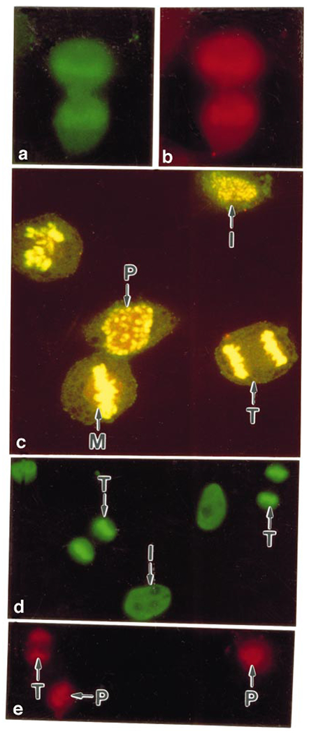
The mixed-lineage leukemia gene (MLL) is associated with more than 25 chromosomal translocations involving band 11q23 in diverse subtypes of human acute leukemia. Conditional expression of a 50 kDa amino terminal fragment spanning the AT hook motifs of MLL (MLL3AT) causes cell cycle arrest, upregulation of p21Cip1 and p27KiP1 and partial monocytic differentiation of the monoblastic U937 cell line, suggesting a major role for MLL3AT in MLL-AF9-induced myelomonocytic differentiation. In this study, we analyzed the subcellular localization of conditionally expressed MLL3AT in both U937 and HeLa cell lines. Immunofluorescence staining, confocal laser scanning microscopy and immunoelectron microscopy indicated that MLL3AT, like endogenous MLL, localized in the nucleoplasm in a punctate pattern of distribution, including regions attached to the nuclear envelope and the periphery of the nucleolus. We found that MLL3AT and endogenous MLL were present in interphase nuclear matrices and colocalized with topoisomerase II to mitotic chromosomal scaffolds. Nucleoplasm and nucleolar localization was observed even for MLL-AF9 and MLL-AF4 conditionally expressed chimeric proteins, suggesting a common target conferred by the amino terminus of MLL to many if not all the chimeric MLL proteins. The nuclear matrix/scaffold association suggests a role for the amino terminus of MLL in the modulation of chromatin structure, leading to epigenetic effects on the maintenance of gene expression.


Figures

Figure 1
Figure 1
(a) Structures of MLL, AF4, MLL-AF4 proteins and MLL3AT mutant. MLL-AF4 contains all regions upstream of the breakpoint cluster region (BCR) of MLL and all but the most amino terminal portion of AF4.,, MLL3AT encodes the amino terminal 410 amino acids of MLL and includes three AT-hook motifs that share homology with minor-groove DNA-binding proteins., Both constructs contain an amino terminal FLAG epitope tag. SNL-1 and SNL-2, speckle nuclear localization domains 1 and 2; trx, short regions of homology to Trithorax Drosophila protein; MT, DNA methyltransferase homology region;,, NLS, nuclear localization signal of AF4; PHD, plant homeodomain zinc-fingers; SET, domain of homology to regions of Su(var)3–9, E(z), and Trx Drosophila proteins. The relative positions of the peptides used to produce antibodies against MLL-AF4 are also shown. (b) Comparison of α-MLL pAb M382 and mAb 2F7 antibodies with α-AF4 polyclonal antibody C15. Lysates from t(4;11) and non-t(4;11) leukemia cell lines (100 μg) were run on low percentage SDS-PAGE (5%), electrophoretically transferred to nitrocellulose membranes, and probed with affinity purified α-MLL pAb M382 and mAb 2F7 or α-AF4 pAb C15. Lane 1, Jurkat; lane 2, RS(4;11); lane 3, ALL-PO(4;11).
Figure 2
Figure 2
Western analysis of FLAG-tagged MLL3AT after conditional expression in HtTA-1 and U937T stably transfected cells. Total cell lysates (50 μg) prepared from each clone grown for 48 h in the presence (+) or absence (−) of transcriptional repressor tetracycline were separated by SDS-PAGE, and transferred on to nitrocellulose membranes. Western analysis was performed using an αFLAG monoclonal antibody. In the absence of tetracycline all clones express a protein of 50 kDa, as predicted for MLL3AT. The band does not show up in parental HtTA-1 and U937T control cells.
Figure 3
Figure 3
Endogenous MLL associates with nucleoli and nucleoplasm through its MLL amino terminal component. (a, b) HtTA-1 and (c, d) U937T cells expressing MLL3AT under the control of a tet-suppressible promoter were grown for 48 h in the absence of tet, and then double-labeled with the (a, c) α-MLL pAb M382 (Texas red) and (b, d) α-FLAG mAb (fluorescein). Some cells showed colabeling of nuclear structures with pAb M382 and α-FLAG mAb (arrows); while others showed labeling with pAb M382 only (arrowheads). (e) Endogenous MLL in HtTA-1 parental cells was detected with α-MLL mAb 2F7. (f–h) HtTA-1 cells expressing MLL3AT were immunostained with (f) α-FLAG mAb and (g) mAb 2F7. The secondary antibodies recognizing murine α-FLAG mAb and hamster mAb 2F7 were conjugated to fluorescein (green) and Texas red (red), respectively. Yellow color in the merged image (h) indicates colocalization of the two antibodies.
Figure 4
Figure 4
Association of the MLL and MLL3AT proteins with chromosomal scaffolds and nuclear matrix. To examine the association of the MLL3AT protein with chromosomal scaffolds, HtTA-1 cells expressing MLL3AT were doubled-labeled with (a) α-FLAG mAb and (b) α-topoisomerase II pAb. (c) Confocal imaging of these cells shows the distribution of α-FLAG mAb and a-topoisomerase II pAb in cells at various stages of the cell cycle (P, prophase; M, metaphase; T, telophase; I, interphase). MLL3AT and topoisomerase II colocalize in telophase and metaphase chromosomes (note the yellow color) with less association in prophase and interphase cells. Nuclear matrices prepared from HtTA-1 cells expressing MLL3AT were double-labeled with (d) α-FLAG mAb and (e) α-MLL mAb 2F7. Note that all antibodies label the chromosomal scaffolds in prophase (P) and telophase (T) cells and interphase (I) nuclear matrices.
Figure 5
Figure 5
Confocal laser scanning microscopy of cells expressing MLL3AT. (a) A merged image of 10 optical sections of HtTA-1 cells expressing MLL3AT reveals a punctate nucleoplasmic and intense nucleolar staining of the protein. Also note the association of MLL3AT with the nuclear envelope (small arrows) and the absence of label over some nucleoli (large arrows). (b) A single optical section through a cell labeled with the α-FLAG mAb and stained with propidium iodide shows perinucleolar localization of the protein (arrowhead) with the core of the nucleolus labeled only by propidium iodide.
Figure 6
Figure 6
Immunoelectron microscopy of cells expressing MLL3AT. HeLa cells induced to express MLL3AT were labeled with the α-FLAG mAb, followed by a gold-conjugated secondary antibody. The clusters (0.1 to 0.2 μm in diameter) of 25–100 gold particles observed by electron microscopy presumably represent the dots seen by fluorescence microscopy, and are located: (A) at the nuclear envelope (arrows); (B) at the periphery of the nucleolus and in the nucleoplasm (arrowheads); and (C) on the metaphase chromosomes (arrowhead, also shown in the inset). N, nucleus; Cy, cytoplasm; NE, nuclear envelope; NU, nucleolus; Ch, chromosome. Original magnifications: A, ×81,000; B, ×37,750; C, ×8,500; C inset, ×46,000.
Figure 7
Figure 7
MLL-AF4 fusion oncoprotein associates with nucleoli and nucleoplasm. Cells transiently transfected with plasmid encoding FLAG-epitope tagged MLL-AF4 under control of a tet-suppressible promoter were grown for 36 h in the absence of tetracycline, and then colabeled with (a) α-FLAG mAb (green) and (b) α-AF4 pAb C15 (red). The HtTA-1 cell in panels a and b was also analyzed by phase contrast microscopy (c) to reveal the nucleoli (Nu). Note the ring-like nucleolar labeling with both α-FLAG and α-AF4 antibodies. As MLL and MLL3AT proteins, MLL-AF4 was localized in a nucleolar pattern and with a granular distribution throughout the nucleus.

References

    1. Ziemin-van der Poel S, McCabe NR, Gill HJ, Espinosa R III, Patel Y, Harden A, Rubinelli P, Smith SD, LeBeau Mm, Rowley JD, Diaz MO. Identification of a gene, MLL, that spans the breakpoint in 11q23 translocations associated with human leukemias. Proc Natl Acad Sci USA 1991; 88: 10735–10739. - PMC - PubMed
    1. Raimondi SC, Peiper SC, Kitchingman G, Behm FG, Williams DL, Hancock ML, Mirro J Jr. Childhood acute lymphoblastic leukemia with chromosomal breakpoints at 11q23. Blood 1989; 73: 1627–1634. - PubMed
    1. Raimondi SC, Kalwinsky DK, Hayashi Y, Behm FG, Mirro J Jr, Williams DL. Cytogenetics of childhood acute nonlymphocytic leukemia. Cancer Genet Cytogenet 1989; 40: 13–27. - PubMed
    1. DeVore R, Whitlock J, Hainsworth JD, Johnson DH. Therapy-related acute nonlymphocytic leukemia with monocytic features and rearrangement of chromosome 11q. Ann Intern Med 1989; 110: 740–742. - PubMed
    1. Pui C- H, Behm FG, Raimondi SC, Dodge RK, George SL, Rivera GK, Mirro J Jr, Kalwinsky DK, Dahl GV, Murphy SB. Secondary acute myeloid leukemia in children treated for acute lymphoid leukemia. New Engl J Med 1989; 321: 136–142. - PubMed

Publication types

MeSH terms

Substances