. 2000 Nov 21;97(24):13045-50.
doi: 10.1073/pnas.230315097.

Multiple quantum solid-state NMR indicates a parallel, not antiparallel, organization of beta-sheets in Alzheimer's beta-amyloid fibrils

Affiliations

Multiple quantum solid-state NMR indicates a parallel, not antiparallel, organization of beta-sheets in Alzheimer's beta-amyloid fibrils

O N Antzutkin et al. Proc Natl Acad Sci U S A. .

Abstract

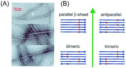
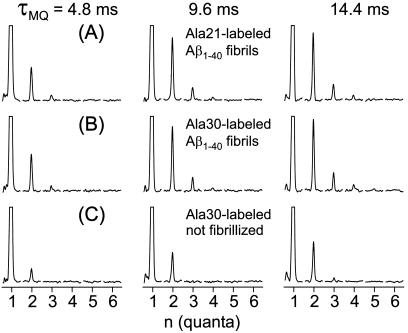
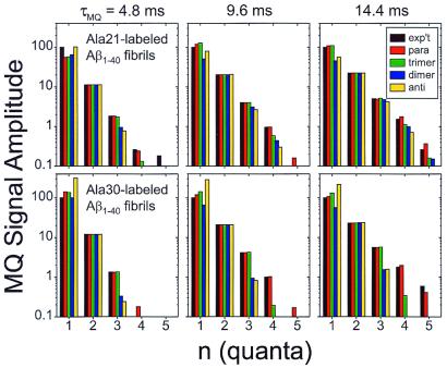
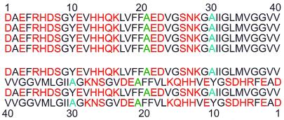
Senile plaques associated with Alzheimer's disease contain deposits of fibrils formed by 39- to 43-residue beta-amyloid peptides with possible neurotoxic effects. X-ray diffraction measurements on oriented fibril bundles have indicated an extended beta-sheet structure for Alzheimer's beta-amyloid fibrils and other amyloid fibrils, but the supramolecular organization of the beta-sheets and other structural details are not well established because of the intrinsically noncrystalline, insoluble nature of amyloid fibrils. Here we report solid-state NMR measurements, using a multiple quantum (MQ) (13)C NMR technique, that probe the beta-sheet organization in fibrils formed by the full-length, 40-residue beta-amyloid peptide (Abeta(1-40)). Although an antiparallel beta-sheet organization often is assumed and is invoked in recent structural models for full-length beta-amyloid fibrils, the MQNMR data indicate an in-register, parallel organization. This work provides site-specific, atomic-level structural constraints on full-length beta-amyloid fibrils and applies MQNMR to a significant problem in structural biology.


Figures

Figure 1
Figure 1
(A) Electron micrographs of negative-stained Aβ1–40 fibrils adsorbed to carbon films from an Aβ1–40 solution after incubation at 24°C and pH 7.4 for 3 days. Typical amyloid fibrils are observed, appearing as single filaments or bundles of filaments with overall diameters ranging from 8 to 20 nm and with twist periodicities between 40 and 150 nm. The same solution, in which the Aβ1–40 peptides were labeled with 13C at the methyl carbon of Ala-21, subsequently was lyophilized for MQNMR measurements shown in Fig. 2. (B) Aβ1–40 fibrils are believed to have a predominantly β-sheet structure with peptide chains (blue arrows) approximately perpendicular to and hydrogen bonds approximately parallel to the long axis of the fibril (green arrow). Four candidates for the supramolecular organization of the fibrils are shown. These can be distinguished experimentally by incorporating 13C labels (red dots) at a single site in the peptide and measuring 13C multiple quantum NMR spectra, because observation of an n-quantum signal requires that at least n 13C nuclei be close enough in space to have significant magnetic dipole-dipole couplings.
Figure 2
Figure 2
13C MQNMR spectra of fibrillized and unfibrillized Aβ1–40 samples, shown in order of increasing MQ excitation time τMQ. Each MQ spectrum is displayed as a series of subspectra for MQ orders from 1 to 6, with a spectral window from −15 kHz to + 15 kHz in each subspectrum. Vertical scales are adjusted so that one-quantum peaks are clipped at 25% of their maximum values. In the fibrillized samples (A and B), the amplitudes of two-, three-, and four-quantum signals increase with increasing τMQ. Spectra of samples with 13C labels at methyl carbons of Ala-21 and Ala-30 are nearly identical. In unfibrillized samples (C), the three-quantum amplitude is small and no four-quantum signal is observed.
Figure 3
Figure 3
Comparison of experimental MQNMR amplitudes (black) with simulations for parallel (red), trimeric (green), dimeric (blue), and antiparallel organizations of β-sheets in Aβ1–40 fibrils, for samples labeled with 13C at methyl carbons of Ala-21 and Ala-30. Experimental MQNMR amplitudes are normalized to a one-quantum amplitude of 100. A logarithmic vertical scale is required because the amplitudes vary over 2 orders of magnitude. The parallel β-sheet model fits all of the experimental data most closely. Experimental amplitudes were determined from MQNMR spectra in Fig. 2 by integrating each subspectrum over the interval from −2 kHz to +3 kHz. Uncertainties in the experimental amplitudes, evaluated as the rms noise integrated over a 5-kHz-wide interval, are ±0.11, ±0.14, and ±0.14 for the Ala-21-labeled Aβ1–40 fibril data, and ±0.15, ±0.17, and ±0.24 for the Ala-30-labeled Aβ1–40 fibril data, for τMQ = 4.8 ms, 9.6 ms, and 14.4 ms, respectively.
Figure 4
Figure 4
The amino acid sequence of Aβ1–40 contains hydrophobic (black) and nonhydrophobic (red) residues. Ala-21 and Ala-30, sites that are shown by the MQNMR data to participate in parallel β-sheets, are colored green and blue, respectively. An in-register, parallel alignment of neighboring peptide chains in a β-sheet (Upper) juxtaposes the hydrophobic segments of neighboring chains, whereas an antiparallel alignment (Lower) does not. Hydrophobic interactions may dictate the β-sheet organization in Aβ1–40 fibrils.

References

    1. Sipe J D. Annu Rev Biochem. 1992;61:947–975. - PubMed
    1. Sunde M, Blake C C F. Q Rev Biophys. 1998;31:1–39. - PubMed
    1. Chiti F, Webster P, Taddei N, Clark A, Stefani M, Ramponi G, Dobson C M. Proc Natl Acad Sci USA. 1999;96:3590–3594. - PMC - PubMed
    1. Jimenez J L, Guijarro J L, Orlova E, Zurdo J, Dobson C M, Sunde M, Saibil H R. EMBO J. 1999;18:815–821. - PMC - PubMed
    1. Blake C, Serpell L. Structure (London) 1996;4:989–998. - PubMed

Publication types

MeSH terms

LinkOut - more resources