. 2000 Nov 21;97(24):13372-7.
doi: 10.1073/pnas.230362997.

Role of the calcium-binding protein parvalbumin in short-term synaptic plasticity

Affiliations

Role of the calcium-binding protein parvalbumin in short-term synaptic plasticity

O Caillard et al. Proc Natl Acad Sci U S A. .

Abstract

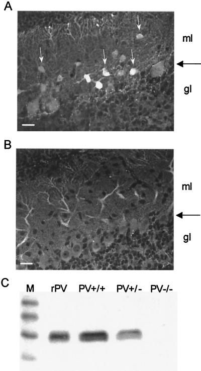
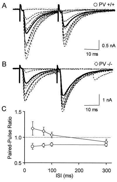
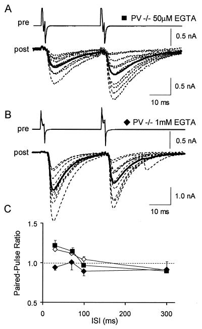
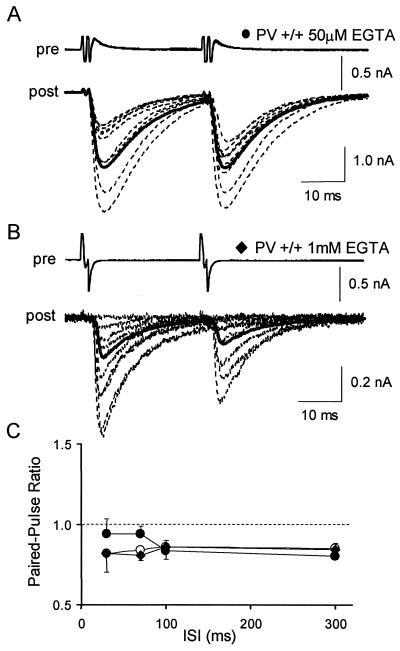
GABAergic (GABA = gamma-aminobutyric acid) neurons from different brain regions contain high levels of parvalbumin, both in their soma and in their neurites. Parvalbumin is a slow Ca(2+) buffer that may affect the amplitude and time course of intracellular Ca(2+) transients in terminals after an action potential, and hence may regulate short-term synaptic plasticity. To test this possibility, we have applied paired-pulse stimulations (with 30- to 300-ms intervals) at GABAergic synapses between interneurons and Purkinje cells, both in wild-type (PV+/+) mice and in parvalbumin knockout (PV-/-) mice. We observed paired-pulse depression in PV+/+ mice, but paired-pulse facilitation in PV-/- mice. In paired recordings of connected interneuron-Purkinje cells, dialysis of the presynaptic interneuron with the slow Ca(2+) buffer EGTA (1 mM) rescues paired-pulse depression in PV-/- mice. These data show that parvalbumin potently modulates short-term synaptic plasticity.


Figures

Figure 1
Figure 1
Expression of PV in the cerebellum. (A) Immunohistochemical staining of a cerebellar slice (sagittal cut) from a PV+/+ mouse (P8). The Purkinje cell layer is marked by a large horizontal arrow. PV-ir interneurons are localized in the molecular layer (ml; white arrows). No PV-ir cells are found in the granule cell layer (gl). Dendritic trees of Purkinje cells extending into two thirds of the molecular layer are also stained. Scale bar = 25 μm. (B) A cerebellar slice from a PV−/− mouse (P11) stained under identical conditions does not show any specific PV immunoreactivity. Contrast was greatly increased in comparison to A. (C) Western blot from cerebellar protein extracts reveal a clear signal in PV+/+ mice, a reduced one in PV+/−, and no detectable signal in PV−/− animals. rPV, recombinant rat PV as positive control; M, marker proteins (sizes in kDa from top to bottom: 31, 21, 14, and 7).
Figure 2
Figure 2
Absence of paired-pulse depression in PV−/− mice. (A) Superimposed IPSCs recorded in a Purkinje cell of a PV+/+ mouse after double extracellular stimulations of GABAergic interneurons (30ms ISI, dotted traces), and average response for the entire experiment (solid trace). (B) Representative results from a similar experiment performed with a PV−/− mouse. (C) Average PPR as a function of ISI in PV+/+ mice (circles, n = 12) and PV−/− mice (diamonds, n = 19). Note that, in this figure and in the following ones, failures are included in the calculation of mean traces.
Figure 3
Figure 3
Paired-pulse depression can be rescued in PV−/− mice by EGTA. (A) Superimposed IPSCs recorded in a Purkinje cell (dotted traces, post) of a PV−/− mouse after induction of two action potentials in a presynaptic interneuron by depolarizing steps (30 ms ISI) and average response for the entire experiment (solid trace). The interneuron was recorded with a solution containing 50 μM EGTA. (B) Representative results from a similar experiment where the interneuron was recorded with a solution containing 1 mM EGTA. (C) Average PPR as a function of ISI in PV−/− mice in conditions where interneurons were extracellularly stimulated (open diamonds, same data as in Fig. 2C), or were recorded with a solution containing 50 μM EGTA (filled squares, n = 13), or were recorded with a solution containing 1 mM EGTA (filled diamonds, n = 11).
Figure 4
Figure 4
Effect of presynaptic dialysis of EGTA in PV+/+ mice. (A) Superimposed IPSCs recorded in a Purkinje cell (dotted traces, post) of a PV+/+ mouse after induction of two action potentials in a presynaptic interneuron by depolarizing steps (30 ms ISI) and average response for the entire experiment (solid trace). The interneuron was recorded with a solution containing 50 μM EGTA. (B) Representative results from a similar experiment where the interneuron was recorded with a solution containing 1 mM EGTA. (C) Average PPR as a function of ISI in PV+/+ mice in conditions where interneurons were extracellularly stimulated (open circles, same data as in Fig. 2C), or were recorded with a solution containing 50 μM EGTA (filled circles, n = 11), or were recorded with a solution containing 1 mM EGTA (filled diamonds, n = 9).
Figure 5
Figure 5
Average PPR at 30 ms ISI in different experimental conditions. (A) Average PPR at 30 ms ISI in PV+/+ (n = 12) and in PV−/− mice (n = 19). The interneurons were stimulated by using an extracellular stimulation. The PPRs are significantly different (asterisks). (B) Average PPR at 30 ms ISI in paired recordings. The interneurons were stimulated by depolarizing steps in whole-cell voltage-clamp configuration. The presynaptic recording solution contained different concentrations of EGTA or Mg2+. Significant differences can be observed between PV+/+ and PV−/− for two different presynaptic solutions (dots and triangles). Both in PV+/+ and in PV−/− mice, the PPR is significantly reduced by increasing the concentration of EGTA (open and filled squares).

References

    1. Debanne D, Guerineau N C, Gähwiler B H, Thompson S M. J Physiol (London) 1996;491:163–176. - PMC - PubMed
    1. Dittman J S, Kreitzer A C, Regehr W G. J Neurosci. 2000;20:1374–1385. - PMC - PubMed
    1. Zucker R S. Annu Rev Neurosci. 1989;12:13–31. - PubMed
    1. Zucker R S. Curr Opin Neurobiol. 1999;9:305–313. - PubMed
    1. Bertram R, Sherman A, Stanley E F. J Neurophysiol. 1996;75:1919–1931. - PubMed

Publication types

MeSH terms

LinkOut - more resources