. 2000 Nov 21;97(24):13103-7.
doi: 10.1073/pnas.240390697.

Regulation of apoptosis at cell division by p34cdc2 phosphorylation of survivin

Affiliations

Regulation of apoptosis at cell division by p34cdc2 phosphorylation of survivin

D S O'Connor et al. Proc Natl Acad Sci U S A. .

Abstract

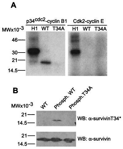
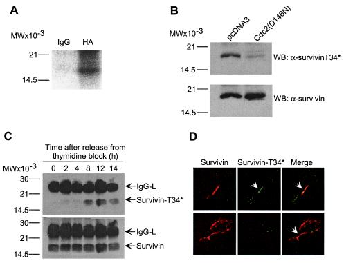
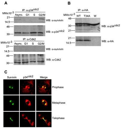
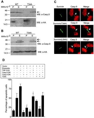
The interface between apoptosis (programmed cell death) and the cell cycle is essential to preserve homeostasis and genomic integrity. Here, we show that survivin, an inhibitor of apoptosis over-expressed in cancer, physically associates with the cyclin-dependent kinase p34(cdc2) on the mitotic apparatus, and is phosphorylated on Thr(34) by p34(cdc2)-cyclin B1, in vitro and in vivo. Loss of phosphorylation on Thr(34) resulted in dissociation of a survivin-caspase-9 complex on the mitotic apparatus, and caspase-9-dependent apoptosis of cells traversing mitosis. These data identify survivin as a mitotic substrate of p34(cdc2)-cyclin B1 and suggest that survivin phosphorylation on Thr(34) may be required to preserve cell viability at cell division. Manipulation of this pathway may facilitate the elimination of cancer cells at mitosis.


Figures

Figure 1
Figure 1
Survivin phosphorylation on Thr34 by p34cdc2-cyclin B1. (A) Kinase assay. Wild-type (WT) survivin, survivin(T34A) (T34A), or histone H1 (H1) were incubated with the indicated baculovirus-expressed kinase complexes, and phosphorylated bands were visualized by autoradiography. (B) Recognition of Thr34 phosphorylated survivin by Western blotting. WT survivin or survivin(T34A) were immunoblotted with affinity-purified antibodies to phosphorylated Thr34 (α-survivinT34*) or survivin (α-survivin) (12), before or after kinase assay (phosph.) with p34cdc2-cyclin B1. Relative molecular weight markers are shown on the left of each panel. WB, Western blotting.
Figure 2
Figure 2
Regulation of survivin phosphorylation by p34cdc2 in vivo. (A) In vivo phosphorylation of survivin. HeLa cells were transfected with HA-survivin, labeled with 200 μCi/ml 32PI and immunoprecipitated with control IgG or anti-HA followed by autoradiography. (B) Requirement of p34cdc2 kinase activity for survivin phosphorylation on Thr34. Endogenous survivin was immunoprecipitated from HeLa cells transfected with control pcDNA3 or kinase-dead p34cdc2 mutant (Cdc2(D146N)), and analyzed with α-survivinT34* or α-survivin, by Western blotting. No significant loss in cell viability was observed in HeLa cells expressing Cdc(D146N). (C) Kinetics of survivin phosphorylation on Thr34. Endogenous survivin was immunoprecipitated from cell cycle-synchronized HeLa cells at the indicated time intervals after thymidine release, and immunoblotted with α-survivinT34* or α-survivin. IgG-L, Ig light chain. (D) Localization of Thr34 phosphorylated survivin to midbodies. Endogenous survivin (Top) or HA-survivin(T34A) transfected in HeLa cells (Bottom) were labeled with mAb 8E2 to survivin (8) or an antibody to HA, respectively, plus α-survivinT34*. Binding of the primary antibodies was detected by addition of Texas Red (TR)-conjugated goat anti-mouse (survivin, red) and FITC-conjugated goat anti-rabbit (Thr34 phosphorylated survivin, green) antibodies. Coverslips were analyzed by confocal laser scanning microscopy. Image merging analysis is shown on the right. (A–C) Relative molecular weight markers are indicated on the left. WB, Western blotting.
Figure 3
Figure 3
Physical association between survivin and p34cdc2. (A) Cdk immunoprecipitation. HeLa cells asynchronously growing (Async.) or synchronized to G1, S, or G2/M were detergent-solubilized and immunoprecipitated (IP) with antibodies to p34cdc2 (Upper), or Cdk2 (Lower) followed by Western blotting with antibodies to survivin, p34cdc2, or Cdk2. (B) Survivin immunoprecipitation. HeLa cells transfected with HA-survivin or HA-survivin(T34A) were immunoprecipitated with an antibody to HA followed by Western blotting with antibodies to p34cdc2 or HA. W, whole extract. (C) Colocalization of survivin and p34cdc2 to the mitotic apparatus. Untransfected HeLa cells at the indicated phases of mitosis were labeled with mAb 8E2 to survivin (FITC, green) and a rabbit antibody to p34cdc2 (TR, red), and analyzed by confocal microscopy. Image merging analysis is shown on the right. (A and B) Relative molecular weight markers are on the left. WB, Western blotting.
Figure 4
Figure 4
Regulation of cell viability at mitosis by survivin phosphorylation on Thr34. (A) Nuclear morphology. HeLa cells transfected with GFP-vector (vector), GFP-survivin(T34A) (T34A), or GFP-caspase-9 (Casp.9) were scored morphologically by DAPI staining after a 48-h culture. The percentages of apoptotic cells were: GFP-vector, 3 ± 2.5; WT-survivin (not shown in the figure), 4.8 ± 5; survivin(T34A), 76.5 ± 1.4; caspase-9, 79.5 ± 9.1 (mean ± SD, n = 2–5). (B) Caspase-dependence. HeLa cells transfected with GFP-survivin or GFP-survivin(T34A) with or without 20 μM of the caspase inhibitor Z-VAD-fmk were analyzed for DNA content by propidium iodide staining and flow cytometry. The percentages of apoptotic cells with hypodiploid DNA content are indicated. Data are representative of one experiment of three independent determinations. (C) Tet-regulated expression of survivin(T34A). YUSAC2/T34A-C4 cells expressing survivin(T34A) on Tet removal (Tet-off system) were analyzed for DNA content after a 3-day culture in the presence or absence of Tet (Tet+/Tet). The percentages of cells with subG1 (apoptotic) DNA content are indicated. (Insets) Internucleosomal DNA fragmentation by TUNEL staining of Tet+ and Tet YUSAC2/T34A-C4 cells. (D) Kinetics of apoptosis and cell cycle progression during Tet-regulated expression of survivin(T34A). Synchronized YUSAC2/T34A-C4 with or without Tet were analyzed for DNA content at the indicated time intervals after thymidine release. The percentages of apoptotic cells under the various conditions tested was 0 h, Tet+ 3.8%/Tet 3.6%; 3 h, Tet+ 2.7%/Tet 10%; 6 h, Tet+ 4.8%/Tet 8.3%; 9 h, Tet+ 3.4%/Tet 9%; 12 h, Tet+ 2.7%/Tet 12.2%; 15 h, Tet+ 5%/Tet 20%; 18 h, Tet+ 4.3%/Tet 25%; 21 h, Tet+ 7%/Tet 32%; 24 h, Tet+ 11%/Tet 40%. (E) Western blotting of caspase-9 cleavage in synchronized Tet+ and Tet YUSAC2/T34A-C4 cells. Arrows, position of ≈46-kDa proform caspase-9 and of ≈35-kDa and ≈37-kDa active caspase-9 subunits. TNFα, Extracts of apoptotic HeLa cells treated with 10 ng/ml TNFα plus 10 μg/ml cycloheximide. Data are representative of one experiment of three independent determinations.
Figure 5
Figure 5
Modulation of survivin-caspase-9 complex by survivin phosphorylation on Thr34. (A) Immunoprecipitation from nonadherent cells. HeLa cells transfected with HA-survivin (WT) or HA-survivin(T34A) (T34A) were harvested after mitotic shake off, immunoprecipitated with anti-HA followed by Western blotting (WB) with an antibody to caspase-9 (casp.9). (B) Immunoprecipitation from synchronized cells. HeLa cells were transfected with HA-survivin (WT) or HA-survivin(T34A) (T34A), immunoprecipitated with anti-HA 12-h after thymidine release, and analyzed for coassociated caspase-9 by Western blotting. Arrows, position of 46-kDa proform caspase-9 and ≈35-kDa active caspase-9. P, pellet; S, supernatant. (A and B) Blots were sequentially immunoblotted with anti-HA. (C) Mislocalization of caspase-9 from midbodies in survivin(T34A)-expressing cells. HeLa cells transfected with HA-survivin (Survivin), survivin(T34A), or survivin(L64A) were labeled for survivin (FITC, green) with a mAb to HA or mAb 8E2 (Bottom), and caspase-9 (Casp.9, TR, red), and analyzed by confocal microscopy. Image-merging analysis is shown on the right. Arrows, differential localization of wild-type survivin, survivin(T34A), or survivin(L64A) with caspase-9 at midbodies. Experiments were repeated at least four times with comparable results. (D) Apoptosis in survivin(T34A)-expressing cells is mediated by caspase-9. HeLa cells transfected with the various indicated combinations of GFP-constructs, with or without etoposide (10 μg/ml) or TNFα (TNF, 10 ng/ml) plus cycloheximide (CHX, 10 μg/ml), were morphologically scored for nuclear fragmentation by DAPI staining. DN, dominant negative. *, P < 0.05; ***, P < 0.0005. Data are the mean ± SEM of four independent experiments.

References

    1. Vaux D L, Korsmeyer S J. Cell. 1999;96:245–254. - PubMed
    1. Green D R. Cell. 1998;94:695–698. - PubMed
    1. Evan G, Littlewood T. Science. 1998;281:1317–1322. - PubMed
    1. Pines J. Nat Cell Biol. 1999;1:E73–E79. - PubMed
    1. Ambrosini G, Adida C, Altieri D C. Nat Med. 1997;3:917–921. - PubMed

Publication types

MeSH terms