. 2000 Nov 1;14(21):2701-11.
doi: 10.1101/gad.184000.

Pax6 activity in the lens primordium is required for lens formation and for correct placement of a single retina in the eye

Affiliations

Pax6 activity in the lens primordium is required for lens formation and for correct placement of a single retina in the eye

R Ashery-Padan et al. Genes Dev. .

Abstract

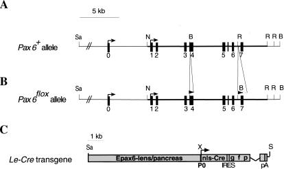
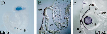
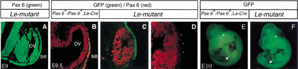
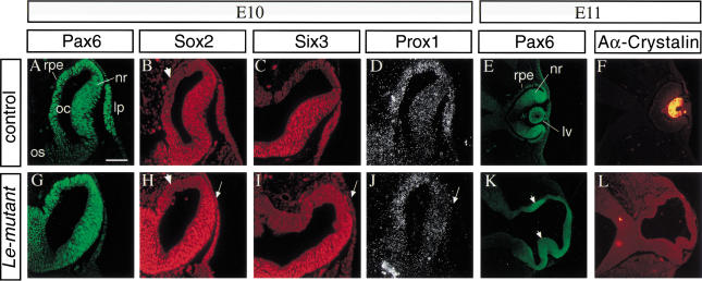
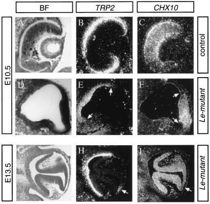
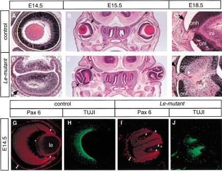
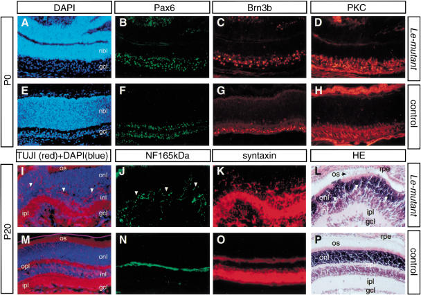
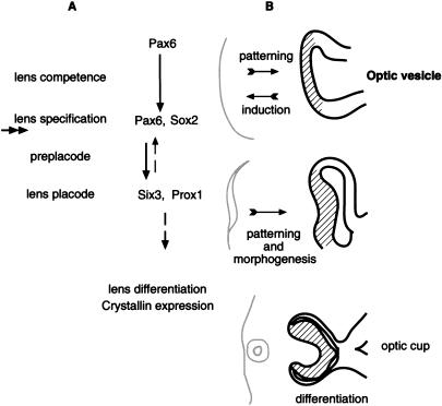
The Pax6 transcription factor plays a key role in ocular development of vertebrates and invertebrates. Homozygosity of the Pax6 null mutation in human and mice results in arrest of optic vesicle development and failure to initiate lens formation. This phenotype obscures the understanding of autonomous function of Pax6 in these tissue components and during later developmental stages. We employed the Cre/loxP approach to inactivate Pax6 specifically in the eye surface ectoderm concomitantly with lens induction. Although lens induction occurred in the mutant, as indicated by Sox2 up-regulation in the surface ectoderm, further development of the lens was arrested. Hence, Pax6 activity was found to be essential in the specified ectoderm for lens placode formation. Furthermore, this mutant model allowed us for the first time to address in vivo the development of a completely normal retina in the absence of early lens structures. Remarkably, several independent, fully differentiated neuroretinas developed in a single optic vesicle in the absence of a lens, demonstrating that the developing lens is not necessary to instruct the differentiation of the neuroretina but is, rather, required for the correct placement of a single retina in the eye.


Figures

Figure 1
Figure 1
Targeted insertion of loxP sites into the Pax6 gene and analysis of Cre activity in the Le-Cre transgenic line. Structure of wild-type (A) and targeted (B) Pax6 loci. One loxP site was introduced in exon 4 upstream of the first ATG. The second loxP was followed by the selection cassette flanked by FRT sequences inserted between exons 6 and 7. The selection cassette was removed to establish the Pax6flox allele (see Materials and Methods). (C) Schematic representation of Le-Cre transgene. A 6.5-kb SacII/XmnI genomic region including the upstream regulatory sequences and the first Pax6 promoter (P0) cloned upstream of sequences encoding the nls-Cre followed by internal ribosome binding sites (IRES) and green fluorescent protein (GFP). The recombination pattern was detected by enzymatic reaction (Lobe et al. 1999) on whole mount (D) or sections (E,F) of Z/AP;Le-Cre embryos at E9.5 (D,E) and E15.5 (F). (Arrows) Transcription start sites; (filled rectangles) exons; (open triangle) FRT; (filled triangles) loxP. (c) Cornea; (con) conjuctiva; (el) eyelid; (le) lens; (nls) nuclear localization signal; (nr) neuroretina; (ov) optic vesicle; (p) pancreas; (pA) poly A; (rpe) retinal pigmented epithelium; (se) surface ectoderm. (B) BamHI; (N) NotI; (R) EcoRV; (Sa) SacII; (S) Sfi; (X) XmnI.
Figure 1
Figure 1
Targeted insertion of loxP sites into the Pax6 gene and analysis of Cre activity in the Le-Cre transgenic line. Structure of wild-type (A) and targeted (B) Pax6 loci. One loxP site was introduced in exon 4 upstream of the first ATG. The second loxP was followed by the selection cassette flanked by FRT sequences inserted between exons 6 and 7. The selection cassette was removed to establish the Pax6flox allele (see Materials and Methods). (C) Schematic representation of Le-Cre transgene. A 6.5-kb SacII/XmnI genomic region including the upstream regulatory sequences and the first Pax6 promoter (P0) cloned upstream of sequences encoding the nls-Cre followed by internal ribosome binding sites (IRES) and green fluorescent protein (GFP). The recombination pattern was detected by enzymatic reaction (Lobe et al. 1999) on whole mount (D) or sections (E,F) of Z/AP;Le-Cre embryos at E9.5 (D,E) and E15.5 (F). (Arrows) Transcription start sites; (filled rectangles) exons; (open triangle) FRT; (filled triangles) loxP. (c) Cornea; (con) conjuctiva; (el) eyelid; (le) lens; (nls) nuclear localization signal; (nr) neuroretina; (ov) optic vesicle; (p) pancreas; (pA) poly A; (rpe) retinal pigmented epithelium; (se) surface ectoderm. (B) BamHI; (N) NotI; (R) EcoRV; (Sa) SacII; (S) Sfi; (X) XmnI.
Figure 2
Figure 2
Elimination of Pax6 protein from the SE is completed at E9.5. (A) Pax6 protein is detected in transverse paraffin section of E9 Le-mutant OV and SE. At E9.5, Pax6 is not detected in the SE of the Le-mutant (C,D, arrowheads), but is present in the SE of Pax6+/Pax6+;Le-Cre control (B). (D) Magnification of Pax6 distribution detected in (C). In the SE, GFP fluorescence is detected in cryosections of E9.5 control (B) and Le-mutant (C) eyes. (E) From E10, GFP fluorescence is detected in the pancreas (open arrowhead) and lens (filled arrowhead) of Pax6+/Pax6+;Le-Cre embryos. (F) In the Le-mutant E10 embryos, GFP is only detected in the pancreas and not in the eyes. The whole embryo appears slightly green because of unspecific fluorescence observed also in completely wild-type littermates. (ov) optic vesicle; (se) surface ectoderm.
Figure 3
Figure 3
The lens ectoderm is specified, but lens placode does not form in the Le-mutant eyes. Molecular analysis of the phenotype of E10 (AD, GJ) and E11 (E,F,K,L) embryos by indirect immunofluorescence. Transverse paraffin sections, double labeled with specific antibodies to Pax6 (A,G) and Sox2 (B,H). Pax6 (G) is not detected above background levels in the SE of the Le-mutant embryos, while Sox2 is detected in the SE (H, arrow) similar to the up-regulation of Sox2 observed in control eyes (B). Adjacent section immunolabeled with antibodies to Six3 (C,I) show that Six3 is not detected above background level in the Le-mutant (I) ectoderm but is expressed in the lens placode of control littermate (C). (D,J) In situ hybridization with 35S labeled Prox1 RNA probe demonstrate that Prox1, which is normally expressed in the lens placode (D), is not detected above background levels in the Le-mutant SE (J). Double immunolabeling with specific antibodies to Pax6 (E,K) and αA-crystallin on E11 control (F) and mutant eyes (L). In the Le-mutant eye, two regions of the OV seem to invaginate, and Pax6 is enhanced at these regions (K, arrowheads). Arrows in HJ, point to the lens ectoderm. Arrowheads in B and H mark regions of presumptive RPE in which Sox2 is not detected above background level. (lp) Lens placode; (lv) lens vesicle; (nr) neuroretina; (oc) optic cup; (os) optic stalk; (rpe) retinal pigmented epithelium. Scale bar 50 μm in AD, GJ and 100μm in E,F,K,L.
Figure 4
Figure 4
The initial delineation of prospective NR and RPE dissolve to several NR and RPE domains. In situ hybridization with 35S-labeled TRP2 or Chx10 RNA probes on adjacent sections through the eye regions of E10.5 and E13.5 eyes. Bright field images (BF) shown in A, D, and G. In the eye of E10.5 control littermate (AC), Trp2 is localized in the prospective RPE located in the outer layer of the cup (B), while Chx10 is expressed in the NR layer (C). Cup formation is delayed in the Le-mutant eyes (D). Trp2, however, is expressed in the proximal (E, between arrows) region, while Chx10 transcripts demarcate the prospective NR located distally (F, between arrows). At E13.5, invagination of the NR forms two folds in the Le-mutant eye (G). Trp2 is expressed not only in the proximal part of the Le-mutant eyecup but also in patches of cells located between the two retina folds (arrow in H). Chx10 is distributed in the two folds and is down-regulated in the region between them (arrow in I).
Figure 5
Figure 5
Several neuroretinas form in the absence of all lens structures in the Le-mutant mice. Hematoxilin-eosin (HE) staining (AF) of transverse (B,C,E,F) or sagittal sections (A,D) through the eye region of the Le-mutant (DF) and control littermates; Paxflox/Pax6lacZ (A,C) or Pax6+/Pax6+ (B). Cell layers including nerve fiber layer (nfl), inner nuclear layer (inl), and outer nuclear layer (onl) are distinguished in the Le-mutant (F) and in the control eye (C) at E18.5. The black arrows point out the RPE (BF). Note that the axons exit each retina fold not from the optic disc but, rather, from an opening between the folds, which in a normal eye would have been occupied by the lens (white arrowheads in E,F). Furthermore, an optic nerve is observed in Le-mutant eyes (F). Coimmunolabeling with antibodies to Pax6 (G,I) and to βIII tubulin (H,J) show that each retina fold develops autonomously in the Le-mutant eye. Enhanced Pax6 expression is marked with arrowheads, RPE with two arrowheads. (le) Lens; (onh) optic nerve head; (*) unspecific fluorescence from blood cells.
Figure 6
Figure 6
Retinal lamination and the major classes of retinal neurons are present in the Le-mutant retina. The Le-mutant neuroretina displays restricted Pax6 distribution (B) and lamination into nbl and gcl (A) similar to the single retina of the control eye (E,F). Immunohistochemistry and histology on newborn (P0, AH) and mature (P20, IP) eyes in adjacent sections of retina. Note the presence of retinal ganglion cells (Brn3b, C,G), bipolar cells (protein kinase C, D,H), horizontal cells (neurofilament 165 kD, J,N), amacrine cells (syntaxin, K,O), photoreceptor cells (TUJI cell bodies and processes in the onl, I,M), and photoreceptor outer segments (HE staining, L,P) in the Le-mutant (AD,IL) and the control (EH,MP) retina. All laminae, onl, inl, gcl, are present in the mature Le-mutant retina, as revealed by HE (L,P) and DAPI+TUJ1 (I,M) staining. However, onl and inl are not separated by a pronounced opl (arrowheads in I,J). The absence of a continuous opl in the Le-mutant retina possibly accounts for the dispersed appearance of horizontal cell processes between inl and onl (J). (gcl) ganglion cell layer; (HE) hematoxilin-eosin; (inl) inner nuclear layer; (ipl) inner plexiform layer; (nbl) neuroblast layer; (onl) outer nuclear layer; (opl) outer plexiform layer; (os) outer segments of photoreceptors; (rpe) retinal pigment epithelium.
Figure 7
Figure 7
A scheme summarizing the possible roles of Pax6 in the SE leading to lens placode formation (A). Three steps precede the formation of the lens placode: lens competence, lens specification, and the preplacode (maintenance) stage. The competence of the ectoderm to respond to the inductive signals from the OV is mediated by Pax6. During lens induction, the upregulation of Sox2 is dependent on Pax6 activity. Inactivation of Pax6 in Le-mutant occurred concomitantly with lens induction and after the up-regulation of Sox2 (the approximate time of inactivation is marked with double arrow). The expression of lens specific genes, Six3 and Prox1, in the preplacode stage is dependent on Pax6 activity. Feedback signals possibly mediated by six3 are required to maintain Pax6 expression in the SE (dashed arrow). The regulatory genes promote lens placode formation and subsequent lens differentiation. (B) Schematic presentation of the influence of the early lens structures (gray) on the patterning and morphology of the OV (black) in mice. The initial patterning of the OV to NR (lines) and RPE progenitors is not dependent on Pax6 activity in the specified ectoderm or on lens placode formation. However, the interaction with the early lens structures is required to define the position of the optic cup and for maintaining the identity of the presumptive NR and RPE domains. Subsequent differentiation of the retina is not dependent on lens structures.

References

    1. Bao ZZ, Cepko CL. The expression and function of Notch pathway genes in the developing rat eye. J Neurosci. 1997;17:1425–1434. - PMC - PubMed
    1. Barnstable CJ. A molecular view of vertebrate retinal development. Mol Neurobiol. 1987;1:9–46. - PubMed
    1. Collinson JM, Hill RE, West JD. Different roles for pax6 in the optic vesicle and facial epithelium mediate early morphogenesis of the murine eye. Development. 2000;127:945–956. - PubMed
    1. Coulombre AJ, Coulombre JL. Lens development. I. Role of the lens in eye growth. J Exp Zool. 1964;156:39–47. - PubMed
    1. Cvekl A, Piatigorsky J. Lens development and crystallin gene expression: Many roles for Pax-6. Bioessays. 1996;18:621–630. - PubMed

Publication types

MeSH terms