Comparative Study
. 2000 Nov 1;14(21):2712-24.
doi: 10.1101/gad.835000.

Regulation of cellular growth by the Drosophila target of rapamycin dTOR

Affiliations
Comparative Study

Regulation of cellular growth by the Drosophila target of rapamycin dTOR

H Zhang et al. Genes Dev. .

Abstract

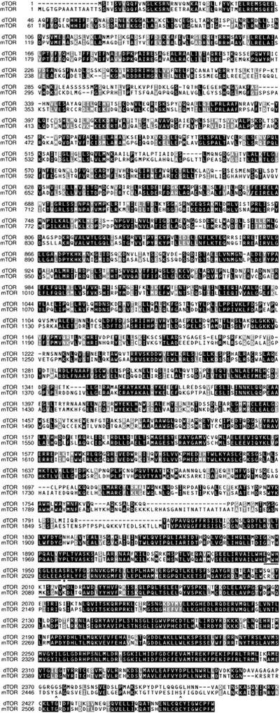
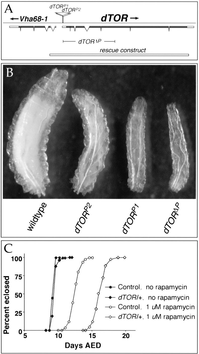
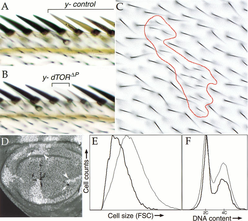
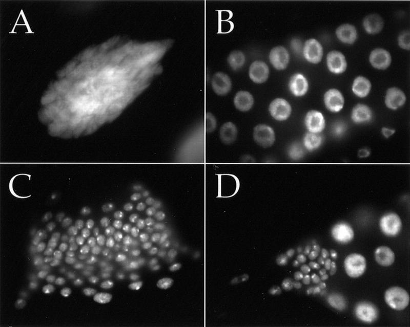
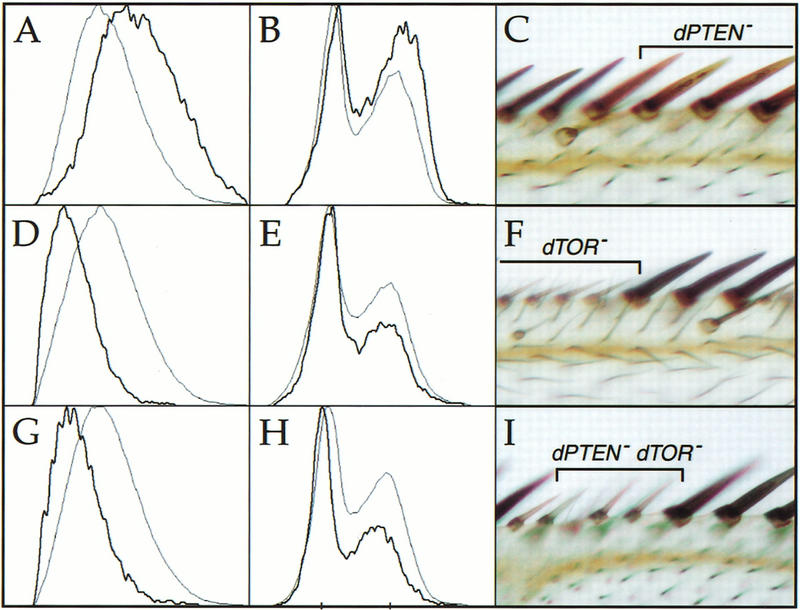
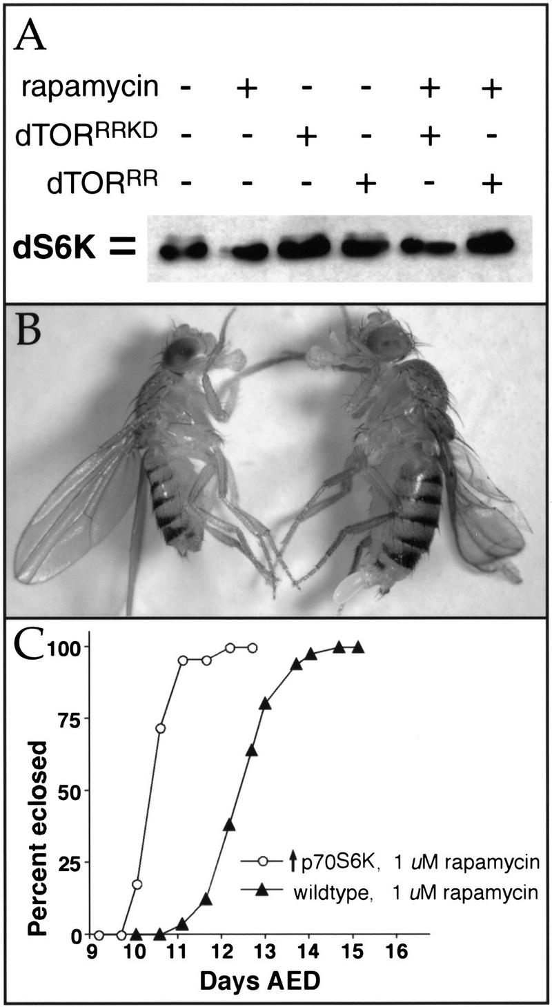
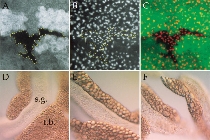
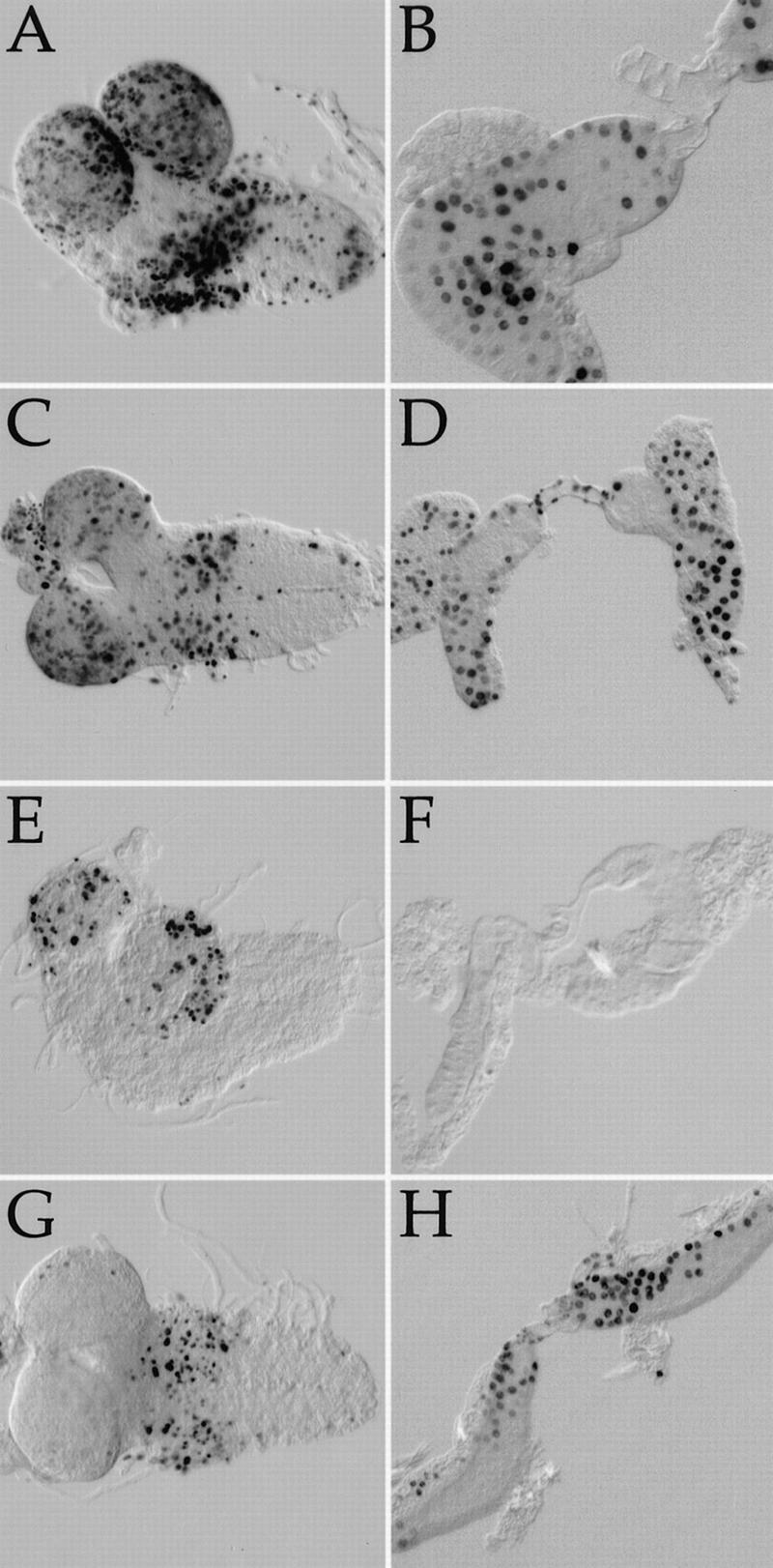
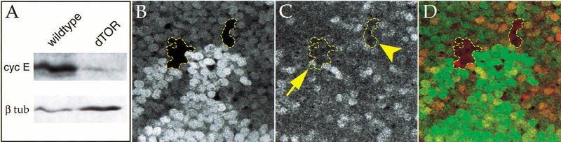
The TOR protein kinases (TOR1 and TOR2 in yeast; mTOR/FRAP/RAFT1 in mammals) promote cellular proliferation in response to nutrients and growth factors, but their role in development is poorly understood. Here, we show that the Drosophila TOR homolog dTOR is required cell autonomously for normal growth and proliferation during larval development, and for increases in cellular growth caused by activation of the phosphoinositide 3-kinase (PI3K) signaling pathway. As in mammalian cells, the kinase activity of dTOR is required for growth factor-dependent phosphorylation of p70 S6 kinase (p70(S6K)) in vitro, and we demonstrate that overexpression of p70(S6K) in vivo can rescue dTOR mutant animals to viability. Loss of dTOR also results in cellular phenotypes characteristic of amino acid deprivation, including reduced nucleolar size, lipid vesicle aggregation in the larval fat body, and a cell type-specific pattern of cell cycle arrest that can be bypassed by overexpression of the S-phase regulator cyclin E. Our results suggest that dTOR regulates growth during animal development by coupling growth factor signaling to nutrient availability.


Figures

Figure 1
Figure 1
Comparison of Drosophila and human TOR proteins. The ClustalW 1.8 algorithm was used to align the amino acid sequences of dTOR and human mTOR/FRAP (GenBank accession no. 744518). Dark boxes indicate identical amino acids; gray boxes indicate similarity. The conserved FKBP12–rapamycin-binding domain (amino acids 2025–2114 in mTOR; Vilella-Bach et al. 1999) is underlined, and the essential serine in this domain is indicated by an asterisk. Sites of mTOR autophosphorylation (Ser-2481) and phosphorylation by Akt/PKB (Ser-2448) are marked with carats.
Figure 2
Figure 2
Mutations in dTOR inhibit larval growth. (A) The structure of the 8.0-kb dTOR transcript is shown. Coding sequence is indicated by filled boxes; 5′ and 3′ untranslated regions are represented by open boxes. Breaks in the boxes indicate introns. Sites of the two P-element insertions in dTOR are indicated, and the range of the dTORΔP deletion is shown. The stippled bar at the bottom of the figure indicates the DNA fragment used for genomic rescue. Vha68-1 encodes a mitochondrial ATPase transcribed in the opposite direction as dTOR. (B) Size comparison of wild-type and dTOR homozygous mutant larvae at 126 h of development. Wild-type larvae pupate within 12 h of this time point, whereas dTOR mutants remain arrested in the larval stage with little or no further growth. (C) Heterozygosity for dTOR sensitizes larvae to rapamycin. On normal food, wild-type (black circles) and dTOR/+ larvae (black diamonds) grow at indistinguishable rates. Addition of 1 μM rapamycin delays development by ∼3 d in wild-type (open circles) and ∼6 d in dTOR/+ larvae (open diamonds).
Figure 3
Figure 3
dTOR clonal phenotypes. (A–C) Loss of dTOR reduces the size of cells in the adult wing. Clones of y-marked wild-type (A) or dTORΔP (B,C) cells were induced during the first larval instar by FLP/FRT-mediated mitotic recombination. Representative clones including bristles of the wing margin (A,B) and epithelial cells of the wing blade (C) are shown. The red trace in C outlines a dTORΔP mutant clone. Each hairlike structure is a trichome emanating from a single epithelial cell. Note the reduced size and increased density of dTOR cells. Genotype of A–C: y,w, HS-FLP122/+; dTORΔP FRT40A/Py+ FRT40A. (D–F) Phenotypes of dTOR mutant cells during proliferation. (D) Confocal image of wing imaginal disc containing dTORΔP clones marked with ubiquitin–GFP. By 72 h after induction, dTORΔP mutant clones (indicated by arrows; cells lack marker) contain fewer cells than their wild-type twinspot clones (indicated by arrowheads; cells carry two copies of GFP marker). (E,F) FACS histograms of dissociated cells from wing discs containing dTORΔP clones, as in D. dTORΔP cells and wild-type control cells are indicated by heavy and light traces, respectively. Cells lacking dTOR have reduced forward light scatter (FSC) values, indicating a smaller cell size (E). Populations of dTOR mutant cells also have a lower fraction of cells in S and G2 phases of the cell cycle (F). Genotype of D–F: y, w, HS-FLP122/+; dTORΔP FRT40A/Ubi-GFP FRT40A.
Figure 4
Figure 4
Both mitotic and endoreplicative cell cycles require dTOR. Salivary gland nuclei stained with Hoechst 33258 are shown for wild type (A,C) and dTORΔP (B,D) at 5–6 d of development. (A–D) Same magnification. (A) A single endoreplicative nucleus of ∼1024 C from a wild-type salivary gland. Endoreplicative salivary gland nuclei from dTORΔP larvae reach a ploidy of only 16–32 C (B). Proliferative diploid cells of the imaginal ring reach ∼fivefold greater numbers in wild-type (C) than dTORΔP (D) salivary glands.
Figure 5
Figure 5
dTOR acts downstream of dPTEN. Clones of cells mutant for null alleles of dPTEN (A–C), dTOR (D–F), or dPTEN and dTOR (G–I) were induced by FLP/FRT-mediated mitotic recombination. Mutant cells were marked by the absence of GFP or y+ as in Fig. 3. The leftmost columns depict histograms of wing imaginal disc cells analyzed by flow cytometry; dark traces indicate mutant cells, and light traces represent wild-type control cells from the same discs. The right column shows photomicrographs of mutant clones in the anterior margin of the adult wing. Loss of dPTEN causes cell enlargement (A,C), and increases the proportion of cells in the S and G2 phases of the cell cycle (B). Loss of dTOR decreases cell size (D,E) and reduces the S and G2 populations (F). Cells lacking dTOR and dPTEN are indistinguishable from cells lacking dTOR alone (G–I). Genotypes: (A,B) y, w, HS-FLP122/+; dPTENDJ189FRT40A/Ubi-GFP FRT40A; (C) y, w, HS-FLP122/+; dPTENDJ189FRT40A/Py+ FRT40A. (D,E) y, w, HS-FLP122/+; dTORΔP FRT40A/Ubi-GFP FRT40A; (F) y, w, HS-FLP122/+; dTORΔP FRT40A/Py+ FRT40A. (G,H) y, w, HS-FLP122/ +; dTORΔP dPTENDJ189 FRT40A/Ubi-GFP FRT40A; (I) y, w, HS-FLP122/+; dTORΔP dPTENDJ189 FRT40A/Py+ FRT40A.
Figure 6
Figure 6
dTOR interacts with dS6K. (A) Immunoblot of extracts from Drosophila S2 cells transfected with HA-tagged dS6K, visualized with anti-HA antibodies. dS6K migrates as a doublet, and the slower migrating band (top), which represents phosphorylated dS6K, is abolished by treatment with rapamycin (lane 2; Stewart et al. 1996). Phosphorylation of dS6K is maintained in the presence of rapamycin when dTORS1956T (dTORRR, lane 6) but not kinase-inactive dTORS1956T (dTORRRKD, lane 5) is cotransfected with dS6K. (B) Constitutive expression of activated human p70S6K1 rescues dTOR flies to viability. Genotypes: (Left) dTORP2 UAS-p70S6K1-D4/dTORP1; Act5c-Gal4/+; (right) dTORP2 UAS-p70S6K1-D4/CyO; Act5c-Gal4/+. Note that the rescued dTOR fly (left) is smaller than the control. (C) Constitutive expression of dS6K provides rapamycin resistance. UAS-dS6K/+; Act5c-Gal4/+ flies cultured with 1 μM rapamycin eclose ∼3 d earlier than wild-type controls.
Figure 7
Figure 7
Loss of dTOR mimics starvation. (A–C) Wing imaginal disc containing a clone of dTORΔP cells marked by the absence of GFP (A) and stained with antifibrillarin to highlight nucleoli (B). The extent of the dTOR mutant clone is outlined in yellow. (C) An overlay of GFP (green) and antifibrillarin (red). Loss of dTOR causes a reduction in nucleolar area. (D–F) Differential interference contrast images of salivary glands (s.g.) and associated fat body (f.b.) from normally fed larvae (D), larvae that were deprived of amino acids for 4 d (E), and normally fed dTORΔP larvae (F). Starvation and loss of dTOR each cause aggregation of lipid vesicles in the fat body, as well as accumulation of proteinaceous granules in salivary gland cells.
Figure 8
Figure 8
Cell cycle behavior of dTOR cells. BrdU incorporation patterns in larval brains (A,C,E,G) and salivary glands (B,D,F,H). Darkly stained nuclei indicate cells that have incorporated BrdU and thus were in S-phase during the experiment. (A,B) Wild type at 3–4 d AED. (C,D) dTORΔP at 3–4 d AED. (E,F) dTORΔP at 5–6 d AED. Note that salivary gland cells no longer incorporate BrdU, whereas CNS neuroblasts continue to cycle. (G,H) dTORΔP carrying a single copy of a HS–cyclin E transgene at 6–7 d AED, after a 2-h heat shock. Ectopic expression of cyclin E drives quiescent dTORΔP salivary gland cells into S-phase.
Figure 9
Figure 9
Cyclin E expression is reduced in dTOR mutants. (A) Immunoblot of total larval extracts from dTORΔP and wild-type second instar larvae, probed with anti-cyclin E. The blot was also probed with anti-β-tubulin as a loading control. (B–D) Shown is a region of a wing imaginal disc containing dTORΔP clones 72 h after induction, marked by the absence of GFP (B) and stained with anti-cyclin E antibody (C). dTOR mutant clones are outlined in yellow. These images are superimposed in D, with GFP shown in green and cyclin E in red. The arrow indicates a single dTOR mutant cell expressing cyclin E; the arrowhead indicates a dTOR clone lacking cyclin E expression.

References

    1. Adams MD, Celniker SE, Holt RA, Evans CA, Gocayne JD, Amanatides PG, Scherer SE, Li PW, Hoskins RA, Galle RF, et al. The genome sequence of Drosophila melanogaster. Science. 2000;287:2185–2195. - PubMed
    1. Aris JP, Blobel G. Identification and characterization of a yeast nucleolar protein that is similar to a rat liver nucleolar protein. J Cell Biol. 1988;107:17–31. - PMC - PubMed
    1. Barbet N-C, Schneider U, Helliwell SB, Stansfield I, Tuite MF, Hall MN. TOR controls translation initiation and early G1 progression in yeast. Mol Biol Cell. 1996;7:25–42. - PMC - PubMed
    1. Beck T, Hall MN. The TOR signalling pathway controls nuclear localization of nutrient-regulated transcription factors. Nature. 1999;402:689–692. - PubMed
    1. Bohni R, Riesgo-Escovar J, Oldham S, Brogiolo W, Stocker H, Andruss BF, Beckingham K, Hafen E. Autonomous control of cell and organ size by CHICO, a Drosophila homolog of vertebrate IRS1–4. Cell. 1999;97:865–875. - PubMed

Publication types

MeSH terms