Comparative Study
. 2000 Nov 1;14(21):2757-70.
doi: 10.1101/gad.832000.

Characterization of fission yeast cohesin: essential anaphase proteolysis of Rad21 phosphorylated in the S phase

Affiliations
Comparative Study

Characterization of fission yeast cohesin: essential anaphase proteolysis of Rad21 phosphorylated in the S phase

T Tomonaga et al. Genes Dev. .

Abstract

Cohesin complex acts in the formation and maintenance of sister chromatid cohesion during and after S phase. Budding yeast Scc1p/Mcd1p, an essential subunit, is cleaved and dissociates from chromosomes in anaphase, leading to sister chromatid separation. Most cohesin in higher eukaryotes, in contrast, is dissociated from chromosomes well before anaphase. The universal role of cohesin during anaphase thus remains to be determined. We report here initial characterization of four putative cohesin subunits, Psm1, Psm3, Rad21, and Psc3, in fission yeast. They are essential for sister chromatid cohesion. Immunoprecipitation demonstrates stable complex formation of Rad21 with Psm1 and Psm3 but not with Psc3. Chromatin immunoprecipitation shows that cohesin subunits are enriched in broad centromere regions and that the level of centromere-associated Rad21 did not change from metaphase to anaphase, very different from budding yeast. In contrast, Rad21 containing similar cleavage sites to those of Scc1p/Mcd1p is cleaved specifically in anaphase. This cleavage is essential, although the amount of cleaved product is very small (<5%). Mis4, another sister chromatid cohesion protein, plays an essential role for loading Rad21 on chromatin. A simple model is presented to explain the specific behavior of fission yeast cohesin and why only a tiny fraction of Rad21 is sufficient to be cleaved for normal anaphase.


Figures

Figure 1
Figure 1
S. pombe cohesin genes and their disruption phenotypes. (A) Schematic representations of cohesin subunits. S. pombe subunits designated Psm1, Psm3, Rad21, and Psc3 are homologs of budding yeast Smc1p, Smc3p, Scc1p, and Scc3p, respectively. The conserved regions are hatched. The arrows indicate the positions of introns in the S. pombe genes. Except for Rad21, cohesin subunits were first identified in this study. (X) Xenopus; (h) human, (Dm) Drosophila; (Mm) mouse. (B) Phenotypes of gene-disrupted Δpsm1, Δpsm3, Δrad21, and Δpsc3. Gene-disrupted cells were observed after germination (33°C for 10 h) using DAPI for DNA and anti-tubulin (TUB) antibody. Aberrant segregation phenotypes of disrupted cells were very similar. Temperature-sensitive rad21-K1 (36°C for 4 h) and wild-type mitotic cells (26°C) are shown as control. The bar, 10 μm. (C) Localization of Mis12–GFP, a GFP-tagged kinetochore protein, in cohesin gene disruption mutant cells. Gene-disrupted cells (Δpsm1, Δrad21) expressing Mis12–GFP were observed after DAPI staining. The GFP dots represent kinetochores, the number of which exceeded more than three, indicating that sister kinetochores were prematurely separated. (D) The LacO repeat was integrated in rad21-K1 mutant about 30 kb apart from the inner centromere region of chromosome I and the repeat was visualized by the LacI–NLS protein tagged with GFP (Straight et al. 1996; Nabeshima et al. 1997). Cells grown at 26°C were shifted to 36°C for 4 h, and interphase cells were measured for the frequencies revealing two GFP signals (two examples shown in the left panel).
Figure 2
Figure 2
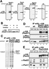
Stable complex formation of cohesin subunits. (A) Detection of Rad21, Psm1, Psm3, and Psc3 by immunoblot using anti-HA(12CA5), anti-Myc(9E10) monoclonal antibodies or polyclonal antibody against the recombinant Psm1 protein. Extracts of the strains chromosomally integrated with Rad21–3HA, Psc3–3HA, or Psm3–8Myc under the native promoter were used. Wild-type control produced no band by anti-HA or anti-Myc antibodies. (B) Extracts of cells expressing tagged Rad21–3HA were treated with (+) or without (−) λ protein phosphatase and a phosphatase inhibitor (10 mM sodium orthovanadate) at 30°C for 30 min. Upper modified bands were diminished and the lowest bands were intensified after treatment. (C) To identify stable complex formation of cohesin subunits, a strain chromosomally integrated with Rad21–3HA and Psm3–8Myc under the native promoter was constructed and employed for immunoprecipitation using antibodies against HA. Silver staining of the precipitated proteins is shown. Proteins present in the precipitates from the integrant strain but not from the wild-type corresponded to Psm3–8Myc and Psm1. (D) Extracts of cells prepared in (C) were examined by immunoblot using antibodies against anti-HA, anti-Myc, and anti-Psm1 antibodies. Psm1 and Psm3–8Myc were coprecipitated with Rad21–3HA. (E) Extracts of cells expressing tagged Psc3–3HA were prepared, and immunoprecipitation was performed using anti-HA antibodies. Precipitates obtained were immunoblotted using anti-HA, anti-Rad21, and anti-Psm1 antibodies. Neither Psm1 nor Rad21 was coprecipitated with Psc3–3HA.
Figure 3
Figure 3
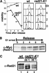
Essential role of Rad21 and its phosphorylation during the S phase. (A) The nitrogen-starved culture of rad21-K1 mutant at 26°C was transferred to the complete medium at 36°C for 8 h. Cell viability and the DNA content were measured by plating and FACScan analysis, respectively. The wild-type control is also shown. Cell viability in the mutant culture initially arrested at the G1 phase decreased during the S phase, which occurred 2–4 h after the shift. (B) Upper phosphorylated bands of Rad21 increased during the S phase. Wild-type cells integrated with the Rad21–8Myc gene were nitrogen starved at 26°C and shifted to the complete medium at 36°C for 8 h as in (A). Cell extracts made were immunoblotted using anti-Myc antibody. Hyperphosphorylated forms of Rad21 were abundant in 2–4 h and subsequently hypophosphorylated form increased (upper panel). Note that the single upper band faintly seen in the arrested cells was not hyperphosphorylated (see text). (C) Upper phosphorylation bands of Rad21 were examined in rad21-K1 mutant. Wild-type cells (lane 1), rad21-K1 at 26°C (lane 2) and at 36°C for 4 h (lane 3). Upper phosphorylated forms were greatly diminished in rad21 mutant cells.
Figure 4
Figure 4
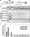
Interaction of Rad21 with centromere and pericentromere. (A) Left panel, schematic representation of cen1, the centromere of chromosome I. Positions of the centromere (cnt1, dg) or pericentromere (lys1) primers are indicated by the vertical lines. Right panel, ten primers derived from c1750, a cosmid covering the cdc2+ gene. (B) CHIP (chromatin immunoprecipitation) was performed using the probes described in A. Extracts of wild-type (wt, no tag), or integrants with Rad21–3HA, Psm1–3HA, Psc3–3HA, or Mis6–8Myc under the native promoter were made. Immunoprecipitation was done using anti-HA antibody (anti-Myc antibody used for the control Mis6), and PCR was done for the precipitates. Control Mis6, a kinetochore protein, was associated only with cnt1 (Saitoh et al. 1997). (C) Quantification of the PCR DNA products is shown. Serial dilutions of the template DNA were made for PCR reactions to optimize the PCR products within linear range. Coprecipitated DNAs with proteins were amplified and the percentage of precipitate DNA compared with total DNA was calculated by measuring the intensity of the PCR products.
Figure 5
Figure 5
Rad21 localizes in the nucleus throughout the cell cycle (A) Fixed wild-type cells expressing integrated Rad21–8Myc were stained by DAPI for DNA, by anti-Sad1 antibody against Sad1, an SPB protein, and by anti-Myc antibodies against Rad21-8Myc. Rad21 was localized in the nuclear chromatin region showing the punctate signals. (B) Localization of Psc3–HA and Rad21 was observed by immunofluorescence microscopy. The merged image is produced by red color Psc3–HA and green color Rad21. They were similar but did not appear to be identical. (C) The detergent-washing procedures (in situ chromatin binding assay; Kearsey et al. 2000) recently developed for visualising the S phase-specific nuclear retention of MCM protein essential for replication (Cdc21 is one of them) were applied to Rad21–GFP. Cdc21–GFP showed the nuclear signal only in the S phase cells, while Rad21–GFP signals were seen in all the cell cycle stages.
Figure 6
Figure 6
Behavior of wild-type and substitution mutant Rad21 proteins during the metaphase–anaphase transition in nda3-KM311. (A) nda3-KM311 strain integrated with the tagged Rad21–3HA gene under the native promoter was arrested at 20°C for 8 h, then shifted to 36°C, the permissive temperature, and aliquots of the culture were taken every 3 min. Extracts prepared were immunoprecipitated using anti-HA antibodies, and coprecipitated DNAs were amplified by PCR using the three different primers indicated. The level of Cdc13 is shown as the control of timing for cyclin destruction. The frequency of cells showing dividing nucleus in early anaphase (filled circles) peaked at 6 min when Cdc13 was rapidly degraded. Open triangles, cells containing a single nucleus; open rectangles, cells containing two nuclei. (B) Schematic drawing of the two putative arginine R cleavage sites in Rad21. (C) Prophase-arrested nda3-KM311 cells integrated with the wild type Rad21–8Myc (wt-Myc), single substitution mutants Rad21 R179E–8Myc or Rad21 R231E–8Myc under the native promoter were released to the permissive temperature as in A. Aliquots of the culture were taken every 3 min. Extracts were then prepared and immunoblotted using anti-Myc, anti-Cdc13 (mitotic cyclin) and anti-PSTAIRE antibodies. Top panel, immunoblot patterns taken after a brief exposure (α-Myc short exposure). Second panel, immunoblot pattern taken after the long exposure (α-Myc long exposure). The arrow and asterisk show the positions of cleaved fragments in wt-Myc, R179E-Myc and R231E-Myc. These cleaved products lack the N-terminal 231 and 179 amino acids, respectively. (D) Wild-type cells carrying plasmid alone (vector), the wild-type rad21+ gene, the single-cleavage mutants (R179E and R231E) or the double mutant R179ER231E under the REP41 promoter were streaked on the synthetic EMM2 plates with (promoter off) or without (promoter on) thiamine. Plates were incubated at 33°C. (E) Plasmid containing the double cleavage mutant gene R179ER231E under the REP81 promoter was introduced in the wild type strain integrated with the tagged Cut2–8Myc gene. Cells were grown in the liquid EMM2 medium in the presence of 2 μM thiamine at 33°C, followed by the removal of thiamine for 14 h in order to induce the expression of the toxic double-mutant protein R179ER231E. Cells were fixed and stained by DAPI for DNA, by anti-Sad1 antibodies for the SPB, and by anti-Myc antibodies for Cut2-Myc. The bar indicates 10 μm.
Figure 7
Figure 7
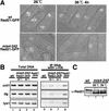
Recruitment and phosphorylation of Rad21 requires fully functional Mis4. (A) Punctate appearance of Rad21–GFP seen in the wild-type nucleus was diminished in mis4-242 at 26°C and completely vanished at 36°C for 4 h. Rad21–GFP was chromosomally integrated and expressed under the native promoter. Punctate staining did not correspond to localization of the centromeres. (B) Extracts of wild type cells (lanes 1,5), mis4-242 mutant cells integrated with Rad21–3HA grown at 26°C or 36°C (lanes 2,3,6,7) or the wild-type cells integrated with Rad21–3HA (lanes 4,8) were prepared and used for CHIP with anti-HA antibodies. Total DNA before immunoprecipitation (lanes 1–4) and coprecipitated DNA (lanes 5–8) were amplified by PCR using the probes, cnt1, dg, and lys1. Association of Rad21 with chromatin was greatly reduced at cnt1, dg locus and almost diminished at lys1 locus in mis4-242 mutant cells even at 26°C, the permissive temperature when compared with the untagged wild type. (C) Upper phosphorylation bands of Rad21 were examined in mis4-242 mutant. Wild-type cells at 26°C (lane 1), mis4-242 mutant cells at 26°C (lane 2), and at 36°C for 4 h (lane 3). Upper phosphorylated forms were greatly diminished in the mutant cells.

Similar articles

Cited by

References

    1. Birkenbihl RP, Subramani S. Cloning and characterization of rad21 an essential gene of Schizosaccharomyces pombe involved in DNA double-strand-break repair. Nucleic Acids Res. 1992;20:6605–6611. - PMC - PubMed
    1. Birkenbihl RP, Subramani S. The rad21 gene product of Schizosaccharomyces pombe is a nuclear, cell cycle-regulated phosphoprotein. J Biol Chem. 1995;270:7703–7711. - PubMed
    1. Castano IB, Brzoska PM, Sadoff BU, Chen H, Christman MF. Mitotic chromosome condensation in the rDNA requires TRF4 and DNA topoisomerase I in Saccharomyces cerevisiae. Genes & Dev. 1996;10:2564–2576. - PubMed
    1. Ciosk R, Zachariae W, Michaelis C, Shevchenko A, Mann M, Nasmyth K. An ESP1/PDS1 complex regulates loss of sister chromatid cohesion at the metaphase to anaphase transition in yeast. Cell. 1998;93:1067–1076. - PubMed
    1. Ciosk R, Shirayama M, Shevchenko A, Tanaka T, Toth A, Shevchenko A, Nasmyth K. Cohesin's binding to chromosomes depends on a separate complex consisting of Scc2 and Scc4 proteins. Mol Cell. 2000;5:243–254. - PubMed

Publication types

MeSH terms