. 2000 Nov 15;20(22):8323-30.
doi: 10.1523/JNEUROSCI.20-22-08323.2000.

R-Type Ca2+ channels are coupled to the rapid component of secretion in mouse adrenal slice chromaffin cells

Affiliations

R-Type Ca2+ channels are coupled to the rapid component of secretion in mouse adrenal slice chromaffin cells

A Albillos et al. J Neurosci. .

Abstract

Patch-clamp measurements of Ca(2+) currents and membrane capacitance were performed on slices of mouse adrenal glands, using the perforated-patch configuration of the patch-clamp technique. These recording conditions are much closer to the in vivo situation than those used so far in most electrophysiological studies in adrenal chromaffin cells (isolated cells maintained in culture and whole-cell configuration). We observed profound discrepancies in the quantities of Ca(2+) channel subtypes (P-, Q-, N-, and L-type Ca(2+) channels) described for isolated mouse chromaffin cells maintained in culture. Differences with respect to previous studies may be attributable not only to culture conditions, but also to the patch-clamp configuration used. Our experiments revealed the presence of a Ca(2+) channel subtype never before described in chromaffin cells, a toxin and dihydropyridine-resistant Ca(2+) channel with fast inactivation kinetics, similar to the R-type Ca(2+) channel described in neurons. This channel contributes 22% to the total Ca(2+) current and controls 55% of the rapid secretory response evoked by short depolarizing pulses. Our results indicate that R-type Ca(2+) channels are in close proximity with the exocytotic machinery to rapidly regulate the secretory process.


Figures

Fig. 1.
Fig. 1.
Voltage dependence of Ca2+currents and secretion in mouse adrenal slice chromaffin cells. Ca2+ currents and capacitance changes were recorded in the presence of 5 μm TTX and 2 mmCa2+ at different voltages. A,Ca2+ currents (Vh = −70 mV), evoked by depolarizing test potentials, as indicated.B, Corresponding Cm traces.C, Plot of ΔCm(ordinate, right) and QCa(ordinate, left) versus the potential. ΔCm reached peak values at the same voltage as QCa, and both declined above that potential.
Fig. 2.
Fig. 2.
P- and “Q-like”-type channels are present in mouse chromaffin cells of adrenal slices. A, Time course of Ca2+ currents measured at the end of 50 msec depolarizing pulses applied every 30 sec to 0 mV from a holding potential of −70 mV. The selective blockade of P-type channels was achieved by perfusion with 20 nm ω-Aga-IVA. Q-type channels were subsequently blocked by perfusion with 2 μmω-Aga-IVA. After wash out, the recovery was hastened by application of two trains of 20 pulses to +110 mV. Blank spaces in the record were caused by ramp voltage protocols applied at those points.B, Reversibility of P-type channel blockade in a different chromaffin cell. After a steady-state was reached in control conditions (Control trace), 20 nmω-Aga-IVA was perfused (ω-Aga-IVA trace), and the subsequent wash out led to an almost complete recovery of the Ca2+ current from the blockade (Wash out trace). C, D, Kinetics of inactivation of ω-Aga-IVA-sensitive Ca2+channels. Traces a (control), b (in the presence of 20 nm ω-Aga-IVA), c (in the presence of 2 μm ω-Aga-IVA), d(after wash out), and e (wash out after two trains of 20 pulses to +110 mV) from the cell of A, are plotted inC. The P channel was obtained as the difference between the control trace and the 20 nm ω-Aga-IVA trace (a, b). The “Q-like” channel was obtained as the difference between b and c traces. The P channel exhibited a slow inactivation, whereas the “Q-like” channel did not inactivate at all.
Fig. 3.
Fig. 3.
N- and L-type Ca2+ channels are present in mouse chromaffin cells in adrenal slices. A,Time course of inhibition and recovery of Ca2+currents during application of 1 μm ω-CTx-GVIA to a voltage-clamped mouse chromaffin cell. Perfusion with control solution but in the absence of Ca2+ (0 mmCa2+-2 mm EGTA) led to a rapid abolition of Ca2+ currents. Blank spaces in the record were caused by ramp voltage protocols applied at those points.B, Original traces before (tracea), in the presence of ω-CTx-GVIA (trace b), and after wash out (tracec), from the same cell as A. Note the partial recovery of the ω-CTx-GVIA blockade. C, Time course of inhibition and recovery of Ca2+ currents during application of 3 μm Nife. D, Original traces before (trace a), at the end of Nife application (trace b), and after wash out (tracec), from the same cell as C.
Fig. 4.
Fig. 4.
A Ca2+ channel current resistant to blockade mediates rapid secretory responses. InA, Ca2+ channel blockers (20 nm ω-Aga-IVA, 1 μm ω-CTx-GVIA, 3 μm ω-CTx-MVIIC, and 3 μm Nife) were added sequentially, as indicated to the right of each trace. No TTX was used in this experiment. Each compound was locally perfused onto the patched cell for ∼10 min. In B, blockers were added in a different cell simultaneously, as indicated by thetop horizontal bar (cocktail: 2 μm ω-Aga-IVA, 1 μm ω-CTx-GVIA, 3 μm ω-CTx-MVIIC, and 3 μm Nife). The temporal course of blockade of Ca2+ currents (Ifinal) and secretion (ΔCm) by the cocktail of blockers is shown in this panel. Pulses were applied every minute.C, Original recordings of both ΔCm, and the correspondingIfinal are shown for the sequential application of control solution, cocktail of blockers, wash out of the cocktail, 5 mm NiCl2, and Ca2+-free–2 mm EGTA in a different cell. D, The residual Ca2+ current exhibited rapid inactivation kinetics. The inactivation phases of control current and the current elicited after addition of the cocktail of blockers were well fitted with a single exponential function with τI = 24 and 14 msec, respectively.E, Voltage ramps from −120 to +60 mV were applied after currents had reached a steady-state with control and cocktail solutions. The ramp duration was 50 msec. They were corrected for leakage currents by subtracting the ramp in the presence of 200 μm CdCl2. The concentration of TTX in the experiments of B–E was 10 μm to ensure the blockade of Na+ channels.
Fig. 5.
Fig. 5.
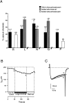
Relative contributions of Ca2+channel subtypes to the whole-cell Ca2+ current. Data from recordings in the whole-cell configuration of the patch-clamp technique in isolated cultured mouse chromaffin cells are compared to those from recordings in the perforated-patch configuration in chromaffin cells of mouse adrenal slices or isolated in culture.A,Black bars represent the data obtained from cells of adrenal medullary slices in the perforated-patch configuration (this study); bars withlines are data from isolated cells kept in culture in the whole-cell configuration (Hernández-Guijo et al., 1998);white bars represent data of R- and P-type channels from isolated cells recorded in the perforated-patch configuration (this study). B, Time course of Ca2+current blockade induced by 20 nm ω-Aga IVA, measured at the end of 50 msec depolarizing pulses to 0 mV, applied to a mouse chromaffin cell cultured for 2 d, under the perforated-patch configuration. The corresponding original traces for control conditions (trace a), in the presence of 20 nm ω-Aga IVA (trace b), or after wash out (trace c) are shown in C.
Fig. 6.
Fig. 6.
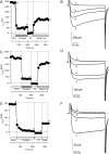
The resistant channel in isolated cultured mouse chromaffin cells. The time course of blockade for each experimental condition is shown in A, C, andD, and the corresponding original traces in the steady-state after superfusion of different solutions (trace a for the control condition, traceb for the cocktail, trace c for cadmium, and traced for wash-out) are shown inB, D, and F. The same cocktail of blockers as in Figure 4B–E was used in these experiments. The perforated-patch configuration and BBS-based solutions were used in A and B. The perforated-patch configuration and HEPES-based solutions were used to record Ca2+ currents in C andD, and the whole-cell configuration and HEPES-based solutions were used in E and F. A residual current slowly disappeared in this cell, which was from the same culture as those cell of C andD.

References

    1. Albillos A, García A, Gandía L. ω-Agatoxin-IVA-sensitive calcium channels in bovine chromaffin cells. FEBS Lett. 1993;336:259–262. - PubMed
    1. Albillos A, Artalejo AR, López MG, Gandía L, García AG, Carbone E. Ca2+ channel subtypes in cat chromaffin cells. J Physiol (Lond) 1994;477:197–213. - PMC - PubMed
    1. Albillos A, García AG, Olivera B, Gandía L. Re-evaluation of the P/Q Ca2+ channel components of Ba2+ currents in bovine chromaffin cells superfused with solutions containing low and high Ba2+ concentrations. Pflügers Arch. 1996;432:1030–1038. - PubMed
    1. Aosaki T, Kasai H. Characterization of two kinds of high-voltage-activated Ca-channel currents in chick sensory neurons. Differential sensitivity to dihydropyridines and ω-conotoxin GVIA. Pflügers Arch. 1989;414:150–156. - PubMed
    1. Artalejo CR, Perlman RL, Fox AP. ω-Conotoxin GVIA blocks a Ca2+ current in chromaffin cells that is not of the “classic” N Type. Neuron. 1992;8:85–95. - PubMed

Publication types

MeSH terms

LinkOut - more resources