. 2000 Nov 15;20(22):8443-51.
doi: 10.1523/JNEUROSCI.20-22-08443.2000.

Dopamine and cAMP-regulated phosphoprotein 32 kDa controls both striatal long-term depression and long-term potentiation, opposing forms of synaptic plasticity

Affiliations

Dopamine and cAMP-regulated phosphoprotein 32 kDa controls both striatal long-term depression and long-term potentiation, opposing forms of synaptic plasticity

P Calabresi et al. J Neurosci. .

Abstract

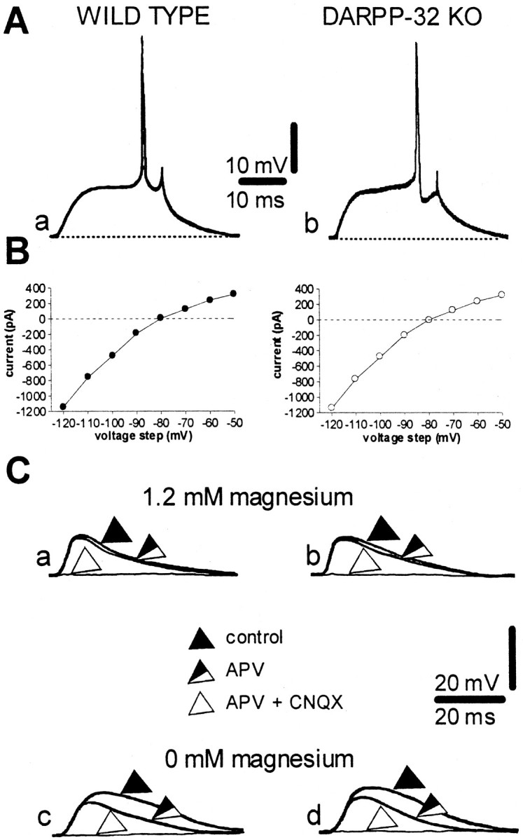
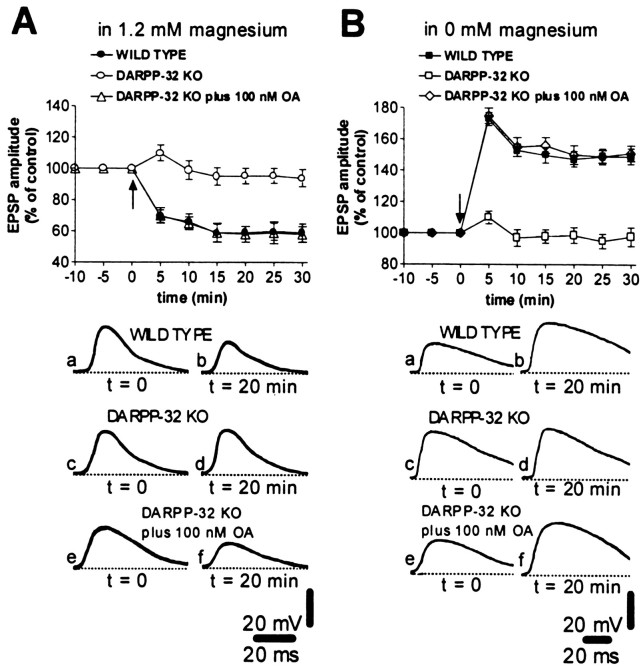
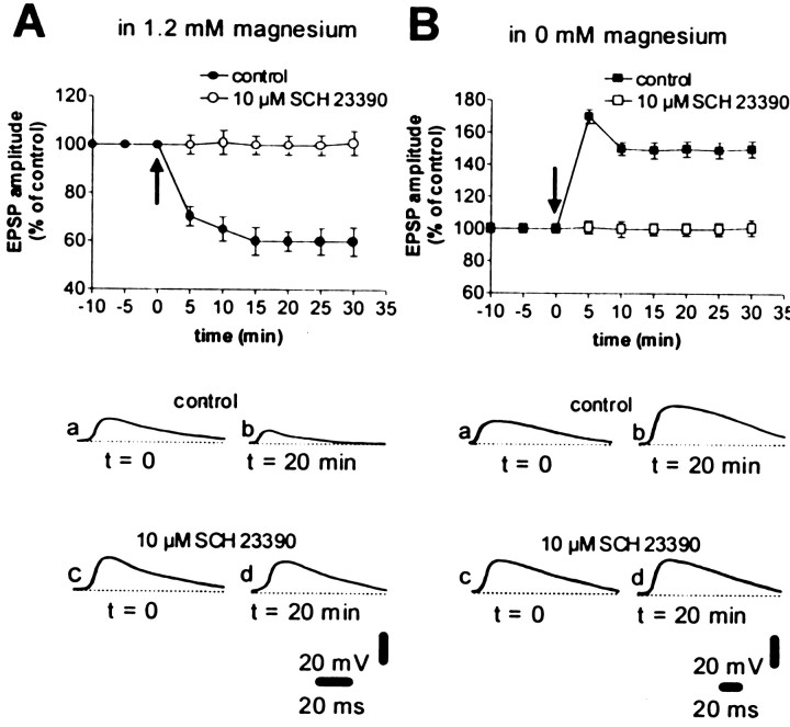
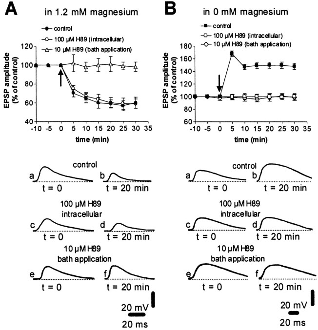
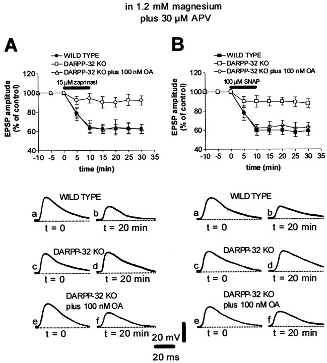
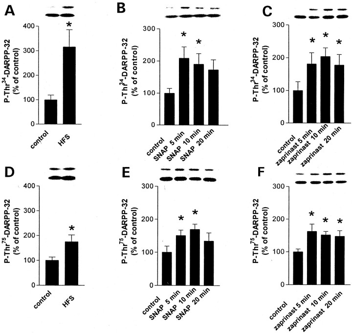
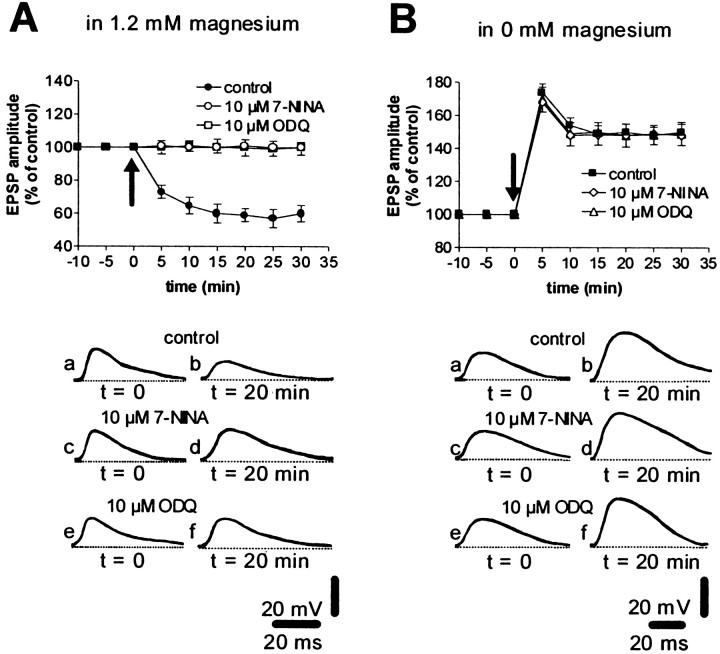
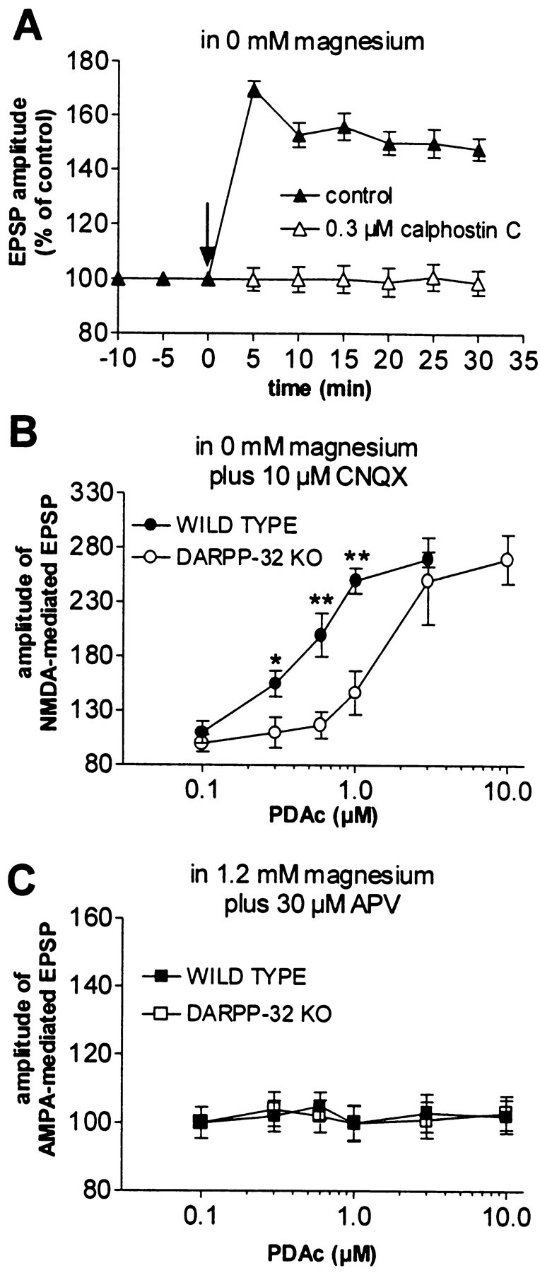
A complex chain of intracellular signaling events, critically important in motor control, is activated by the stimulation of D1-like dopamine (DA) receptors in striatal neurons. At corticostriatal synapses on medium spiny neurons, we provide evidence that the D1-like receptor-dependent activation of DA and cyclic adenosine 3',5' monophosphate-regulated phosphoprotein 32 kDa is a crucial step for the induction of both long-term depression (LTD) and long-term potentiation (LTP), two opposing forms of synaptic plasticity. In addition, formation of LTD and LTP requires the activation of protein kinase G and protein kinase A, respectively, in striatal projection neurons. These kinases appear to be stimulated by the activation of D1-like receptors in distinct neuronal populations.


Figures

Fig. 1.
Fig. 1.
Intrinsic and synaptic properties of striatal spiny neurons recorded from WT and DARPP-32 KO mice. A, Action potential discharge was induced by depolarizing current steps (+0.9 nA) both in WT (a, RMP = −86 mV) and in DARPP-32 KO (b, RMP = −85 mV) neurons.B, Current–voltage plots obtained during single-microelectrode voltage-clamp experiments from WT (left) and DARPP-32 KO (right) neurons. The membrane potential of both neurons was held at −85 mV.C, In 1.2 mm magnesium, the blockade of glutamate NMDA receptors by 30 μm APV did not affect the EPSP recorded from striatal neurons from either WT (a) or DARPP-32 KO (b) mice. By contrast, in both experimental groups, the EPSP was fully suppressed by coadministration of 30 μm APV plus 10 μm CNQX. The RMP of both neurons was −86 mV. In magnesium-free medium, an APV-sensitive component was present in striatal neurons in slices from both WT (c) and DARPP-32 KO (d) mice. The RMP of both neurons was −86 mV.
Fig. 2.
Fig. 2.
Role of DARPP-32 and PP-1 in the expression of HFS-induced corticostriatal LTD and LTP. The graphs summarize the results from intracellular experiments performed in the presence of physiological concentrations of magnesium (A) and in the absence of magnesium (B) from WT slices (filled circles and squares), DARPP-32 KO slices in control medium (open circles andsquares), and DARPP-32 KO slices bathed in medium containing okadaic acid (OA) (open triangles and diamonds). Okadaic acid was applied 10 min before HFS was delivered. The arrowsindicate when HFS was delivered. The bottom part of the figure shows EPSPs recorded in six neurons from WT slices (a, b), DARPP-32 KO slices perfused in control medium (c, d), and DARPP-32 KO slices bathed in okadaic acid (e, f) immediately before (a, c, e) and 20 min after (b, d, f) HFS. RMPs were (in mV): −86 (A, WT and KO; B, WT), −84 (B, KO), and −82 (A, B, KO plus okadaic acid).
Fig. 3.
Fig. 3.
The D1 DA receptor antagonist SCH 23390 blocks both corticostriatal LTD and LTP in WT mice. The graphs summarize the results obtained from intracellular experiments performed in WT mice in the presence of a physiological concentration of magnesium (A) and in the absence of magnesium (B) in control medium (filled circles and squares) and in the presence of 10 μm SCH 23390 (applied 10 min before HFS was delivered,open circles and squares). Thearrows indicate when HFS was delivered. Thebottom part of the figure shows EPSPs recorded in four neurons in control condition (a, b) and in the presence of 10 μm SCH 23390 (c, d), immediately before (a, c) and 20 min after (b, d) HFS. RMPs were (in mV): −82 (A, control), −85 (A, SCH 23390), −87 (B, control) and −84 (B, SCH 23390).
Fig. 4.
Fig. 4.
Role of PKA in the expression of HFS-induced corticostriatal LTP and LTD. The graphs summarize the results obtained in WT mice from intracellular experiments performed in the presence of physiological concentrations of magnesium (A) and in the absence of magnesium (B) in control condition (filled circles andsquares), after intracellular injection of 100 μm H89, a PKA inhibitor (open circles andsquares), and after bath application of 10 μm H89 (10 min before HFS was delivered, open triangles and diamonds). Thearrows indicate when HFS was delivered. Thebottom part of the figure shows EPSPs recorded in six neurons in control condition (a, b), after intracellular injection of 100 μm H89 (c, d), and in the presence of bath-applied H89 (10 μm) (e, f) immediately before (a, c, e) and 20 min after (b, d, e) HFS. RMPs were (in mV): −83 (A, control, intracellular, and bath-applied H89), −84 (B, bath-applied H89), and −88 (B, control and intracellular H89).
Fig. 5.
Fig. 5.
Stimulation of the NO/cGMP pathway induces AMPA receptor-mediated corticostriatal LTD in WT but not in DARPP-32 KO mice. A, In the presence of 1.2 mm magnesium plus 30 μm APV, the cGMP phosphodiesterase inhibitor zaprinast induced LTD of AMPA-mediated EPSPs in WT mice (filled circles) but not in DARPP-32 KO mice (open circles). In these mutant animals, the pharmacological LTD was restored after the application (10 min before zaprinast application) of the PP-1 inhibitor okadaic acid (100 nm, open triangles). The bottom part of the figure shows EPSPs recorded in neurons from WT (a, b) and DARPP-32 KO (c–f) mice immediately before (a, c, e) and 20 min after (b, d, f) the application of zaprinast. The RMP of neurons was −86 mV. B, In the presence of 1.2 mmmagnesium plus 30 μm APV, the NO donor SNAP induced LTD of AMPA-mediated EPSPs in WT (filled squares) but not DARPP-32 KO (open squares) mice. In these mutant animals the SNAP-induced LTD was restored after the bath application (10 min before SNAP application) of 100 nm okadaic acid (open diamonds). The bottom part of the figure shows EPSPs recorded in neurons from WT (a, b) and DARPP-32 KO (c–f) mice immediately before (a, c, e) and 20 min after (b, d, f) the application of SNAP). RMPs were (in mV): −85 (WT) and −84 (DARPP-32 KO).
Fig. 6.
Fig. 6.
Effect of HFS (A, D), SNAP (B, E), and zaprinast (C, F) on the level of phospho[Thr34]-DARPP-32 (A–C) and phospho[Thr75]-DARPP-32 (D–F) in striatal slices. The amount of phospho-DARPP-32 was normalized to the level of total DARPP-32 in each sample and is expressed as a percentage of that measured in control slices. Data represent means ± SEM for four experiments performed in duplicate or triplicate (n = 9–12). *p < 0.05 versus the control group; ANOVA was followed by Dunnett's test. The top lanes above each graph illustrate phospho-DARPP-32. The bottom lanesillustrate the corresponding total DARPP-32.
Fig. 7.
Fig. 7.
Inhibition of the NO/cGMP pathway blocks corticostriatal LTD but not LTP in WT mice. The graphs summarize the results obtained in WT mice in 1.2 mm magnesium (A) and in the absence of magnesium (B) in control condition (filled circles and squares), in the presence of the neuronal NOS inhibitor 7-NINA (10 μm, open circles and diamonds), and in the presence of the soluble guanylyl cyclase inhibitor ODQ (10 μm,open squares and triangles). 7-NINA and ODQ were applied 10 min before HFS was delivered. Thearrows indicate when HFS was delivered. Thebottom part of the figure shows EPSPs recorded from six neurons in control condition (a, b), in the presence of 10 μm 7-NINA (c, d) and in the presence of 10 μm ODQ (e, f) immediately before (a, c, e) and 20 min after (b, d, f) HFS of the corticostriatal pathway. RMPs were (in mV): −86 (A, control), −85 (A, 7-NINA), −80 (A, ODQ; B, 7-NINA), −81 (B, control), and −88 (B, ODQ).
Fig. 8.
Fig. 8.
PKC activity is required for striatal LTP.A, Blockade of PKC function by the selective inhibitor calphostin C (0.3 μm) prevented LTP in WT mice. Thearrow indicates when HFS was delivered.B, In magnesium-free solution plus 10 μmCNQX, the stimulation of PKC activity by PDAc (applied for 10 min) produces a dose-dependent enhancement of the cortically evoked NMDA-mediated EPSPs in both WT (filled circles) and DARPP-32 KO (open circles) mice. Note that disruption of the DARPP-32 gene significantly reduced the PDAc-induced enhancement of NMDA-mediated EPSPs (*p < 0.05; **p < 0.01). C, In both WT (filled squares) and DARPP-32 KO (open squares) mice, the AMPA component of corticostriatal EPSPs, recorded in the presence of a physiological concentration of external magnesium plus 30 μm APV, was not modified by PDAc.

Similar articles

Cited by

References

    1. Altar CA, Boyar WC, Kim HS. Discriminatory roles for D1 and D2 dopamine receptor subtypes in the in vivo control of neostriatal cyclic GMP. Eur J Pharmacol. 1990;181:17–21. - PubMed
    1. Bibb JA, Snyder GL, Nishi A, Yan Z, Meijer L, Fienberg AA, Tsai LH, Kwon YT, Girault JA, Czernik AJ, Huganir RL, Hemmings HC, Jr, Nairn AC, Greengard P. Phosphorylation of DARPP-32 by Cdk5 modulates dopamine signalling in neurons. Nature. 1999;402:669–671. - PubMed
    1. Calabresi P, Mercuri NB, Bernardi G. Synaptic and intrinsic control of membrane excitability of neostriatal neurons. II. An in vitro analysis. J Neurophysiol. 1990;63:663–675. - PubMed
    1. Calabresi P, Maj R, Pisani A, Mercuri NB, Bernardi G. Long-term synaptic depression in the striatum: physiological and pharmacological characterization. J Neurosci. 1992a;12:4224–4233. - PMC - PubMed
    1. Calabresi P, Pisani A, Mercuri NB, Bernardi G. Long-term potentiation in the striatum is unmasked by removing the voltage-dependent block of NMDA receptor channels. Eur J Neurosci. 1992b;4:929–935. - PubMed

Publication types

MeSH terms

LinkOut - more resources