

Characterization of human herpesvirus 8 ORF59 protein (PF-8) and mapping of the processivity and viral DNA polymerase-interacting domains
- PMID: 11069986
- PMCID: PMC113171
- DOI: 10.1128/jvi.74.23.10920-10929.2000
Characterization of human herpesvirus 8 ORF59 protein (PF-8) and mapping of the processivity and viral DNA polymerase-interacting domains
Abstract
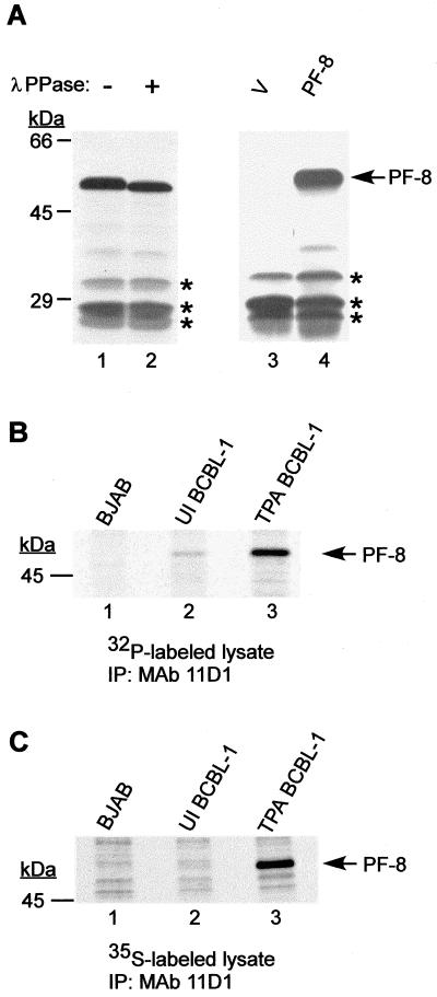
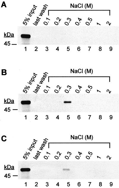
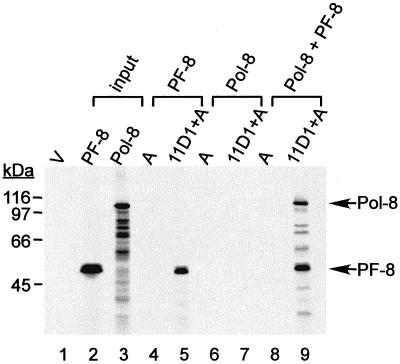
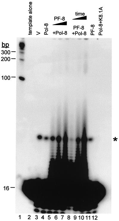
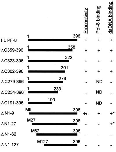
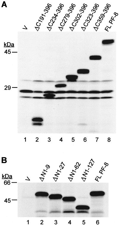
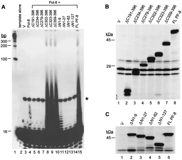
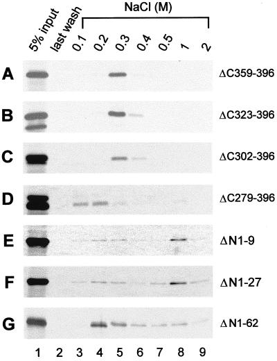
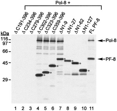
Human herpesvirus 8 (HHV-8) or Kaposi's sarcoma-associated herpesvirus (KSHV) ORF59 protein (PF-8) is a processivity factor for HHV-8 DNA polymerase (Pol-8) and is homologous to processivity factors expressed by other herpesviruses, such as herpes simplex virus type 1 UL42 and Epstein-Barr virus BMRF1. The interaction of UL42 and BMRF1 with their corresponding DNA polymerases is essential for viral DNA replication and the subsequent production of infectious virus. Using HHV-8-specific monoclonal antibody 11D1, we have previously identified the cDNA encoding PF-8 and showed that it is an early-late gene product localized to HHV-8-infected cell nuclei (S. R. Chan, C. Bloomer, and B. Chandran, Virology 240:118-126, 1998). Here, we have further characterized PF-8. This viral protein was phosphorylated both in vitro and in vivo. PF-8 bound double-stranded DNA (dsDNA) and single-stranded DNA independent of DNA sequence; however, the affinity for dsDNA was approximately fivefold higher. In coimmunoprecipitation reactions, PF-8 also interacted with Pol-8. In in vitro processivity assays with excess poly(dA):oligo(dT) as a template, PF-8 stimulated the production of elongated DNA products by Pol-8 in a dose-dependent manner. Functional domains of PF-8 were determined using PF-8 truncation mutants. The carboxyl-terminal 95 amino acids (aa) of PF-8 were dispensable for all three functions of PF-8: enhancing processivity of Pol-8, binding dsDNA, and binding Pol-8. Residues 10 to 27 and 279 to 301 were identified as regions critical for the processivity function of PF-8. Interestingly, aa 10 to 27 were also essential for binding Pol-8, whereas aa 1 to 62 and aa 279 to 301 were involved in binding dsDNA, suggesting that the processivity function of PF-8 is correlated with both the Pol-8-binding and the dsDNA-binding activities of PF-8.
Figures
Similar articles
-
Kaposi's Sarcoma-Associated Herpesvirus Processivity Factor, ORF59, Binds to Canonical and Linker Histones, and Its Carboxy Terminus Is Dispensable for Viral DNA Synthesis.J Virol. 2021 Feb 24;95(6):e02169-20. doi: 10.1128/JVI.02169-20. Print 2021 Feb 24. J Virol. 2021. PMID: 33361421 Free PMC article.
-
Processivity factor of KSHV contains a nuclear localization signal and binding domains for transporting viral DNA polymerase into the nucleus.Virology. 2005 Sep 30;340(2):183-91. doi: 10.1016/j.virol.2005.06.017. Virology. 2005. PMID: 16043206
-
DNA-Binding Activities of KSHV DNA Polymerase Processivity Factor (PF-8) Complexes.Viruses. 2025 Jan 29;17(2):190. doi: 10.3390/v17020190. Viruses. 2025. PMID: 40006945 Free PMC article.
-
Inhibiting KSHV replication by targeting the essential activities of KSHV processivity protein, PF-8.J Med Virol. 2024 Oct;96(10):e29958. doi: 10.1002/jmv.29958. J Med Virol. 2024. PMID: 39370884 Free PMC article. Review.
-
Herpesvirus DNA polymerase processivity factors: Not just for DNA synthesis.Virus Res. 2021 Jun;298:198394. doi: 10.1016/j.virusres.2021.198394. Epub 2021 Mar 26. Virus Res. 2021. PMID: 33775751 Review.
Cited by
-
The crystal structure of PF-8, the DNA polymerase accessory subunit from Kaposi's sarcoma-associated herpesvirus.J Virol. 2009 Dec;83(23):12215-28. doi: 10.1128/JVI.01158-09. Epub 2009 Sep 16. J Virol. 2009. PMID: 19759157 Free PMC article.
-
Structure and mutagenesis reveal essential capsid protein interactions for KSHV replication.Nature. 2018 Jan 25;553(7689):521-525. doi: 10.1038/nature25438. Epub 2018 Jan 17. Nature. 2018. PMID: 29342139 Free PMC article.
-
The FAT10 post-translational modification is involved in the lytic replication of Kaposi's sarcoma-associated herpesvirus.J Virol. 2021 Apr 26;95(10):e02194-20. doi: 10.1128/JVI.02194-20. Epub 2021 Feb 24. J Virol. 2021. PMID: 33627385 Free PMC article.
-
Phosphorylation of Kaposi's sarcoma-associated herpesvirus processivity factor ORF59 by a viral kinase modulates its ability to associate with RTA and oriLyt.J Virol. 2013 Jul;87(14):8038-52. doi: 10.1128/JVI.03460-12. Epub 2013 May 15. J Virol. 2013. PMID: 23678174 Free PMC article.
-
Downregulation of Poly(ADP-Ribose) Polymerase 1 by a Viral Processivity Factor Facilitates Lytic Replication of Gammaherpesvirus.J Virol. 2015 Sep;89(18):9676-82. doi: 10.1128/JVI.00559-15. Epub 2015 Jul 8. J Virol. 2015. PMID: 26157130 Free PMC article.
References
-
- Ambroziak J A, Blackbourn D J, Herndier B G, Glogau R G, Gullett J H, McDonald A R, Lennette E T, Levy J A. Herpes-like sequences in HIV-infected and uninfected Kaposi's sarcoma patients. Science. 1995;268:582–583. - PubMed
-
- Ausubel F M, Brent R, Kingston R E, Moore D D, Seidman J G, Smith J A, Struhl K, editors. Current protocols in molecular biology. New York, N.Y: John Wiley & Sons, Inc.; 1987.
-
- Bambara R A, Fay P J, Mallaber L M. Methods of analyzing processivity. Methods Enzymol. 1995;262:270–280. - PubMed
-
- Beral V. Epidemiology of Kaposi's sarcoma. Cancer Surv. 1991;10:5–22. - PubMed
-
- Chan S R, Bloomer C, Chandran B. Identification and characterization of human herpesvirus-8 lytic cycle-associated ORF 59 protein and the encoding cDNA by monoclonal antibody. Virology. 1998;240:118–126. - PubMed
Publication types
MeSH terms
Substances
Grants and funding
LinkOut - more resources
Full Text Sources
