. 2000 Dec;74(23):10984-93.
doi: 10.1128/jvi.74.23.10984-10993.2000.

The level of CD4 expression limits infection of primary rhesus monkey macrophages by a T-tropic simian immunodeficiency virus and macrophagetropic human immunodeficiency viruses

Affiliations

The level of CD4 expression limits infection of primary rhesus monkey macrophages by a T-tropic simian immunodeficiency virus and macrophagetropic human immunodeficiency viruses

N Bannert et al. J Virol. 2000 Dec.

Abstract

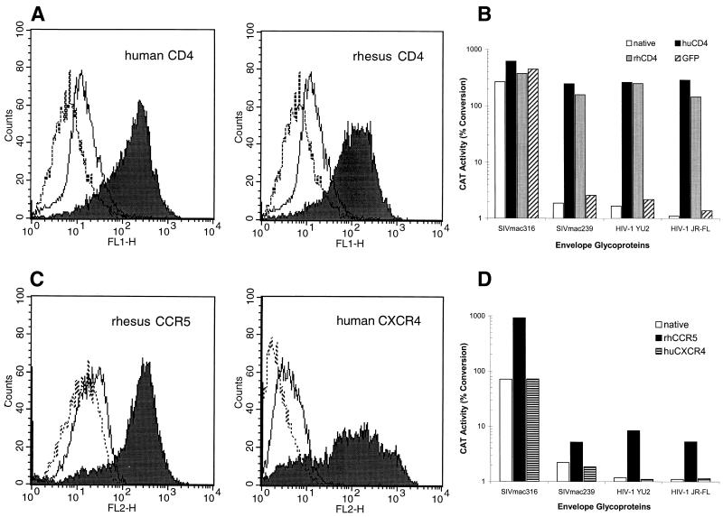
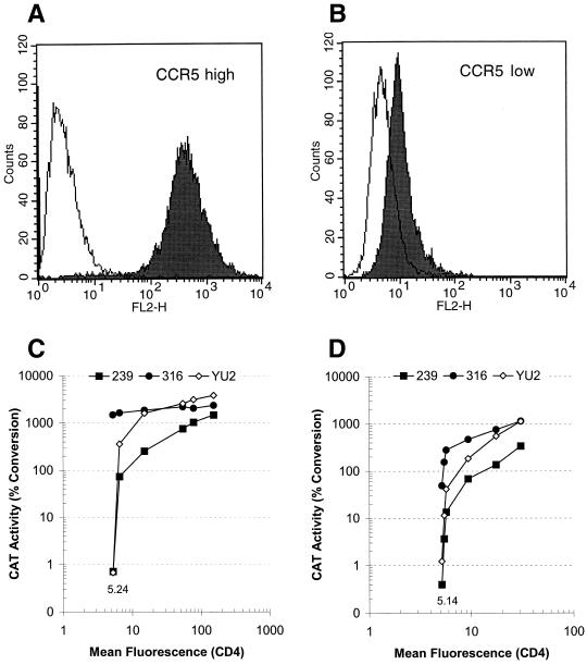
The entry of primate immunodeficiency viruses into cells is dependent on the interaction of the viral envelope glycoproteins with receptors, CD4, and specific members of the chemokine receptor family. Although in many cases the tropism of these viruses is explained by the qualitative pattern of coreceptor expression, several instances have been observed where the expression of a coreceptor on the cell surface is not sufficient to allow infection by a virus that successfully utilizes the coreceptor in a different context. For example, both the T-tropic simian immunodeficiency virus (SIV) SIVmac239 and the macrophagetropic (M-tropic) SIVmac316 can utilize CD4 and CCR5 as coreceptors, and both viruses can infect primary T lymphocytes, yet only SIVmac316 can efficiently infect CCR5-expressing primary macrophages from rhesus monkeys. Likewise, M-tropic strains of human immunodeficiency virus type 1 (HIV-1) do not infect primary rhesus monkey macrophages efficiently. Here we show that the basis of this restriction is the low level of CD4 on the surface of these cells. Overexpression of human or rhesus monkey CD4 in primary rhesus monkey macrophages allowed infection by both T-tropic and M-tropic SIV and by primary M-tropic HIV-1. By contrast, CCR5 overexpression did not specifically compensate for the inefficient infection of primary monkey macrophages by T-tropic SIV or M-tropic HIV-1. Apparently, the limited ability of these viruses to utilize a low density of CD4 for target cell entry accounts for the restriction of these viruses in primary rhesus monkey macrophages.


Figures

FIG. 1
FIG. 1
Infectability of primary rhesus monkey cells by viruses with different envelope glycoproteins. Recombinant CAT-expressing viruses containing the envelope glycoproteins of SIVmac316, SIVmac239, or HIV-1 strain YU2, or lacking functional envelope glycoproteins (the ΔKS control), were incubated overnight with either rhesus monkey PBMC (A) or rhesus monkey monocyte-derived macrophages (B). Three days later, CAT activity was measured in cell lysates. Samples in which the conversion of chloramphenicol to acetylated forms was greater than 70% were diluted and reassayed to bring the conversion value within range.
FIG. 2
FIG. 2
Inhibition of SIVmac316 entry by TAK-779. Cf2Th cells transiently expressing rhesus monkey CD4 and CCR5 proteins (A) or primary rhesus monkey macrophages (B and C) were infected with recombinant CAT-expressing viruses with the envelope glycoproteins of SIVmac316 (A and B) or with VSV G (C) in the presence of increasing TAK-779 concentrations. The results are reported as percentages of chloramphenicol conversion to acetylated forms and represent the means and standard deviations of three samples for each TAK-779 concentration. A representative experiment of three is shown.
FIG. 3
FIG. 3
Effect of CD4 and CCR5 overexpression on infection of rhesus monkey macrophages with recombinant viruses. Primary rhesus monkey macrophages were transduced with retroviral vectors encoding either CD4 (A and B) or chemokine receptors (C and D). As a control, cells were transduced in parallel with a similar vector encoding GFP. (A) FACS profile of cells transduced with the human CD4 (left) and rhesus monkey CD4 (right) genes. The anti-CD4 antibody OKT4 was used to generate the unbroken curve (untransduced macrophages) and the filled curve (transduced macrophages). The broken curve was generated using a control antibody of the same isotype. (B) CAT activity in native rhesus monkey macrophages and in macrophages transduced with a vector expressing either human or rhesus monkey CD4 (huCD4 or rhCD4) or GFP after infection with CAT reporter viruses containing the indicated envelope glycoproteins. (C) FACS profiles of untransduced macrophages and macrophages transduced with rhesus monkey CCR5 (left) or human CXCR4 (right). The broken curves were obtained using isotype-matched irrelevant antibodies. In the left panel, the anti-CCR5 antibody 45531.111 was used to stain untransduced macrophages (unbroken curve) or macrophages transduced with the rhesus CCR5 gene (filled curve). In the right panel, the 12G5 anti-CXCR4 antibody was used to stain untransduced macrophages (unbroken curve) or macrophages transduced with the human CXCR4 gene (filled curve). (D) CAT activity in native rhesus monkey macrophages and in macrophages transduced with the gene for either rhesus monkey CCR5 or human CXCR4 following infection with CAT reporter viruses containing the indicated envelope glycoproteins.
FIG. 4
FIG. 4
Replication of SIVmac239 and SIVmac316 in rhesus monkey macrophages overexpressing human or monkey CD4. Primary rhesus monkey macrophages were transduced with VSV G-pseudotyped vectors containing the gene for human (hu) CD4, rhesus monkey (rh) CD4, or GFP. Three days after transduction, the 14-day-old cells were incubated with wild-type SIVmac316 or SIVmac239 (50,000 RT units/ml) for 12 h. Culture supernatants were harvested and assayed for p27 protein concentration every 2 days thereafter. The experiment was repeated four times with macrophages obtained from different rhesus monkeys, and the results were similar to those shown here.
FIG. 5
FIG. 5
Influence of CD4 expression in Cf2Th canine thymocytes expressing high or low levels of CCR5. FACS profiles of Cf2Th cells stably expressing high (A) or low (B) levels of human CCR5 were determined using the anti-CCR5 antibody 2D7 or an isotype-matched control antibody. These cells were transfected with increasing amounts of a human CD4-expressing plasmid (pcDNA/CD4). The total DNA was kept constant in each transfection by the addition of empty vector DNA. After 48 h, the cells were infected with CAT reporter viruses containing the envelope glycoproteins of SIVmac316, SIVmac239, or HIV-1 strain YU2. In parallel, an aliquot of the same cells was analyzed for CD4 surface expression using the FITC-conjugated monoclonal antibody OKT4. The mean channel fluorescence of CD4-negative CF2Th cells was set at 5.24 for high (C) and 5.14 for low (D) CCR5 expressors. The CAT activity in the infected cells is plotted against the mean fluorescence intensity.
FIG. 6
FIG. 6
Effect of an anti-CD4 MAb and sCD4 on infection of rhesus monkey macrophages. Rhesus monkey macrophages were incubated with recombinant CAT reporter viruses (50,000 RT units/ml) with the envelope glycoproteins of SIVmac316 in the presence of increasing concentrations of the anti-CD4 antibody OKT4a (A) or sCD4 (C). VSV G-pseudotyped reporter virus was used as a control for any nonspecific effects of the OKT4a antibody (B). A representative experiment is shown. The data are means and standard deviations of three samples for each concentration.

Similar articles

Cited by

References

    1. Adamson D C, Dawson T M, Zink M C, Clements J E, Dawson V L. Neurovirulent simian immunodeficiency virus infection induces neuronal, endothelial, and glial apoptosis. Mol Med. 1996;2:417–428. - PMC - PubMed
    1. Alkhatib G, Combadiere C, Broder C C, Feng Y, Kennedy P E, Murphy P M, Berger E A. CC CKR5: a RANTES, MIP-1α, MIP-1β receptor as a fusion cofactor for macrophage-tropic HIV-1. Science. 1996;272:1955–1958. - PubMed
    1. Alkhatib G, Liao F, Berger E A, Farber J M, Peden K W. A new SIV co-receptor, STRL33. Nature. 1997;388:238. - PubMed
    1. Anderson M G, Hauer D, Sharma D P, Joag S V, Narayan O, Zink M C, Clements J E. Analysis of envelope changes acquired by SIVmac239 during neuroadaption in rhesus macaques. Virology. 1993;195:616–626. - PubMed
    1. Baba M, Nishimura O, Kanzaki N, Okamoto M, Sawada H, Iizawa Y, Shiraishi M, Aramaki Y, Okonogi K, Ogawa Y, Meguro K, Fujino M. A small-molecule, nonpeptide CCR5 antagonist with highly potent and selective anti-HIV-1 activity. Proc Natl Acad Sci USA. 1999;96:5698–5703. - PMC - PubMed

Publication types