. 2000 Dec;74(23):11001-7.
doi: 10.1128/jvi.74.23.11001-11007.2000.

Dynamics of CCR5 expression by CD4(+) T cells in lymphoid tissues during simian immunodeficiency virus infection

Affiliations

Dynamics of CCR5 expression by CD4(+) T cells in lymphoid tissues during simian immunodeficiency virus infection

R S Veazey et al. J Virol. 2000 Dec.

Abstract

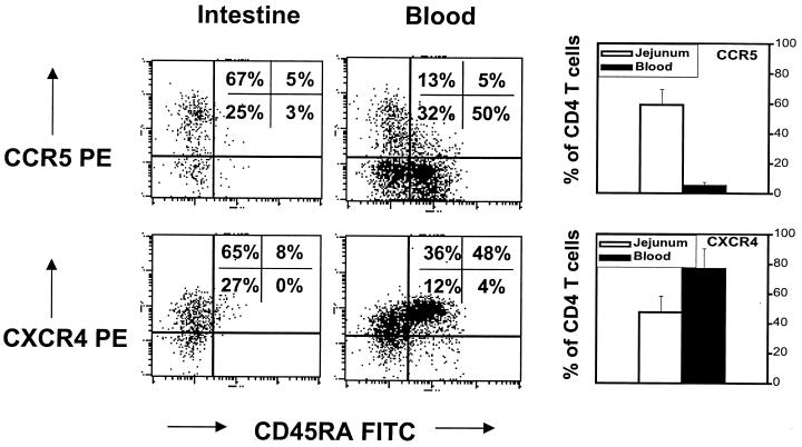
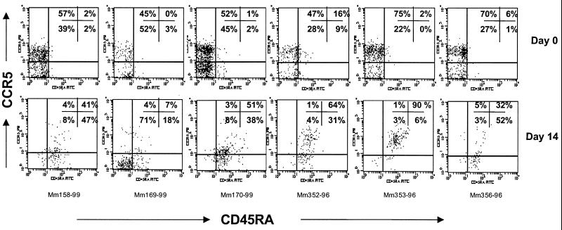
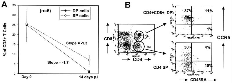
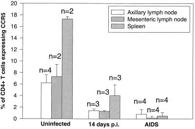
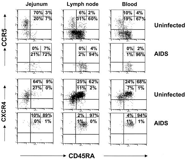
Early viral replication and profound CD4(+) T-cell depletion occur preferentially in intestinal tissues of macaques infected with simian immunodeficiency virus (SIV). Here we show that a much higher percentage of CD4(+) T cells in the intestine express CCR5 compared with those found in the peripheral blood, spleen, or lymph nodes. In addition, the selectivity and extent of the CD4(+) T-cell loss in SIV infection may depend upon these cells coexpressing CCR5 and having a "memory" phenotype (CD45RA(-)). Following intravenous infection with SIVmac251, memory CD4(+) CCR5(+) T cells were selectively eliminated within 14 days in all major lymphoid tissues (intestine, spleen, and lymph nodes). However, the effect on CD4(+) T-cell numbers was most profound in the intestine, where cells of this phenotype predominate. The CD4(+) T cells that remain after 14 days of infection lacked CCR5 and/or were naive (CD45RA(+)). Furthermore, when animals in the terminal stages of SIV infection (with AIDS) were examined, virtually no CCR5-expressing CD4(+) T cells were found in lymphoid tissues, and all of the remaining CD4(+) T cells were naive and coexpressed CXCR4. These findings suggest that chemokine receptor usage determines which cells are targeted for SIV infection and elimination in vivo.


Figures

FIG. 1
FIG. 1
Comparison of CCR5 and CXCR4 expression on normal (uninfected) CD4+ T lymphocytes from the jejunum to those of peripheral blood. Note that very few (∼5%) peripheral blood CD4+ lymphocytes express CCR5, whereas most (∼60%) intestinal CD4+ lymphocytes express CCR5. Also note that CCR5 expression and CD45RA expression are relatively mutually exclusive; most naive CD4 cells (CD45RAHI) do not express CCR5, whereas CCR5HI cells are CD45RALO. In contrast, most peripheral blood CD4 cells express CXCR4. CD45RAHI cells are usually CXCR4HI. The dot plots shown were generated by first gating through CD4+ T lymphocytes and are representative of uninfected animals (except that the macaque with the highest CCR5 expression in the peripheral blood was selected for illustration of CD45RA coexpression). Graphs represent the mean of six animals examined ± standard deviation.
FIG. 2
FIG. 2
Dot plots showing CD4 T-cell depletion due to SIV infection in the intestinal mucosa of three macaques. Intestinal lymphocytes were obtained from endoscopic pinch biopsies from the jejunum of the same animals at different time points (prospective studies). Plots were generated by gating through CD3+ lymphocytes. From left to right, note the rapid loss of CD4+ T cells within 14 days after infection. Also note that CD4+ CD8+ DP cells (upper right quadrants) are more profoundly eliminated than the CD4+ SP population. DP cells were essentially eliminated by 14 days p.i. and did not return in the time points examined, whereas some SP CD4+ T cells return by 35 days p.i. (lower right quadrants).
FIG. 3
FIG. 3
Dot plots demonstrating selective depletion of the CD45RALO CCR5HI subset of CD4+ T cells in the intestine within 14 days p.i. All plots were generated by first gating through CD4+ T lymphocytes; top panels are from intestinal biopsies taken before infection (day 0), and bottom panels correspond to the same animals and region 14 days after SIV infection (prospective analysis). Before infection, 45 to 75% of the CD4+ T cells are both memory (CD45RALO) and CCR5HI (upper left quadrants). After infection, virtually all of this memory CCR5HI subset of CD4+ T cells has been selectively depleted in all six animals examined. The remaining CD4+ T cells are either CD45RAHI CCR5HI (upper right) or lack CCR5 expression (lower left and right).
FIG. 4
FIG. 4
(A) Comparison of the rate of DP CD4+ CD8+ to SP (CD4+) intestinal T-cell depletion after 14 days of SIV infection. Note that approximately equal percentages of DP and SP cells are present in the jejunum of uninfected animals, but the DP cells are virtually eliminated by day 14 p.i. A few (mean, 7%) SP cells remain, indicating that a selective depletion of DP cells is occurring, as indicated by the slope of the lines. Points represent the mean of six animals ± standard deviation. Percentages were determined by gating through CD3+ lymphocytes. (B) Four-color flow cytometry demonstrating that DP intestinal T cells from normal (uninfected) macaques are virtually all CCR5HI, whereas a significant proportion of the SP T cells lack CCR5 expression. Also note that both DP and SP T cells lack significant CD45RA expression. Plots were generated by gating through intestinal lymphocytes.
FIG. 5
FIG. 5
Comparison of CCR5 expression on CD4+ T cells from peripheral lymphoid tissues of uninfected macaques to those sacrificed at 14 days after SIV infection and those sacrificed with advanced SIVmac251 infection (AIDS). Note that there are markedly fewer CCR5-expressing CD4+ T cells in tissues from acutely infected macaques, and animals with AIDS show profound depletion of CCR5-expressing CD4+ T cells in all lymphoid tissues. Graphs represent the mean of two to four different animals per group (as indicated) ± standard deviation. Data were generated by gating through CD4+ T lymphocytes.
FIG. 6
FIG. 6
Dot plots demonstrating profound loss of CCR5-expressing CD4+ T cells in macaques sacrificed with advanced SIV disease (AIDS). All plots were generated by gating through CD4+ T lymphocytes. Top panels are representative of uninfected macaques demonstrating CCR5HI cells in (from left to right) the jejunum, axillary lymph node, and peripheral blood. Note that in animals with AIDS, essentially all of the CCR5HI as well as the memory (CD45RALO) CD4+ T cells have been eliminated, leaving only CXCR4HI and CD45RAHI cells. Plots are representative of four animals with AIDS examined.

References

    1. Berger E A, Doms R W, Fenyo E-M, Korber T M, Littman D R, Moore J P, Sattentau Q J, Schuitemaker H, Soedroski J, Weiss R A. A new classification for HIV-1. Nature. 1998;391:240. - PubMed
    1. Borvak J, Chou C-S, Bell K, Van Dyke G, Zola H, Ramilo O, Vitetta E S. Expression of CD25 defines peripheral blood mononuclear cells with productive versus latent HIV infection. J Immunol. 1995;155:3196–3204. - PubMed
    1. Chou C-S, Ramilo O, Vitetta E S. Highly purified CD25− resting T cells cannot be infected de novo with HIV-1. Proc Natl Acad Sci USA. 1997;94:1361–1365. - PMC - PubMed
    1. Chun T-W, Engel D, Mizell S B, Ehler L A, Fauci A S. Induction of HIV-1 replication in latently infected CD4+ T cells using a combination of cytokines. J Exp Med. 1998;188:83–91. - PMC - PubMed
    1. De Maria R, Fais S, Testi R. Persistent in vivo activation and transient anergy to TCR/CD3 stimulation of normal human intestinal lymphocytes. In: Mestecky J, editor. Advances in mucosal immunology. New York, N.Y: Plenum Press; 1995. pp. 43–46. - PubMed

Publication types

MeSH terms