. 2000 Dec;74(23):11017-26.
doi: 10.1128/jvi.74.23.11017-11026.2000.

Respiratory syncytial virus can tolerate an intergenic sequence of at least 160 nucleotides with little effect on transcription or replication in vitro and in vivo

Affiliations

Respiratory syncytial virus can tolerate an intergenic sequence of at least 160 nucleotides with little effect on transcription or replication in vitro and in vivo

A Bukreyev et al. J Virol. 2000 Dec.

Abstract

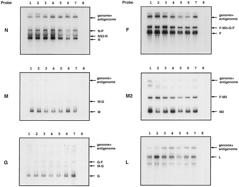
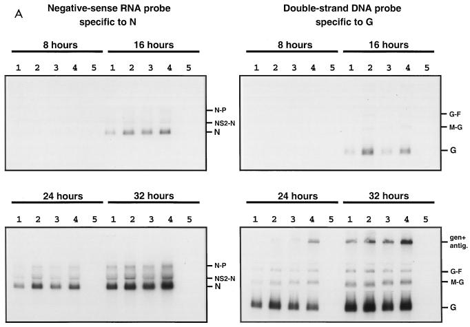
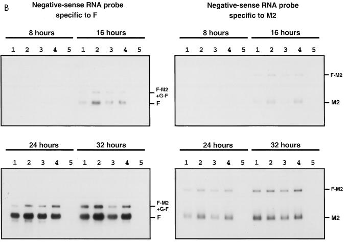
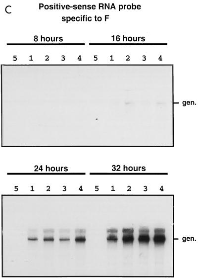
The intergenic sequences (IGS) between the first nine genes of human respiratory syncytial virus (RSV) vary in length from 1 to 56 nucleotides and lack apparent conserved sequence motifs. To investigate their influence on sequential transcription and viral growth, recombinant RSV strain A2, from which the SH gene had been deleted to facilitate manipulation, was further modified to contain an M-G IGS of 16, 30, 44, 58, 65, 72, 86, 100, 120, 140, or 160 nucleotides. All of the viruses were viable. For viruses with an M-G IGS of 100 nucleotides or more, plaque size decreased with increasing IGS length. In this same length range, increasing IGS length was associated with modest attenuation during single-step, but not multistep, growth in HEp-2 cells. Surprisingly, Northern blot analysis of the accumulation of six different mRNAs indicated that there was little or no change in transcription with increasing IGS length. Thus, the RSV polymerase apparently can readily cross IGS of various lengths, including unnaturally long ones, with little or no effect on the efficiency of termination and reinitiation. This finding supports the view that the IGS do not have much effect on sequential transcription and provides evidence from infectious virus that IGS length is not an important regulatory feature. To evaluate replication in vivo, BALB/c mice were infected intranasally with RSV containing an M-G IGS of 65, 140, or 160 nucleotides. Replication of the latter two viruses was decreased up to 5- and 25-fold in the upper and lower respiratory tracts, respectively, on day 3 following infection. However, the level of replication at both sites on days 4 and 5 was very similar to that of the virus with an IGS of 65 nucleotides. Thus, the modest attenuation in vivo associated with the longer IGS was additive to that conferred by deletion of the SH gene and might be useful to incrementally increase the level of attenuation of a live-attenuated vaccine virus.


Figures

FIG. 1
FIG. 1
Structures of artificial IGS inserted between the M and G genes of recombinant RSVs lacking the SH gene. (A) Deletion of the SH gene. The ScaI-PacI restriction fragment bearing most of the SH gene was replaced by a synthetic double-stranded DNA that restored the downstream noncoding region of the M gene and restored a GE signal. The sequence below the diagram shows the restored ScaI and PacI sites (underlined) and GE signal (overlined), and the dashes in the sequence indicate the boundary between sequence originally derived from the M (left) and SH (right) genes. The restored signal contains a single nucleotide substitution (boxed) that creates a PacI site and makes the GE signal identical to the naturally occurring SH GE signal. GS and GE signals are shown as white and gray boxes, respectively, and the number of nucleotides in the IGS is indicated. (B) Structures of IGS between the M and G genes of rRSV/M-G-16, -30, -44, -58, -72, -86, and -65. rRSV/M-G-44 contains the parental 44-nucleotide IGS modified by nucleotide substitution at four positions (lowercase letters) to create a KpnI site (underlined). To create rRSV/M-G-16 and -30, the indicated deletions (dashed line) were made in the 44-nucleotide IGS between the M and G genes. The KpnI site introduced into this IGS was used to accept addition sequence segments, all of which were derived from increasing increments of the naturally occurring 52-nucleotide G-F IGS, which is shown. The inserts had an additional GTAC (underlined) at the downstream end that represents an incomplete KpnI site. Thus, the introduction of incrementally longer segments of the G-F IGS resulted in the creation of rRSV/M-G-58, -72, and -86. rRSV/M-G-65, constructed in previous work (4), contains a 21-nucleotide insertion with an XmaI site (underlined) and was placed immediately downstream of the M GE signal. (C) Structures of IGS between the M and G genes of rRSV/M-G-100, -120, -140, and -160. The diagram shows the downstream end of the M gene on the left, the upstream end of the G gene on the right, and the intervening M-G IGS. The open reading frames (ORF) and GS and GE transcription signals are shown as filled rectangles; nontranslated gene regions and the M-G IGS are shown as thin lines. The set of synthetic M-G IGS was assembled from sequence segments copied from the naturally occurring G-F and SH-G IGS. These are identified according to their original sequence position in the complete 15,223-nucleotide recombinant wt rRSV antigenome, and the nucleotide length of each segment is indicated above the lines. The left-hand KpnI site is indicated as 5 nucleotides because it overlaps with the last nucleotide of the upstream G-F intergenic region; the second KpnI site, also indicated as 5 nucleotides, is in parentheses because the complete site was not regenerated by the design of the construction.
FIG. 2
FIG. 2
Plaque morphology in HEp-2 cells of recombinant RSVs bearing M-G IGS of increasing length. HEp-2 cell monolayers were infected with wt rRSV (A), rRSV/M-G-65 (B), rRSV/M-G-100 (C), rRSV/M-G-120 (D), rRSV/M-G-140 (E), or rRSV/M-G-160 (F). Note that rRSV/M-G-65 (B), rather than wt rRSV, should be used as the parent for comparison because the latter virus differs from the others in containing the SH gene. The cells were incubated for 6 days at 32°C and fixed with methanol; plaques were visualized by immunostaining with F-specific monoclonal antibodies.
FIG. 3
FIG. 3
RT-PCR analysis of the M-G gene junction from recovered recombinant RSV. Total intracellular RNA isolated from HEp-2 cells after the fourth or fifth passage of the indicated recombinant RSVs was subjected to RT-PCR using primers spanning positions 4158 to 4782 of antigenomic RNA, which includes the M-G IGS. The products were analyzed on a 2.5% agarose gel and visualized with ethidium bromide. The lengths of DNA fragments of a commercial DNA molecular size marker are indicated in nucleotides at the left. The length of the RT-PCR product produced from rRSV/M-G-65 is predicted to be 226 nucleotides.
FIG. 4
FIG. 4
Kinetics of replication in vitro of recombinant RSVs bearing M-G IGS of increasing length. Confluent HEp-2 cell monolayers were infected in duplicate with the indicated viruses at an input MOI of 5 (A) or 0.01 (B) PFU per cell. The cells were washed three times and incubated at 37°C. Aliquots of the medium overlying the cells were taken at 8 (A)- or 24 (B)-h intervals, replaced with an equal volume of fresh medium, flash-frozen, and analyzed later in a single assay by plaque titration.
FIG. 5
FIG. 5
Northern blot analysis of RNAs synthesized by recombinant RSVs bearing M-G IGS of increasing length. HEp-2 cells were infected with the indicated virus (MOI of 5 PFU), incubated for 48 h, and harvested; then total intracellular RNA was extracted. The RNA was subjected to electrophoresis in a formaldehyde-containing agarose gel, transferred onto nitrocellulose membranes, and hybridized with a double-stranded cDNA probe specific to the N, M, G, F, M2 or L mRNA, as indicated to the left. Lanes: 1, rRSV/M-G-44; 2, rRSV/M-G-65; 3, rRSV/M-G-100; 4, rRSV/M-G-120; 5, rRSV/M-G-140; 6, rRSV/M-G-160; 7, wt rRSV; 8, noninfected. Positions of the various RSV and readthrough mRNAs, as well as genomic and antigenomic RNAs, are indicated to the right.
FIG. 6
FIG. 6
Kinetics of genome transcription of recombinant RSVs bearing M-G IGS of 44, 65, 140, or 160 nucleotides in length. The accumulation viral RNAs in infected HEp-2 cells (MOI of 5 PFU) was evaluated by Northern blot hybridization of total intracellular RNA harvested 8, 16, 24, and 32 h postinfection. The blots were hybridized with a negative-sense RNA probe specific to the N, F, or M2 mRNA, a double-stranded cDNA probe specific to the G mRNA, or a positive-sense RNA probe specific to RSV genomic RNA. Lanes: 1, rRSV/M-G-44; 2, rRSV/M-G-65; 3, rRSV/M-G-140; 4, rRSV/M-G-160; 5, uninfected. Positions of the individual mRNAs and readthrough mRNAs, and of genomic and antigenomic RNAs, are indicated to the right.
FIG. 6
FIG. 6
Kinetics of genome transcription of recombinant RSVs bearing M-G IGS of 44, 65, 140, or 160 nucleotides in length. The accumulation viral RNAs in infected HEp-2 cells (MOI of 5 PFU) was evaluated by Northern blot hybridization of total intracellular RNA harvested 8, 16, 24, and 32 h postinfection. The blots were hybridized with a negative-sense RNA probe specific to the N, F, or M2 mRNA, a double-stranded cDNA probe specific to the G mRNA, or a positive-sense RNA probe specific to RSV genomic RNA. Lanes: 1, rRSV/M-G-44; 2, rRSV/M-G-65; 3, rRSV/M-G-140; 4, rRSV/M-G-160; 5, uninfected. Positions of the individual mRNAs and readthrough mRNAs, and of genomic and antigenomic RNAs, are indicated to the right.

References

    1. Barr J N, Whelan S P, Wertz G W. Role of the intergenic dinucleotide in vesicular stomatitis virus RNA transcription. J Virol. 1997;71:1794–1801. - PMC - PubMed
    1. Bermingham A, Collins P L. The M2–2 protein of human respiratory syncytial virus is a regulatory factor involved in the balance between RNA replication and transcription. Proc Natl Acad Sci USA. 1999;96:11259–11264. - PMC - PubMed
    1. Bukreyev A, Camargo E, Collins P L. Recovery of infectious respiratory syncytial virus expressing an additional, foreign gene. J Virol. 1996;70:6634–6641. - PMC - PubMed
    1. Bukreyev A, Whitehead S S, Murphy B R, Collins P L. Recombinant respiratory syncytial virus from which the entire SH gene has been deleted grows efficiently in cell culture and exhibits site-specific attenuation in the respiratory tract of the mouse. J Virol. 1997;71:8973–8982. - PMC - PubMed
    1. Collins P L, Dickens L E, Buckler-White A, Olmsted R A, Spriggs M K, Camargo E, Coelingh K V. Nucleotide sequences for the gene junctions of human respiratory syncytial virus reveal distinctive features of intergenic structure and gene order. Proc Natl Acad Sci USA. 1986;83:4594–4598. - PMC - PubMed

LinkOut - more resources