. 2000 Dec;74(23):11027-39.
doi: 10.1128/jvi.74.23.11027-11039.2000.

The carboxy-terminal fragment of nucleolin interacts with the nucleocapsid domain of retroviral gag proteins and inhibits virion assembly

Affiliations

The carboxy-terminal fragment of nucleolin interacts with the nucleocapsid domain of retroviral gag proteins and inhibits virion assembly

E Bacharach et al. J Virol. 2000 Dec.

Abstract

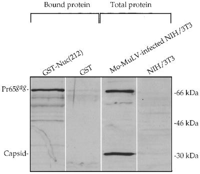
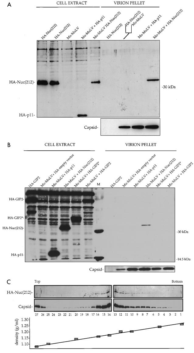
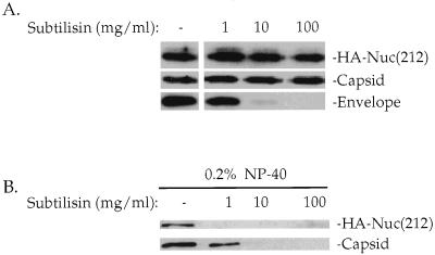
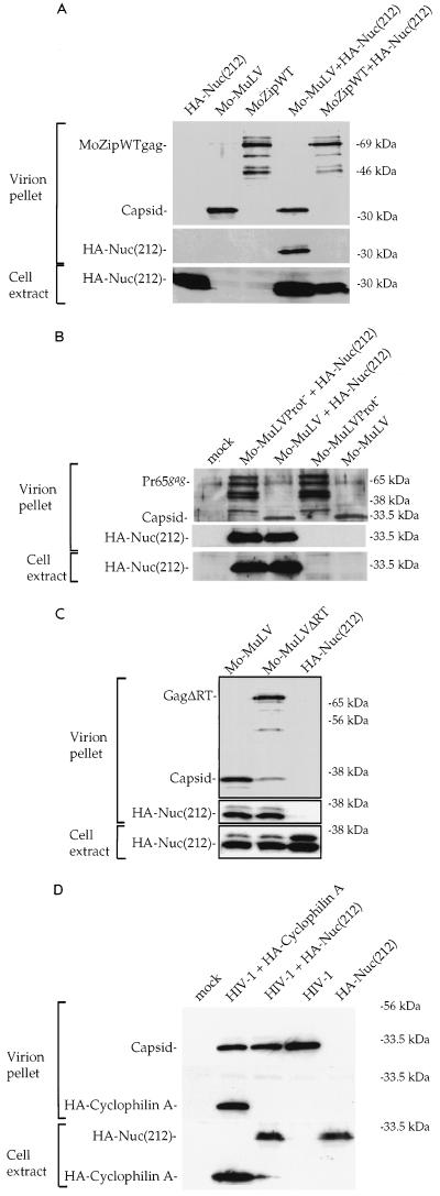
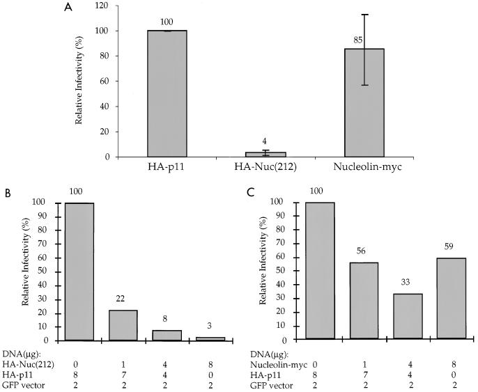
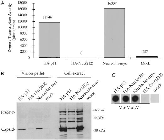
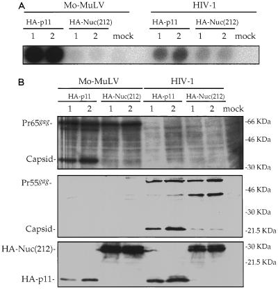
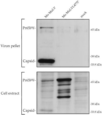
A yeast two-hybrid screen for cellular proteins that interact with the murine leukemia virus (MuLV) Gag protein resulted in the identification of nucleolin, a host protein known to function in ribosome assembly. The interacting fusions contained the carboxy-terminal 212 amino acids of nucleolin [Nuc(212)]. The nucleocapsid (NC) portion of Gag was necessary and sufficient to mediate the binding to Nuc(212). The interaction of Gag with Nuc(212) could be demonstrated in vitro and was manifested in vivo by the NC-dependent incorporation of Nuc(212) inside MuLV virions. Overexpression of Nuc(212), but not full-length nucleolin, potently and specifically blocked MuLV virion assembly and/or release. A mutant of MuLV, selected to specifically disrupt the binding to Nuc(212), was found to be severely defective for virion assembly. This mutant harbors a single point mutation in capsid (CA) adjacent to the CA-NC junction, suggesting a role for this region in Moloney MuLV assembly. These experiments demonstrate that selection for proteins that bind assembly domain(s) can yield potent inhibitors of virion assembly. These experiments also raise the possibility that a nucleolin-Gag interaction may be involved in virion assembly.


Figures

FIG. 1
FIG. 1
In vitro binding of Mo-MuLV Gag to Nuc(212). An extract of Mo-MuLV-infected NIH 3T3 cells was divided and mixed with extracts of bacteria expressing either GST or GST-Nuc(212); the GST proteins were recovered on glutathione-coated beads. Proteins in whole lysates and proteins bound to the beads and eluted were analyzed by Western blotting using a polyclonal antibody against capsid protein. Migration of size marker is shown on the right. Positions of Pr65gag and capsid are indicated on the left.
FIG. 2
FIG. 2
In vivo binding of Mo-MuLV Gag to Nuc(212). Five micrograms of plasmid expressing Mo-MuLV and/or 5 μg of plasmid expressing the indicated HA-tagged protein were transiently expressed in COS-7 cells. All plasmids contained the SV40 origin of replication. Two days later the cells were extracted, and the virions were purified from supernatants, analyzed by Western blotting with a monoclonal antibody against the HA epitope (top panels), and reprobed with a polyclonal antibody against the capsid protein (bottom panels). (A and B) Virions purified through 25 and 45% sucrose step gradients, followed by a second purification step through a 25% sucrose cushion. About 2% of each cell extract and 30% of each virion pellet were analyzed by Western blotting. The fork-like line indicates that the supernatants of the indicated transfections were mixed prior to virion purification. M, protein size marker. Positions of migration of size marker bands (right) and of capsid and HA-tagged proteins (left) are shown. (C) Virions purified through a 25% sucrose cushion, followed by purification through a 20 to 60% continuous sucrose gradient. 1 to 27, gradient fraction numbers. Top and Bottom designate the top and bottom gradient fractions, respectively. The locations of HA-Nuc(212) and capsid are shown on the left. The plot of the gradient density is shown at the bottom.
FIG. 3
FIG. 3
Subtilisin treatment of Mo-MuLV virions. COS-7 cells were transiently transfected with plasmids expressing Mo-MuLV and HA-Nuc(212) protein, and virions were purified as for Fig. 2A. Twenty percent of the pellet was digested with the indicated amount of subtilisin, after which PMSF and aprotinin were added to terminate the digestion. (A) Subtilisin digestion in the absence of detergent for 17 h at room temperature, followed by particle purification through 25% sucrose cushion. Thirty percent of the pellet was analyzed by Western blotting. (B) Subtilisin digestion in the presence of 0.2% NP-40 for 1 h at room temperature. Twenty percent of the sample was analyzed by Western blotting. The membranes were probed with a monoclonal anti-HA epitope antibody reprobed with a polyclonal anticapsid serum, and probed again with a monoclonal antienvelope antibody, as indicated on the left. The locations of HA-Nuc(212) and capsid are shown on the right.
FIG. 4
FIG. 4
In vivo incorporation of HA-Nuc(212) by different Gag proteins. The ability to incorporate HA-Nuc(212) by different viruses was compared to that of Mo-MuLV. COS-7 transfection, virion purification, and Western blot analysis were done as described for Fig. 2A. The lower panel shows analysis of the cell extracts for the expression of HA-Nuc(212), and the upper panels show analysis of the virion pellets for the expression of HA-Nuc(212) and Gag proteins. Shown is comparison between Mo-MuLV and Mo-MuLV with a modified Gag protein in which the NC domain was replaced with the BZip domain from human CREB (MoZipWT) (A), Mo-MuLV with an inactivated protease (Mo-MuLVProt) (B), Mo-MuLV lacking RT (Mo-MuLVΔRT) (C), and HIV-1 (D). Note that the deletion in Mo-MuLVΔRT causes poor Gag-Pol processing although this virus contains a wild-type protease. HIV-1 was expressed from plasmid pHIV-HSA.
FIG. 5
FIG. 5
Overexpression of HA-Nuc(212) in a helper cell line reduces the infectivity of the released virions. (A) Phoenix helper cells were transfected with 8 μg of HA-p11, HA-Nuc(212), or nucleolin-Myc expression plasmid, together with 2 μg of a GFP-containing retroviral vector. The HA- and Myc-tagged proteins, but not the GFP vector, were expressed from plasmids containing the SV40 origin of replication. The graph represents the average of three independent experiments. In each experiment, normalized infectivity was calculated (see Materials and Methods) for each transfection. Infectivity is reported relative to the HA-p11 control. (B and C) Phoenix helper cells were transfected with the indicated plasmid mixtures, and the normalized infectivity was calculated. Infectivity is reported relative to the control, where no HA-Nuc(212) (B) or nucleolin-Myc (C) was expressed.
FIG. 6
FIG. 6
Overexpression of HA-Nuc(212) in producer cells reduces Mo-MuLV release. 293T cells were transiently transfected with 8 μg of HA-p11, HA-Nuc(212), or nucleolin-Myc expression plasmid, together with 2 μg of plasmid expressing Mo-MuLV. The HA- and Myc-tagged proteins, but not Mo-MuLV, were expressed from plasmids containing the SV40 origin of replication. Levels of viral protein expression were determined 2 days later. (A) Quantitative RT assay (see Materials and Methods) of unpurified virions in supernatants of transfected cells. The HA- or Myc-tagged proteins that were coexpressed with Mo-MuLV are indicated below the columns. RT activity is represented in arbitrary pixel units, quantitated by a PhosphorImager, divided by the reaction time in minutes. Mock, transfection without adding plasmid DNA. (B) Cell extracts and purified virions from the experiment described for panel A were analyzed by the Western blot procedure as described for Fig. 2A. The membrane was probed with polyclonal antibodies against capsid. Positions of migration of size marker bands (right) and of full-length Pr65gag and capsid (left) are shown. (C) Exogenous RT assay of unpurified virions from supernatants of transfected cells. The HA- or Myc-tagged proteins indicated at the top were coexpressed with Mo-MuLV. MoZipWT virus that lacks RT was used as a mock control.
FIG. 7
FIG. 7
Overexpression of HA-Nuc(212) strongly reduces Mo-MuLV, but not HIV-1, particle release. 293T cells were transiently transfected in duplicates (1 and 2) with 0.1 μg of plasmid expressing luciferase, 2 μg of plasmid expressing either Mo-MuLV or HIV-1, and 8 μg of plasmid expressing either HA-p11 or HA-Nuc(212). All plasmids except those expressing the viruses contained the SV40 origin of replication. HIV-1 was expressed from pNLENV-1. (A) Exogenous RT assay with unpurified virions in supernatants of transfected cells. Mock, transfection without adding plasmid DNA. (B) Cell extracts were analyzed by Western blotting, and the membrane was probed with a polyclonal anti-MuLV capsid serum (top), monoclonal anti-HIV-1 capsid antibody (middle), and monoclonal anti-HA epitope antibody (bottom). Positions of migration of size marker bands (right) and of full-length Mo-MuLV Gag (Pr65gag), Mo-MuLV capsid, full-length HIV-1 (Pr55gag), HIV-1 capsid, HA-Nuc(212), and HA-p11 (left) are shown.
FIG. 8
FIG. 8
Mo-MuLV harboring the L477P mutation fails to assemble. 293T cells were transfected with 10 μg of plasmid expressing either Mo-MuLV or L477P mutant. Mock, transfection without adding plasmid DNA. Cell extracts (bottom) and purified virions (top) were analyzed by Western blotting as described for Fig. 2A. The membrane was probed with polyclonal anticapsid serum. Positions of migration of size marker bands (right) and of full-length Gag (Pr65gag) and capsid (left) are shown.

References

    1. Accola M A, Hoglund S, Gottlinger H G. A putative alpha-helical structure which overlaps the capsid-p2 boundary in the human immunodeficiency virus type 1 Gag precursor is crucial for viral particle assembly. J Virol. 1998;72:2072–2078. - PMC - PubMed
    1. Accola M A, Strack B, Gottlinger H G. Efficient particle production by minimal Gag constructs which retain the carboxy-terminal domain of human immunodeficiency virus type 1 capsid-p2 and a late assembly domain. J Virol. 2000;74:5395–5402. - PMC - PubMed
    1. Alin K, Goff S P. Amino acid substitutions in the CA protein of Moloney murine leukemia virus that block early events in infection. Virology. 1996;222:339–351. - PubMed
    1. Alin K, Goff S P. Mutational analysis of interactions between the Gag precursor proteins of murine leukemia viruses. Virology. 1996;216:418–424. - PubMed
    1. Bacharach E, Goff S P. Binding of the human immunodeficiency virus type 1 Gag protein to the viral RNA encapsidation signal in the yeast three-hybrid system. J Virol. 1998;72:6944–6949. - PMC - PubMed

Publication types

LinkOut - more resources