. 2000 Dec;74(23):11081-7.
doi: 10.1128/jvi.74.23.11081-11087.2000.

Lentivirus Nef specifically activates Pak2

Affiliations

Lentivirus Nef specifically activates Pak2

V K Arora et al. J Virol. 2000 Dec.

Abstract

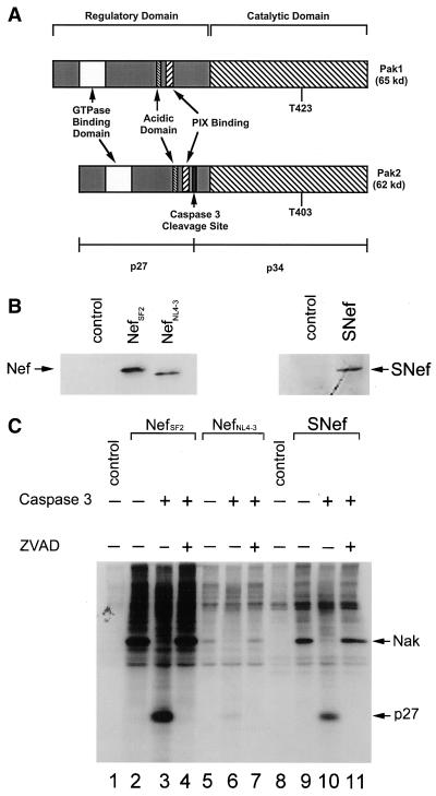
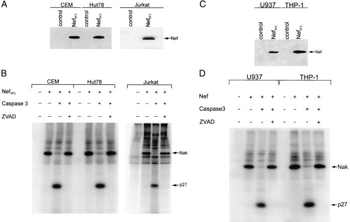
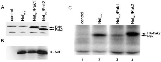
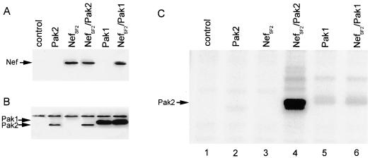
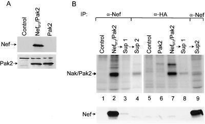
Nef proteins from human immunodeficiency virus type 1 (HIV-1) and simian immunodeficiency virus (SIV) have been found to associate with an active cellular serine/threonine kinase designated Nef-associated kinase (Nak). The exact identity of Nak remains controversial, with two recent studies indicating that Nak may be either Pak1 or Pak2. In this study, we investigated the hypothesis that such discrepancies arise from the use of different Nef alleles or different cell types by individual investigators. We first confirm that Pak2 but not Pak1 is cleaved by caspase 3 in vitro and then demonstrate that Nak is caspase 3 sensitive, regardless of Nef allele or cell type used. We tested nef alleles from three lentiviruses (HIV-1 SF2, HIV-1 NL4-3, and SIVmac239) and used multiple cell lines of myeloid, lymphoid, and nonhematopoietic origin to evaluate the identity of Nak. We demonstrate that ectopically expressed Pak2 can substitute for Nak, while ectopically expressed Pak1 cannot. We then show that Nef specifically mediates the robust activation of ectopically expressed Pak2, directly demonstrating that Nef regulates Pak2 activity and does not merely associate with activated Pak2. We report that most of the active Pak2 is found bound to Nef, although a fraction is not. In contrast, only a small amount of Nef is found associated with Pak2. We conclude that Nak is Pak2 and that Nef specifically mediates Pak2 activation in a low-abundance complex. These results will facilitate both the elucidation of the role of Nef in pathogenesis and the development of specific inhibitors of this highly conserved function of Nef.


Figures

FIG. 1
FIG. 1
Caspase 3-mediated cleavage of Nak is nef allele independent. (A) Diagrammatic representation of human Pak1 and Pak2. The two highly homologous kinases contain an N-terminal regulatory region and a C-terminal catalytic domain. Threonine phosphorylation of either residue 423 in Pak1 or residue 403 in Pak2 activates the kinases and primes them for autophosphorylation in vitro. Particularly germane to this study is the caspase 3 cleavage site found in Pak2 but not Pak1. Cleavage at this site generates a 27-kDa N-terminal fragment and a 34-kDa C-terminal fragment. Serine phosphorylation of the 27-kDa N-terminal fragment increases its apparent molecular mass to 32 kDa (51). Also indicated are the p21 GTPase binding domain, the acidic domain, and the PIX binding domain. (B) Western blot analysis of HIV-1 and SIV Nef expression in lysates of transfected 293T cells. (C) In vitro kinase assay and caspase 3 treatments of the same lysates. Note that all three Nefs associate with a caspase 3-sensitive Nak. Positions of Nak and its 32-kDa cleavage product (p27) are indicated on the right. Lanes 1 and 8, samples from 293T cells transfected with control (empty) expression plasmid; lanes 2 to 4, 5 to 7, and 9 to 11, samples from 293T cells transfected with plasmids expressing NefSF2, NefNL4-3, and SNef, respectively. Samples in lanes 3, 6, and 10 were treated with caspase 3 following the kinase assay; samples in lanes 4, 7, and 11 were treated with caspase 3 in the presence of the inhibitor ZVAD.
FIG. 2
FIG. 2
Nef binds Pak2 in human T cells and monocytic cells. (A) Western blot analysis for NefSF2 expression in the human T-cell lines CEM, HuT-78, and Jurkat. (B) In vitro kinase assay followed by caspase 3 cleavage of anti-Nef immunoprecipitates from cell lysates obtained from each cell line. In all three cases the Nak immunoprecipitated was sensitive to caspase 3 treatment, and this cleavage was inhibited by ZVAD. (C) Western blot analysis for NefSF2 expression in two human monocytic cell lines, U937 and THP-1. (D) In vitro kinase assay followed by caspase 3 treatment of Nef immunoprecipitates from cell lysates obtained from each cell line. Nak was found to be caspase 3 sensitive in both monocytic cell lines.
FIG. 3
FIG. 3
Exogenous Pak2 substitutes for endogenous Nak. (A) Western blot analysis demonstrating expression of both Pak1 and Pak2 (arrows at right) in 293T cells transfected with NefSF2 and either Pak1 or Pak2, as indicated; (B) Western blot analysis demonstrating Nef expression in the transfected cells; (C) in vitro kinase assay of anti-Nef immunoprecipitates from the transfected cells. Positions of the phosphorylated HA-tagged Pak2 and endogenous Nak are indicated on the right in panel C.
FIG. 4
FIG. 4
Specific activation of Pak2 by Nef. (A and B) Western blot analyses of Nef and Pak1/Pak2 expression, respectively, in transfected 293T cells; (C) in vitro kinase assays of anti-HA immunoprecipitates from the transfected cells. Note that Pak2 activity was clearly increased in the presence of NefSF2 (compare lanes 2 and 4), whereas Nef had no significant effect on basal Pak1 activity (lanes 5 and 6).
FIG. 5
FIG. 5
Nef-activated Pak2 is found mostly in a low-abundance Nef-Pak2 complex, but a fraction of active Pak2 is free. (A) Western blot analysis to confirm Nef (top) and Pak2 (bottom) expression in transfected 293T cells. (B) In vitro kinase assays (top) and Nef Western blot analysis (bottom) of immunoprecipitates (IP) from Nef/Pak2 expression plasmid (or control) transfections, using antibodies to Nef and HA, as indicated. Samples in lanes 2 to 4 represent successive immunoprecipitations of the same lysate, as do samples in lanes 7 to 9. In lanes 2 and 3 (top), immunoprecipitation with anti-Nef depletes all of the Nef-associated Pak2 and most, but not all, of the total active Pak2 (lane 4); also, depletion of HA-tagged Pak2 (lanes 7 and 8, top) depletes the majority of Nak but does not deplete endogenous Pak2 (lane 9). Interestingly, HA-Pak2 immunodepletion did not remove an appreciable amount of the total Nef in the lysates (lanes 7 to 9, bottom), indicating that only a small amount of Nef interacts with Pak2 even in the presence of overexpressed Pak2. Sup, supernatant.

Similar articles

Cited by

References

    1. Aldrovandi G M, Gao L, Bristol G, Zack J A. Regions of human immunodeficiency virus type 1 nef required for function in vivo. J Virol. 1998;72:7032–7039. - PMC - PubMed
    1. Alexander L, Du Z, Howe A Y, Czajak S, Desrosiers R C. Induction of AIDS in rhesus monkeys by a recombinant simian immunodeficiency virus expressing nef of human immunodeficiency virus type 1. J Virol. 1999;73:5814–5825. - PMC - PubMed
    1. Anderson S, Shugars D C, Swanstrom R, Garcia J V. Nef from primary isolates of human immunodeficiency virus type 1 suppresses surface CD4 expression in human and mouse T cells. J Virol. 1993;67:4923–4931. - PMC - PubMed
    1. Benson R E, Sanfridson A, Ottinger J S, Doyle C, Cullen B R. Downregulation of cell-surface CD4 expression by simian immunodeficiency virus Nef prevents viral super infection. J Exp Med. 1993;177:1561–1566. - PMC - PubMed
    1. Brown A, Wang X, Sawai E, Cheng-Mayer C. Activation of the PAK-related kinase by human immunodeficiency virus type 1 Nef in primary human peripheral blood lymphocytes and macrophages leads to phosphorylation of a PIX-p95 complex. J Virol. 1999;73:9899–9907. - PMC - PubMed

Publication types