Naturally occurring V1-env region variants mediate simian immunodeficiency virus SIVmac escape from high-titer neutralizing antibodies induced by a protective subunit vaccine
- PMID: 11070011
- PMCID: PMC113200
- DOI: 10.1128/jvi.74.23.11145-11152.2000
Naturally occurring V1-env region variants mediate simian immunodeficiency virus SIVmac escape from high-titer neutralizing antibodies induced by a protective subunit vaccine
Abstract
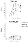
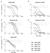
Macaques which developed high-titer neutralizing antibodies (htNAb) after immunization with a virion-derived oligomeric envelope glycoprotein subunit vaccine were protected against a homologous simian immunodeficiency virus SIVmac challenge. Here we demonstrate that the htNAb could be overcome by V1-env region variants isolated ex vivo from an SIVmac-infected macaque. The results further suggest that the development of V1-env region neutralization escape mutants is also necessary for survival of the virus in infected macaques. The immunological capacity of a single variable region to induce neutralizing antibodies in vaccinated and infected macaques initiate new ideas for a successful vaccine strategy.
Figures


Similar articles
-
Correlation between env V1/V2 region diversification and neutralizing antibodies during primary infection by simian immunodeficiency virus sm in rhesus macaques.J Virol. 2004 Apr;78(7):3561-71. doi: 10.1128/jvi.78.7.3561-3571.2004. J Virol. 2004. PMID: 15016879 Free PMC article.
-
Identification of the V1 region as a linear neutralizing epitope of the simian immunodeficiency virus SIVmac envelope glycoprotein.J Virol. 1997 Dec;71(12):9475-81. doi: 10.1128/JVI.71.12.9475-9481.1997. J Virol. 1997. PMID: 9371609 Free PMC article.
-
Breakthrough of SIV strain smE660 challenge in SIV strain mac239-vaccinated rhesus macaques despite potent autologous neutralizing antibody responses.Proc Natl Acad Sci U S A. 2015 Aug 25;112(34):10780-5. doi: 10.1073/pnas.1509731112. Epub 2015 Aug 10. Proc Natl Acad Sci U S A. 2015. PMID: 26261312 Free PMC article.
-
Antibody responses elicited in macaques immunized with human immunodeficiency virus type 1 (HIV-1) SF162-derived gp140 envelope immunogens: comparison with those elicited during homologous simian/human immunodeficiency virus SHIVSF162P4 and heterologous HIV-1 infection.J Virol. 2006 Sep;80(17):8745-62. doi: 10.1128/JVI.00956-06. J Virol. 2006. PMID: 16912322 Free PMC article.
-
Control of Heterologous Simian Immunodeficiency Virus SIVsmE660 Infection by DNA and Protein Coimmunization Regimens Combined with Different Toll-Like-Receptor-4-Based Adjuvants in Macaques.J Virol. 2018 Jul 17;92(15):e00281-18. doi: 10.1128/JVI.00281-18. Print 2018 Aug 1. J Virol. 2018. PMID: 29793957 Free PMC article.
Cited by
-
SIVsm quasispecies adaptation to a new simian host.PLoS Pathog. 2005 Sep;1(1):e3. doi: 10.1371/journal.ppat.0010003. Epub 2005 Sep 30. PLoS Pathog. 2005. PMID: 16201015 Free PMC article.
-
A dominant role for CD8+-T-lymphocyte selection in simian immunodeficiency virus sequence variation.J Virol. 2004 Dec;78(24):14012-22. doi: 10.1128/JVI.78.24.14012-14022.2004. J Virol. 2004. PMID: 15564508 Free PMC article.
-
Envelope variable region 4 is the first target of neutralizing antibodies in early simian immunodeficiency virus mac251 infection of rhesus monkeys.J Virol. 2012 Jul;86(13):7052-9. doi: 10.1128/JVI.00107-12. Epub 2012 Apr 24. J Virol. 2012. PMID: 22532675 Free PMC article.
-
Neuropathogenic SIVsmmFGb genetic diversity and selection-induced tissue-specific compartmentalization during chronic infection and temporal evolution of viral genes in lymphoid tissues and regions of the central nervous system.AIDS Res Hum Retroviruses. 2010 Jun;26(6):663-79. doi: 10.1089/aid.2009.0168. AIDS Res Hum Retroviruses. 2010. PMID: 20518690 Free PMC article.
-
Role of microglial cells in selective replication of simian immunodeficiency virus genotypes in the brain.J Virol. 2003 Jan;77(1):208-16. doi: 10.1128/jvi.77.1.208-216.2003. J Virol. 2003. PMID: 12477826 Free PMC article.
References
-
- Almond N, Jenkins A, Heath A B, Kitchin P. Sequence variation in the env gene of simian immunodeficiency virus recovered from immunized macaques is predominantly in the V1 region. J Gen Virol. 1993;74:865–871. - PubMed
-
- Babas T, Belhadj-Jrad B, Le Grand R, Dormont D, Montagnier L, Bahraoui E. Specificity and neutralizing capacity of the three monoclonal antibodies produced against the envelope glycoprotein of simian immunodeficiency virus isolate 251. Virology. 1995;211:339–344. - PubMed
-
- Benichou S, Le Grand R, Nakagawa N, Faure T, Traincard F, Vogt G, Dormont D, Tiollais P, Kieny M-P, Madaule P. Identification of a neutralizing domain in the external envelope glycoprotein of simian immunodeficiency virus. AIDS Res Hum Retroviruses. 1992;8:1165–1170. - PubMed
-
- Bruck C, Thiriart C, Fabry L, Francotte M, Pala P, VanOpstal O, Culp J, Rosenberg M, DeWilde M, Heidt P, Heeney J. HIV-1 envelope elicited neutralizing antibody titers correlate with protection and virus load in chimpanzees. Vaccine. 1994;12:1141–1148. - PubMed
Publication types
MeSH terms
Substances
LinkOut - more resources
Full Text Sources

