. 2000 Dec;74(23):11278-85.
doi: 10.1128/jvi.74.23.11278-11285.2000.

DNA-Dependent protein kinase is not required for efficient lentivirus integration

Affiliations

DNA-Dependent protein kinase is not required for efficient lentivirus integration

V Baekelandt et al. J Virol. 2000 Dec.

Abstract

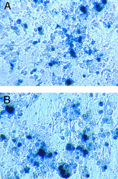
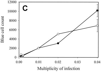
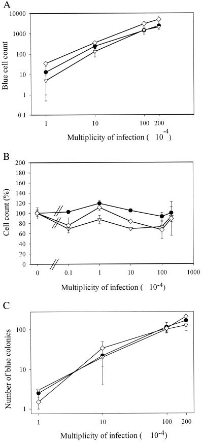
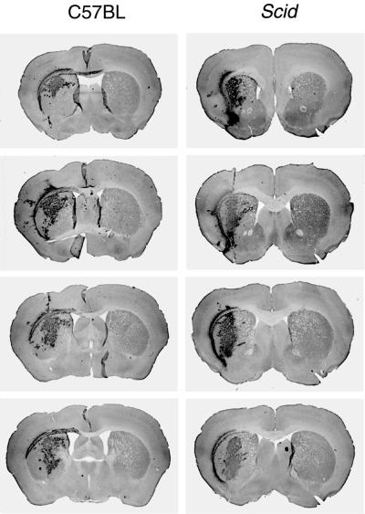
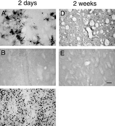
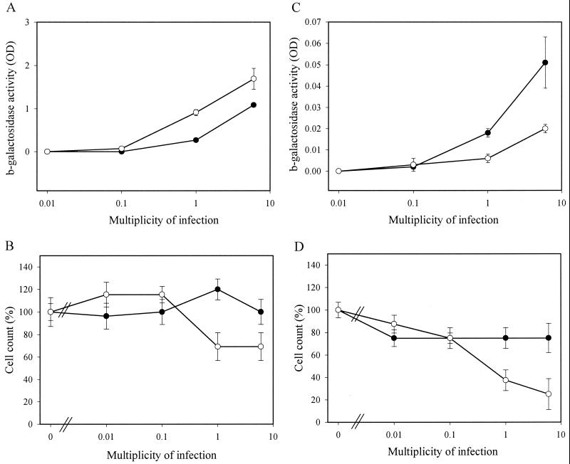
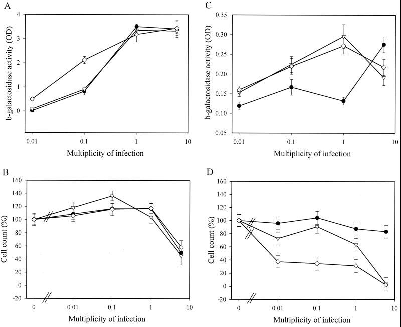
How DNA is repaired after retrovirus integration is not well understood. DNA-dependent protein kinase (DNA-PK) is known to play a central role in the repair of double-stranded DNA breaks. Recently, a role for DNA-PK in retroviral DNA integration has been proposed (R. Daniel, R. A. Katz, and A. M. Skalka, Science 284:644-647, 1999). Reduced transduction efficiency and increased cell death by apoptosis were observed upon retrovirus infection of cultured scid cells. We have used a human immunodeficiency virus (HIV) type 1 (HIV-1)-derived lentivirus vector system to further investigate the role of DNA-PK during integration. We measured lentivirus transduction of scid mouse embryonic fibroblasts (MEF) and xrs-5 or xrs-6 cells. These cells are deficient in the catalytic subunit of DNA-PK and in Ku, the DNA-binding subunit of DNA-PK, respectively. At low vector titers, efficient and stable lentivirus transduction was obtained, excluding an essential role for DNA-PK in lentivirus integration. Likewise, the efficiency of transduction of HIV-derived vectors in scid mouse brain was as efficient as that in control mice, without evidence of apoptosis. We observed increased cell death in scid MEF and xrs-5 or xrs-6 cells, but only after transduction with high vector titers (multiplicity of infection [MOI], >1 transducing unit [TU]/cell) and subsequent passage of the transduced cells. At an MOI of <1 TU/cell, however, transduction efficiency was even higher in DNA-PK-deficient cells than in control cells. Taken together, the data suggest a protective role of DNA-PK against cellular toxicity induced by high levels of retrovirus integrase or integration. Another candidate cellular enzyme that has been claimed to play an important role during retrovirus integration is poly(ADP-ribose) polymerase (PARP). However, no inhibition of lentivirus vector-mediated transduction or HIV-1 replication by 3-methoxybenzamide, a known PARP inhibitor, was observed. In conclusion, DNA-PK and PARP are not essential for lentivirus integration.


Figures

FIG. 1
FIG. 1
Lentivirus transduction efficiency in scid cells. Different amounts of a second-generation HIV-1-derived lentivirus vector (devoid of Vpr, Nef, Vif, and Vpu) encoding β-galactosidase were used to transduce subconfluent WT and scid MEF. At confluence, WT (A) and scid (B) MEF were stained with X-Gal, and positive WT (●) or scid (○) MEF were counted microscopically (C). Means and standard deviations of duplicate experiments are shown.
FIG. 1
FIG. 1
Lentivirus transduction efficiency in scid cells. Different amounts of a second-generation HIV-1-derived lentivirus vector (devoid of Vpr, Nef, Vif, and Vpu) encoding β-galactosidase were used to transduce subconfluent WT and scid MEF. At confluence, WT (A) and scid (B) MEF were stained with X-Gal, and positive WT (●) or scid (○) MEF were counted microscopically (C). Means and standard deviations of duplicate experiments are shown.
FIG. 2
FIG. 2
Lentivirus transduction efficiency in Ku-deficient cell lines. Subconfluent CHO-K1 (●), xrs-5/2 (▿), and xrs-6/1 (◊) cells were transduced with different amounts of a first-generation lentivirus vector and stained after 3 days. The number of β-galactosidase-positive cells (A) and the cell count (B) were determined microscopically. After passage (1/5) of the transduced CHO-K1 (●), xrs-5/2 (▿), and xrs-6/1 (□) cells, β-galactosidase-positive colonies (≥10 blue cells) were counted at confluence (C). Means and standard deviations of duplicate experiments are shown.
FIG. 3
FIG. 3
Efficient lentivirus transduction in scid mouse brain. (Top) Expression of the GFP transgene in the striatum of C57BL and scid mice 2 weeks after transduction with the same batch of lentivirus vectors. Serial sections with an interval of 250 μm showed equal transduction efficiency in both mouse strains. (Bottom) Quantification of GFP expression in serial sections over a distance of 2 mm around the injection site in the striatum of scid and C57BL mice 11 weeks after lentivirus vector transduction. The y axis indicates the area of positive GFP expression. Although there was some interindividual variation in transgene expression, no significant difference between scid and C57BL mice was observed.
FIG. 4
FIG. 4
No evidence for increased apoptosis after lentivirus transduction in scid mouse brain. (A and D) GFP expression around the needle track in the striatum of scid mice 2 days (A) and 2 weeks (D) after lentivirus transduction. (B and E) TUNEL staining of adjacent sections shows no apoptotic cells in the area of the striatum positive for GFP expression 2 days (B) and 2 weeks (E) after lentivirus transduction. (C) DNase treatment of a section like that in the positive control results in numerous labeled nuclei. Scale bar, 100 μm.
FIG. 5
FIG. 5
Titer- and passage-dependent cellular toxicity after lentivirus transduction of scid cells. Subconfluent WT (●) or scid (○) MEF-T were transduced in duplicate with different amounts of a second-generation lentivirus vector devoid of Vpr, Nef, Vif, and Vpu. Half of the cells were analyzed at confluence (A and B), and half of the cells were passaged (1/5) and analyzed at confluence (C and D). β-Galactosidase activity (OD, optical density) was measured in the cell lysate (A and C), and the cell count was determined by trypan blue dye exclusion (B and D). Reporter gene activity was not normalized for differential cell growth. Means and standard deviations of duplicate experiments are shown.
FIG. 6
FIG. 6
Titer- and passage-dependent cellular toxicity after lentivirus transduction of Ku-deficient cells. Subconfluent CHO-K1 (●), xrs-5/2 (▿), and xrs-6/1 (◊) cells were transduced in duplicate with different amounts of a second-generation lentivirus vector. Half of the cells were analyzed at confluence (A and B), and half of the cells were passaged (1/5) and analyzed at confluence (C and D). β-Galactosidase activity (OD, optical density) was measured in the cell lysate (A and C), and the cell count was determined by trypan blue dye exclusion (B and D). Reporter gene activity was not normalized for differential cell growth. Means and standard deviations of duplicate experiments are shown.

References

    1. Abdallah B, Hassan A, Benoist C, Goula D, Behr J P, Demeneix B A. A powerful nonviral vector for in vivo gene transfer into the adult mammalian brain: polyethylenimine. Hum Gene Ther. 1997;7:1947–1954. - PubMed
    1. Acel A, Udashkin B E, Wainberg M A, Faust E A. Efficient gap repair catalyzed in vitro by an intrinsic DNA polymerase activity of human immunodeficiency virus type 1 integrase. J Virol. 1998;72:2062–2071. - PMC - PubMed
    1. Ausubel F M, Brent R, Kingston R E, Moore D D, Seidman J G, Smith J A, Struhl K, editors. Current protocols in molecular biology. New York, N.Y: John Wiley & Sons, Inc.; 1987.
    1. Baekelandt, V., B. De Strooper, B. Nuttin, and Z. Debyser. Gene therapeutic strategies for neurodegenerative diseases. Curr. Opin. Mol. Ther., in press. - PubMed
    1. Blier P R, Griffith A J, Craft J, Hardin J A. Binding of Ku protein to DNA. Measurement of affinity for ends and demonstration of binding to nicks. J Biol Chem. 1993;268:7594–7601. - PubMed

Publication types

MeSH terms