. 2000 Dec;74(23):11311-21.
doi: 10.1128/jvi.74.23.11311-11321.2000.

Open reading frame S/L of varicella-zoster virus encodes a cytoplasmic protein expressed in infected cells

Affiliations
Free PMC article

Open reading frame S/L of varicella-zoster virus encodes a cytoplasmic protein expressed in infected cells

G W Kemble et al. J Virol. 2000 Dec.
Free PMC article

Abstract

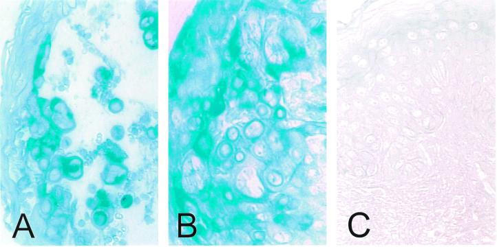
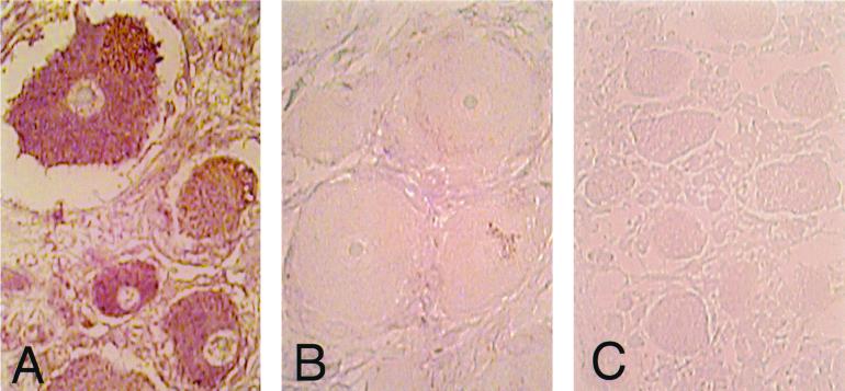
We report the discovery of a novel gene in the varicella-zoster virus (VZV) genome, designated open reading frame (ORF) S/L. This gene, located at the left end of the prototype VZV genome isomer, expresses a polyadenylated mRNA containing a splice within the 3' untranslated region in virus-infected cells. Sequence analysis reveals significant differences between the ORF S/Ls of wild-type and attenuated strains of VZV. Antisera raised to a bacterially expressed portion of ORF S/L reacted specifically with a 21-kDa protein synthesized in cells infected with a VZV clinical isolate and with the original vaccine strain of VZV (Oka-ATCC). Cells infected with other VZV strains, including a wild-type strain that has been extensively passaged in tissue culture and commercially produced vaccine strains of Oka, synthesize a family of proteins ranging in size from 21 to 30 kDa that react with the anti-ORF S/L antiserum. MeWO cells infected with recombinant VZV harboring mutations in the C-terminal region of the ORF S/L gene lost adherence to the stratum and adjacent cells, resulting in an altered plaque morphology. Immunohistochemical analysis of VZV-infected cells demonstrated that ORF S/L protein localizes to the cytoplasm. ORF S/L protein was present in skin lesions of individuals with primary or reactivated infection and in the neurons of a dorsal root ganglion during virus reactivation.


Figures

FIG. 1
FIG. 1
Location of ORF S/L in the VZV genome. (A) Schematic representation of the linear VZV genome. The repeat sequences TRL/IRL (■) and IRS/TRS (░⃞) are depicted. The four overlapping cosmids are indicated relative to their map positions beneath the genomic diagram. (B) The VZV genome is circularized by adjoining TRL and TRS and accounting for the one unpaired nucleotide. The region of the genome near the TRL/TRS junction is expanded to show the presence of ORFs and selected restriction sites in both Dumas and Oka. The ORF 1 and ORF 2 designations are consistent with those of Davison and Scott (12). ORF S/L Dumas and ORF S/L Oka initiate at different positions.
FIG. 2
FIG. 2
Sequence comparison of VZV genomes near ORF S/L. The nucleotide sequences of Oka-ATCC, Oka-Merck, Oka-SK, Dumas, and Ellen as displayed all begin within TRS; the TRS/TRL boundary is identified with a vertical line, and nucleotide deletions (−) are indicated. Sequence differences between the strains are shaded. The predicted amino acid sequences are displayed beneath the nucleotide sequences. Consensus# represents the Oka-Merck and Oka-SK consensus amino acids. Positions of the transcription initiation site (∗∗∗), the 130-nt intron (⩵⩵), potential termination sites (underline), and primer sites used to generate anti-ORF S/L serum (overline) are indicated. The terminator at position 567 would truncate the S/L protein encoded by Oka-Merck if it were to initiate at the same ATG as Dumas.
FIG. 2
FIG. 2
Sequence comparison of VZV genomes near ORF S/L. The nucleotide sequences of Oka-ATCC, Oka-Merck, Oka-SK, Dumas, and Ellen as displayed all begin within TRS; the TRS/TRL boundary is identified with a vertical line, and nucleotide deletions (−) are indicated. Sequence differences between the strains are shaded. The predicted amino acid sequences are displayed beneath the nucleotide sequences. Consensus# represents the Oka-Merck and Oka-SK consensus amino acids. Positions of the transcription initiation site (∗∗∗), the 130-nt intron (⩵⩵), potential termination sites (underline), and primer sites used to generate anti-ORF S/L serum (overline) are indicated. The terminator at position 567 would truncate the S/L protein encoded by Oka-Merck if it were to initiate at the same ATG as Dumas.
FIG. 3
FIG. 3
Mapping the ORF S/L mRNA. (A) Depiction of pGS/LC3, a plasmid clone of a PCR product spanning the TRL/TRS junction of Oka-ATCC. Directions of the T7 and Sp6 transcripts generated from this insert are indicated; the Sp6-transcribed RNA would be the same sense as ORF S/L mRNA. (B) Northern analysis of RNA from ORF S/L. Ten micrograms of whole-cell (Tot) or 1 μg of poly(A)+ (pA+) RNA isolated from uninfected (HF) or VZV-infected (VZV) fibroblasts was subjected to Northern analysis with either the T7/XhoI (T7) or Sp6/EcoRI (Sp6) probe. The autoradiographic image of this analysis is shown, with sizes of the RNA markers indicated in kilobases. (C) RNase protection analysis of ORF S/L RNA. The T7/XhoI (T7) in vitro-transcribed probe was hybridized to whole-cell RNA isolated from VZV-infected (VZV) or uninfected (HF) fibroblasts and digested with RNase ONE for 60 min. The products were separated by electrophoresis in a denaturing polyacrylamide gel, and the gel was exposed to X-ray film. Positions and sizes (in nucleotides) of the molecular weight standards (lane M) are indicated at the left. The closed circle adjacent to the lane marked probe identifies the position of unhybridized probe. (D) 3′ RACE analysis of ORF S/L RNA. Whole-cell RNAs from uninfected (HF) and infected (VZV) cells were reverse transcribed and subjected to RT-PCR. The products of these reactions and a control reaction that was done without reverse transcriptase (−RT) were electrophoresed on a 4% 3:1 agarose gel and subsequently stained with ethidium bromide. The RT-PCR product shown in the VZV lane was directly sequenced. The junction of the Oka ORF S/L mRNA and the poly(A) region are depicted and aligned with the Dumas sequence (accession no. X04370). The numbers refer to Dumas genomic DNA. Sizes of the DNA markers (lane M) are shown in nucleotides at the left. Autoradiograms and negatives of stained gels were scanned with a GS-250 imaging densitometer (Bio-Rad, Hercules, Calif.). Photographic quality images were generated using Molecular Analyst (Bio-Rad) and Adobe Photoshop (Adobe Systems, Mountain View, Calif.) software.
FIG. 4
FIG. 4
Southern analysis of recombinant VZVs. (A) Ethidium bromide-stained gel and Southern analysis of EcoRV-digested DNA prepared from Vero cells infected with either Oka-ATCC, Oka-R, or the three recombinant viruses Oka-gB2, Oka-gB3, and Oka-Δ4-3. Positions of molecular weight markers are shown in kilobases at the left. (B) Diagram depicting the ORF S/L region from the Oka-ATCC, Oka-Δ4-3, Oka-gB2, and Oka-gB3 viruses. Oka-ATCC is depicted on the top line, and the 1.2-kbp XhoI-to-PmeI probe is indicated. OkaΔ4 has a deletion of 400 nt between the ApaLI and BspEI sites (▵); and the gB mutants have a 60-nt CMV gB sequence (formula image
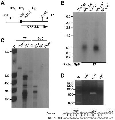
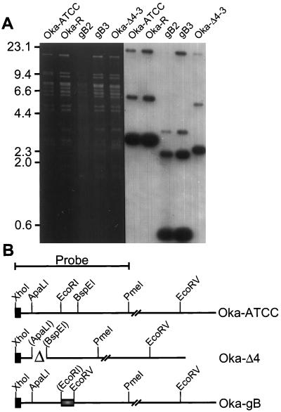
) containing a novel EcoRV site replacing the 120 bp between the EcoRI and BspEI sites of Oka. Images of the autoradiograms and stained gels were generated as for Fig. 3.
FIG. 5
FIG. 5
Plaque morphology of Oka-R and Oka-gB2 on MeWO cells. Syncytial plaques formed by Oka-R are shown at 36 and 60 hpi on MeWO cells. Plaques formed by Oka-gB2 were photographed at the same points in time as the Oka-R-infected cells. Note the cleared area at 60 hpi on the Oka-gB2-infected MeWO cells.
FIG. 6
FIG. 6
Identification of S/L proteins in extracts of infected cells. Whole-cell extracts of uninfected HELF (M) or HELF infected with the VZV strains noted above the lanes were subjected to Western blot analysis using an anti-S/L rabbit serum diluted 1:500, followed by goat anti-rabbit antibody conjugated to horseradish peroxidase. The molecular masses of prestained size markers (Amersham Pharmacia Biotech, Piscataway, N.J.) are indicated at the left. The 20.1-kDa marker was too faint to see in this reproduction.
FIG. 7
FIG. 7
Immunohistochemical detection of VZV proteins in HELF. Cells were infected with VZV strains Ellen and Oka-SK and the recombinant viruses Oka-Δ4-3 and Oka-R. Immunohistochemical analysis of mock-infected cells is also shown. The products of ORF S/L and gC were detected using anti-ORF S/L or anti-gC antibodies. The signal was visualized after addition of AP-conjugated goat anti-rabbit immunoglobulin secondary antibody and developed with AP substrate in the presence of levamisole. The viruses infecting the monolayers are noted at the left; VZV proteins that were analyzed are identified at the top.
FIG. 8
FIG. 8
Immunohistochemical analysis of ORF S/L protein in skin biopsy specimens. Sections of formalin-fixed, paraffin-embedded skin biopsy specimens from patients with chickenpox (A), zoster (B), and Grover's disease (C) were deparaffinized with xylene and rehydrated with successive alcohol washes. ORF S/L protein was detected using anti-ORF S/L antibody. The signal was visualized as described in the legend to Fig. 7.
FIG. 9
FIG. 9
Immunohistochemical analysis of ORF S/L in DRG. Sections of formalin-fixed, paraffin-embedded DRG harboring reactivated (A) or latent (B) VZV and a control uninfected fetal DRG (C) were processed as described in the legend to Fig. 8. ORF S/L was detected by immunohistochemistry essentially as described in the legend to Fig. 7 except that levamisole was omitted from the AP detection buffer and nitroblue tetrazolium chloride was the substrate.

Similar articles

Cited by

References

    1. Annunziato P W, Lungu O, Panagiotidis C, Zhang J H, Silvers D N, Gershon A A, Silverstein S J. Varicella-zoster virus proteins in skin lesions: implications for a novel role of ORF29p in chickenpox. J Virol. 2000;74:2005–2010. - PMC - PubMed
    1. Bohenzky R A, Papavassiliou A G, Gelman I H, Silverstein S. Identification of a promoter mapping within the reiterated sequences that flank the herpes simplex virus type 1 ULregion. J Virol. 1993;67:632–642. - PMC - PubMed
    1. Cai W, Schaffer P A. Herpes simplex virus type 1 ICP0 regulates expression of immediate-early, early, and late genes in productively infected cells. J Virol. 1992;66:2904–2915. - PMC - PubMed
    1. Centers for Disease Control and Prevention. Varicella-related deaths among adults—United States 1997. Morbid Mortal Wkly Rep. 1997;46:409–412. - PubMed
    1. Chen J, Silverstein S. Herpes simplex viruses with mutations in the gene encoding ICP0 are defective in gene expression. J Virol. 1992;66:2916–2927. - PMC - PubMed

Publication types

Associated data

LinkOut - more resources