. 2000 Dec;74(23):11322-8.
doi: 10.1128/jvi.74.23.11322-11328.2000.

Interaction between herpes simplex virus type 1 IE63 protein and cellular protein p32

Affiliations

Interaction between herpes simplex virus type 1 IE63 protein and cellular protein p32

H E Bryant et al. J Virol. 2000 Dec.

Abstract

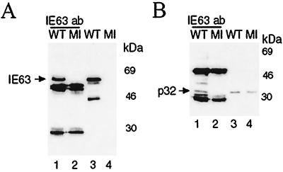
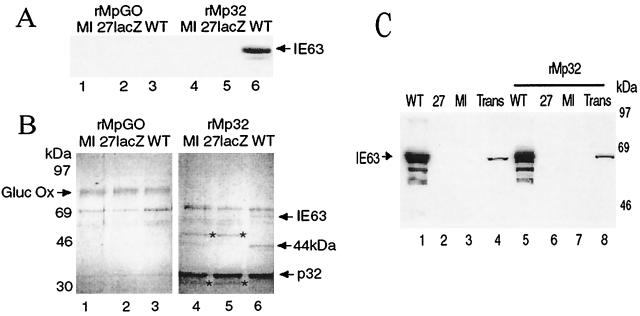
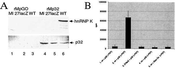
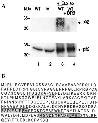
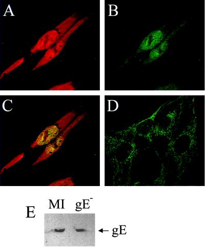
The herpes simplex virus type 1 (HSV-1) immediate-early gene IE63 (ICP27), the only HSV-1 regulatory gene with a homologue in every mammalian and avian herpesvirus sequenced so far, is a multifunctional protein which regulates transcriptional and posttranscriptional processes. One of its posttranscriptional effects is the inhibition of splicing of viral and cellular transcripts. We previously identified heterogeneous nuclear ribonucleoprotein (hnRNP) K and casein kinase 2 (CK2) as two protein partners of IE63 (H. Bryant et al., J. Biol. Chem. 274:28991-28998, 1999). Here, using a yeast two-hybrid assay, we identify another partner of IE63, the cellular protein p32. Confirmation of this interaction was provided by coimmunoprecipitation from virus-infected cells and recombinant p32 binding assays. A p32-hnRNP K-CK2 complex, which required IE63 to form, was isolated from HSV-1-infected cells, and coimmunoprecipitating p32 was phosphorylated by CK2. Expression of IE63 altered the cytoplasmic distribution of p32, with some now colocalizing with IE63 in the nuclei of infected and transfected cells. As p32 copurifies with splicing factors and can inhibit splicing, we propose that IE63 together with p32, possibly with other IE63 partner proteins, acts to disrupt or regulate pre-mRNA splicing. As well as contributing to host cell shutoff, this effect could facilitate splicing-independent nuclear export of viral transcripts.


Figures

FIG. 1
FIG. 1
Schematic representation (not to scale) of IE63 protein showing the different functional regions as described elsewhere (4). NLS, nuclear localization signal; NES, leucine-rich nuclear export signal; R1 and R2, arginine-rich regions. Shown below are the IE63 truncations used in the yeast two-hybrid assay to map the region involved in interaction with p32.
FIG. 2
FIG. 2
In vivo coimmunoprecipitation of IE63 and p32, using antibodies directed against IE63. HSV-1 wt-infected (WT) or mock-infected (MI) HeLa cell extracts were immunoprecipitated with IE63 MAb. Aliquots of the precipitated proteins were separated by SDS-PAGE, transferred to nitrocellulose, and analyzed by Western blotting using p32 MAb (B, lanes 1 and 2) or IE63 MAb (A, lanes 1 and 2). A 100-μg aliquot of total protein was added to each immunoprecipitation, and half was loaded in lanes 1 and 2; 20 μg of total protein from extracts was loaded in lanes 3 and 4.
FIG. 3
FIG. 3
Interaction of IE63 with p32 Sepharose column. HSV-1 wt- or 27-lacZ-infected, pCMV63-transfected, or mock-infected HeLa cell extracts were mixed with rMpGO-Sepharose or rMp32-Sepharose. After washing, proteins were boiled off the Sepharose, separated by SDS-PAGE, transferred to nitrocellulose, and Coomassie blue stained or analyzed by Western blotting using IE63 MAb. (A) Western blot using IE63 MAb of proteins from wt-infected (WT), 27-lacZ-infected, or mock-infected (MI) cell extracts, bound and then removed from an rMpGO (lanes 1 to 3) or rMp32 (lanes 4 to 6) column. (B) Coomassie blue staining of the bound proteins shown in panel A. (C) Western blot using IE63 MAb of proteins from wt-infected (WT), mock-infected (MI), or pCMV63-transfected (Trans) cell extracts, bound and then removed from an rMp32 column (lanes 5 to 8); cell extracts used (lanes 1 to 4).
FIG. 4
FIG. 4
CK2 and hnRNP K from HSV-1-infected extracts interact with p32 attached to a Sepharose column. Aliquots of bound and subsequently eluted proteins, as shown in Fig. 3, were separated by SDS-PAGE, transferred to nitrocellulose, and analyzed by Western blotting using anti-hnRNP K serum or p32 MAb and also analyzed for CK2 activity using a peptide assay. (A) Western blot analysis using anti-hnRNP K serum (top) or p32 MAb (bottom) of proteins from wt-infected (WT), 27-lacZ-infected, or mock-infected (MI) cell extracts. Proteins were bound and then removed from an rMpGO (lanes 1 to 3) or rMp32 (lanes 4 to 6) column. (B) CK2 activity, as measured by phosphorylation of a specific peptide substrate, by proteins which bound to rMpGO from wt-infected extracts (lane 5) and to rMp32 from wt-infected (lanes 1 and 2), 27-lacZ-infected (lane 3), and mock-infected (lane 4) extracts. CK2 assays were performed in the presence (lanes 2 to 5) or absence (lane 1) of peptide substrate.
FIG. 5
FIG. 5
The CK2 inhibitor DRB inhibits phosphorylation of p32. (A) Using IE63 MAb, coimmunoprecipitation was performed as for Fig. 2. The coimmunoprecipitate from wt-infected cell extract (WT) was incubated with [γ-32P]ATP, with (lane 4) or without (lane 3) the CK2 inhibitor DRB. Proteins were separated by SDS-PAGE and transferred to nitrocellulose; the same gel was analyzed by phosphorimaging (top) and then Western blotted for p32 (bottom). Cell extracts were used in lanes 1 and 2. (B) CK2 phosphorylation consensus sites in p32. The sequence of a 282-aa protein was obtained from the GenBank database. CK2 consensus sites (8) are alternately underlined and boxed to distinguish between overlapping sites; serine and threonine (S/T) residues potentially phosphorylated by CK2 are in boldface.
FIG. 6
FIG. 6
IE63 causes p32 to redistribute in cells. Immunofluorescence was performed on HeLa cells infected with HSV-1 gE and on mock-infected cells using anti-IE63 MAb and anti-p32 serum. A, B, and C show the same field of vision. (A) HSV-1 gE-infected HeLa cells 6 h postinfection, stained for IE63; (B) HSV-1 gE-infected HeLa cells 6 h postinfection, stained for p32; (C) HSV-1 gE-infected HeLa cells 6 h postinfection, stained for IE63 and p32; (D) mock-infected cells stained for p32; (E) extracts of HSV-1 gE-infected and mock-infected cells Western blotted for p32.

References

    1. Ackerman M, Braun D K, Pereira L, Roizman B. Characterization of herpes simplex 1 α proteins 0, 4, and 27 with monoclonal antibodies. J Virol. 1984;52:108–118. - PMC - PubMed
    1. Bell S, Cranage M, Borysiewicz L, Minson T. Induction of immunoglobulin G Fc receptors by recombinant vaccinia viruses expressing glycoproteins E and I of herpes simplex type 1. J Virol. 1990;64:2181–2186. - PMC - PubMed
    1. Bruni R, Roizman B. Open reading frame P—a herpes simplex virus gene repressed during productive infection encodes a protein that binds a splicing factor and reduces synthesis of viral proteins made from spliced mRNA. Proc Natl Acad Sci USA. 1996;93:10423–10427. - PMC - PubMed
    1. Bryant H, Wadd S, Filhol O, Scott J E, Hsieh T, Everett R, Clements J B. The multifunctional herpes simplex virus IE63 protein interacts with heterogeneous ribonucleoprotein K and with casein kinase 2. J Biol Chem. 1999;274:28991–28998. - PubMed
    1. Chen M R, Yang J F, Wu C W, Middeldorp J M, Chen J Y. Physical association between the EBV protein EBNA 1 and P32/TAP/hyaluronectin. J Biomed Sci. 1998;5:173–179. - PubMed

Publication types