. 2000 Dec;74(23):11398-406.
doi: 10.1128/jvi.74.23.11398-11406.2000.

The host range phenotype displayed by a Sindbis virus glycoprotein variant results from virion aggregation and retention on the surface of mosquito cells

Affiliations

The host range phenotype displayed by a Sindbis virus glycoprotein variant results from virion aggregation and retention on the surface of mosquito cells

K W Boehme et al. J Virol. 2000 Dec.

Abstract

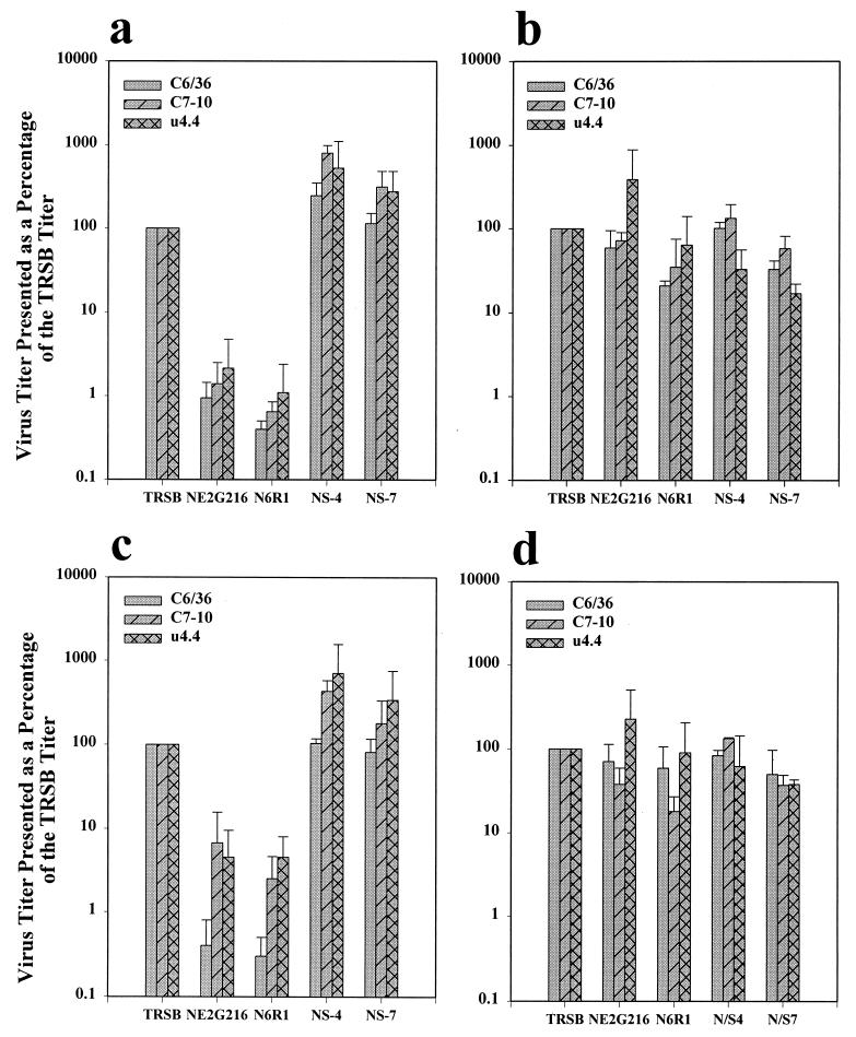
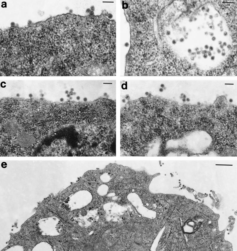
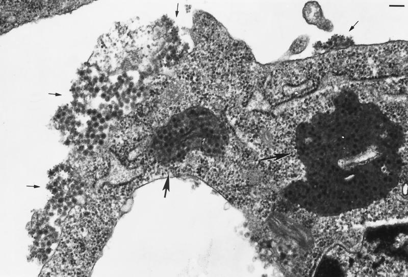
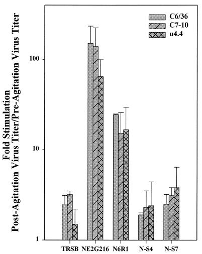
The Sindbis virus variant NE2G216 is a PE2-containing host range mutant that is growth restricted in cultured mosquito cells (C6/36) due to inefficient release of virions from this cell type. The maturation defect of NE2G216 has been linked to the structures of N-linked oligosaccharides synthesized by arthropod cells. Analysis of C6/36 cells infected with NE2G216 by transmission electron microscopy revealed the presence of dense virus aggregates within cytoplasmic vacuoles and virus aggregates adhered to the cell surface. The virus aggregation phenotype of NE2G216 was reproduced in vertebrate cells (Pro-5) by the addition of 1-deoxymannojirimycin, an inhibitor of carbohydrate processing which limits the processing of N-linked oligosaccharides to structures that are structurally similar, albeit not identical, to those synthesized in C6/36 cells. We conclude that defective maturation of NE2G216 in mosquito cells is due to virion aggregation and retention on the cell surface and that this phenotype is directly linked to the carbohydrate-processing properties of these cells.


Figures

FIG. 1
FIG. 1
Comparisons between extracellular and cell-associated virus titers from infected mosquito cell lines. Titers are presented as a percentage of that obtained for TRSB (100%). Extracellular virus titers (a and c) and cell-associated virus titers (b and d) were determined at 12 h (a and b) and 20 h (c and d) postinfection as described in the text. Virus used for infection is shown on the x axis. Results represent the averages of values obtained in two independent experiments.
FIG. 2
FIG. 2
C6/36 cells infected with TRSB (a, b, and e) and NS-4 (c and d). In cells infected with these viruses, virions were most commonly seen attached to the cell surface or in the process of budding at this site (a, c, d, and e). Virions were also seen within cytoplasmic vacuoles (b). Cells were processed for TEM analysis at 20 h postinfection. Bar, 100 nM (a to d) or 0.5 μM (e). Magnifications: ×78,375 (a and b), ×57,750 (c and d), and ×24,475 (e).
FIG. 3
FIG. 3
C6/36 cell infected with N6R1. Virions are present at a high density within cytoplasmic vacuoles (large arrows) and within virus aggregates adhered to the plasma membrane (small arrows). Cells were processed for TEM analysis at 20 h postinfection. Bar, 100 nM. Magnification, ×62,700.
FIG. 4
FIG. 4
C6/36 cells infected with NE2G216 (a, c, and d), and N6R1 (b). Virion aggregates are seen covering large areas of the infected cell and appear to be associated with a matrix material. A vacuole filled with virions appears to be in the final stage of fusion with the plasma membrane (arrow in panel c). Panel d presents a higher-power magnification of a surface-bound virus aggregate from panel c (aggregate on right). Cells were processed for TEM analysis at 20 h postinfection. Bar, 100 nM. Magnifications: ×62,700 (a), ×94,000 (b and d), and ×31,550 (c).
FIG. 5
FIG. 5
Effect of cell agitation on extracellular virus titers. Extracellular virus titers were determined prior to and following mechanical agitation of infected mosquito cells as described in the text. Results are presented as the fold enhancement of virus titers as a result of cell agitation. Virus used for infection is shown on the x axis. Results represent the averages of values obtained in two independent experiments.
FIG. 6
FIG. 6
Pro-5 cells infected with TRSB (a and b) and NE2G216 (c, d, and e). Cells were infected and maintained in the absence of 1-dMM (a and c) or in the presence of 2.5 mM 1-dMM (b, d, and e). TRSB and NE2G216 virions were observed budding individually at the plasma membrane in the absence of 1-dMM (a and c) and in TRSB-infected cells maintained in the presence of 1-dMM (b). NE2G216 virions formed aggregates at the plasma membrane of Pro-5 cells in the presence of 1-dMM, and budding virions appeared to assemble into aggregates concomitantly with the budding process (d and e, arrows). Cells were processed for TEM analysis at 14 h postinfection. Bar, 100 nM. Magnifications: ×85,250 (a and b), ×90,000 (c and d), and ×94,000 (e).

Similar articles

Cited by

References

    1. Bischoff J, Liscum L, Kornfeld R. The use of 1-deoxymannojirimycin to evaluate the role of various α-mannosidases in oligosaccharide processing in intact cells. J Biol Chem. 1986;261:4766–4774. - PubMed
    1. Boehme K W, Williams J C, Johnston R E, Heidner H W. Linkage of an alphavirus host-range restriction to the carbohydrate processing phenotypes of the host cell. J Gen Virol. 2000;81:161–170. - PubMed
    1. Burge B W, Pfefferkorn E R. Isolation and characterization of conditional-lethal mutants of Sindbis virus. Virology. 1966;30:204–213. - PubMed
    1. Doms R W, Lamb R A, Rose J K, Helenius A. Folding and assembly of viral membrane proteins. Virology. 1993;193:545–562. - PubMed
    1. Fiedler K, Simons K. The role of N-glycans in the secretory pathway. Cell. 1995;81:309–312. - PubMed

Publication types

Substances