. 2000 Nov 7;97(23):12513-8.
doi: 10.1073/pnas.97.23.12513.

Repression of human papillomavirus oncogenes in HeLa cervical carcinoma cells causes the orderly reactivation of dormant tumor suppressor pathways

Affiliations

Repression of human papillomavirus oncogenes in HeLa cervical carcinoma cells causes the orderly reactivation of dormant tumor suppressor pathways

E C Goodwin et al. Proc Natl Acad Sci U S A. .

Abstract

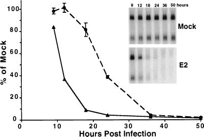
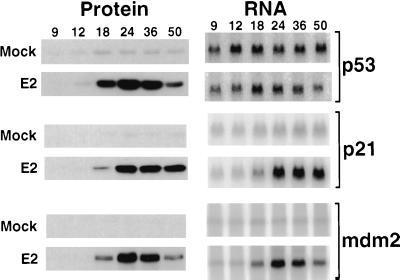
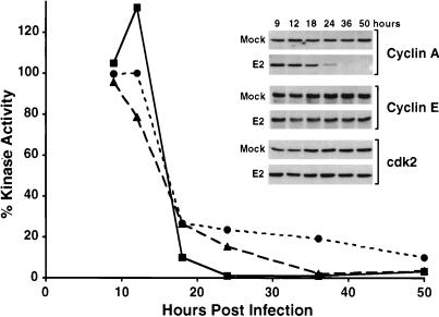
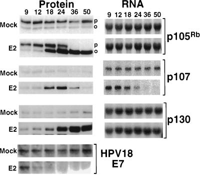
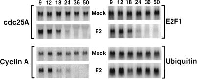
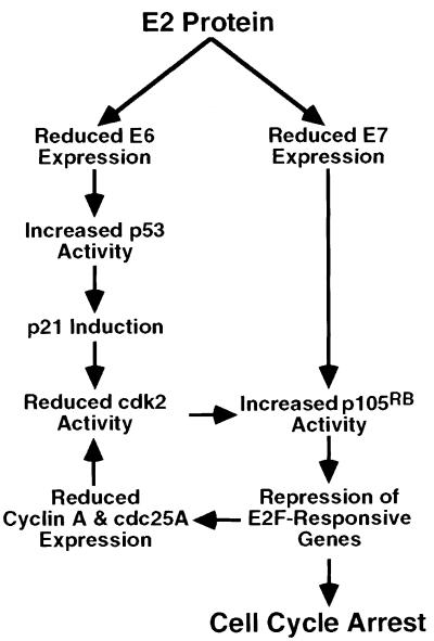
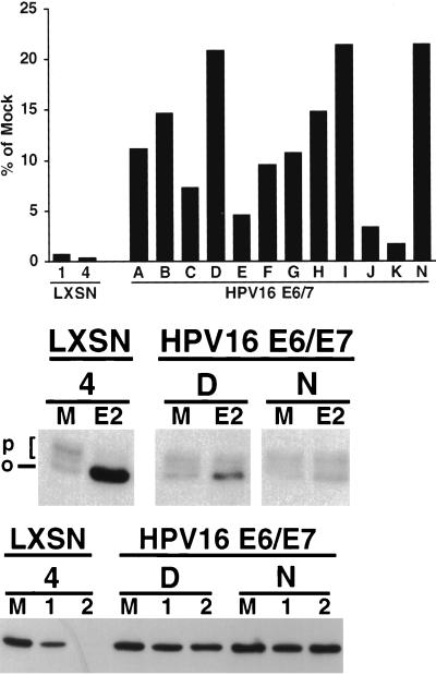
Most cervical carcinomas express high-risk human papillomaviruses (HPVs) E6 and E7 proteins, which neutralize cellular tumor suppressor function. To determine the consequences of removing the E6 and E7 proteins from cervical cancer cells, we infected HeLa cells, a cervical carcinoma cell line that contains HPV18 DNA, with a recombinant virus that expresses the bovine papillomavirus E2 protein. Expression of the E2 protein resulted in rapid repression of HPV E6 and E7 expression, followed approximately 12 h later by profound inhibition of cellular DNA synthesis. Shortly after E6/E7 repression, there was dramatic posttranscriptional induction of p53. Two p53-responsive genes, mdm2 and p21, were induced with slightly slower kinetics than p53 and appeared to be functional, as assessed by inhibition of cyclin-dependent kinase activity and p53 destabilization. There was also dramatic posttranscriptional induction of p105(Rb) and p107 after E6/E7 repression, followed shortly thereafter by induction of p130. By 24 h after infection, only hypophosphorylated p105(Rb) was detectable and transcription of several Rb/E2F-regulated genes was dramatically repressed. Constitutive expression of the HPV16 E6/E7 genes alleviated E2-induced growth inhibition and impaired activation of the Rb pathway and repression of E2F-responsive genes. This dynamic response strongly suggests that the p53 and Rb tumor suppressor pathways are intact in HeLa cells and that repression of HPV E6 and E7 mobilizes these pathways in an orderly fashion to deliver growth inhibitory signals to the cells. Strikingly, the major alterations in the cell cycle machinery underlying cervical carcinogenesis can be reversed by repression of the endogenous HPV oncogenes.


Figures

Figure 1
Figure 1
Time course of HPV E6/E7 repression and growth inhibition. HeLa cells were infected or mock-infected, and, at the indicated time after infection, RNA was analyzed for HPV E6/E7 expression by Northern blotting (solid line), and cellular DNA synthesis was determined by incorporation of tritiated thymidine (dashed line). The error bars indicate two standard deviations of the mean. (Inset) Repression of HPV E6/E7 expression. Northern blot described above. RNA was isolated at the indicated hours after E2 infection (Lower) or mock-infection (Upper), electrophoresed, transferred, and probed with a radiolabeled HPV18 E6/E7 DNA fragment. The signal obtained was quantitated, normalized for the signal obtained with a ubiquitin probe, and expressed as the percentage of the normalized signal obtained with RNA from mock-infected cells.
Figure 2
Figure 2
(Left) Western analysis of p53 pathway. HeLa cell proteins were harvested at the indicated hours after E2 infection or mock-infection, electrophoresed, transferred, and probed with antibodies specific for p53, p21, or human mdm2, as indicated. (Right) Northern analysis of p53 pathway. HeLa cell RNA was isolated at the indicated hours after E2 infection or mock-infection. After electrophoresis and transfer, p53, p21, and mdm2 mRNA was detected by hybridization to the appropriate radiolabeled cDNA probe.
Figure 3
Figure 3
Cyclin-dependent kinase activity. HeLa cell extracts were prepared at the indicated hours after E2 infection or mock-infection. After immunoprecipitation with control antibodies or antibodies specific for cyclin E (dotted line), cdk2 (dashed line), or cyclin A (solid line), kinase activity toward histone H1 was measured. The signal obtained was quantitated and expressed as the percentage of the corrected signal obtained with mock-infected samples. (Inset) Western analysis of cdk components. HeLa cell proteins were harvested at the indicated times from mock-infected or E2-infected cells. After electrophoresis and transfer, samples were probed with antibodies specific for cyclin A, cyclin E, and cdk2, as indicated.
Figure 4
Figure 4
(Left) Western analysis of retinoblastoma family members. HeLa cell protein was prepared at the indicated hours after E2 infection or mock-infection. After electrophoresis and transfer, specific antibodies were used to detect HPV18 E7, p105Rb, p107, and p130. The hyperphosphorylated (p) and hypophosphorylated (o) form of p105Rb are indicated. (Right) Northern analysis of retinoblastoma family members. HeLa cell RNA was prepared at the indicated hours after mock-infection or E2 infection. After electrophoresis and transfer, p105Rb, p107, and p130 mRNA were detected by hybridization to the appropriate radiolabeled cDNA probe.
Figure 5
Figure 5
Northern analysis of E2F-responsive genes. HeLa cell RNA was prepared at the indicated hours after mock-infection or E2 infection. After electrophoresis and transfer, E2F1, cyclin A, cdc25A, and ubiquitin mRNA were detected by hybridization to the appropriate radiolabeled cDNA probe.
Figure 6
Figure 6
Model for the growth regulatory pathway activated by the E2 protein. See text for details.
Figure 7
Figure 7
Effect of HPV16 E6/E7 expression on the acute response to the E2 protein. Cell lines derived from individual clones of cells infected with control retrovirus (LXSN) or HPV16 E6/E7 retrovirus were analyzed. (Top) DNA synthesis by the indicated cell lines was measured by incorporation of tritiated thymidine 48 h after infection with the E2 virus, expressed as the percentage of DNA synthesis by each clone after mock-infection. (Middle) Expression of p105Rb in the indicated cell lines after mock-infection or 1 day after infection with the E2 virus. (Bottom) Expression of cyclin A in the indicated cell lines after mock-infection or 1 or 2 days after infection with the E2 virus.

References

    1. Villa L L. Adv Cancer Res. 1997;71:321–341. - PubMed
    1. Prives C. Cell. 1998;95:5–8. - PubMed
    1. Nevins J R. Cell Growth Differ. 1998;9:585–593. - PubMed
    1. Dyson N. Genes Dev. 1998;12:2245–2262. - PubMed
    1. Hiebert S, Chellappan S, Horowitz J, Nevins J. Genes Dev. 1992;6:177–185. - PubMed

Publication types

MeSH terms

Substances