Comparative Study
. 2000 Nov 7;97(23):12729-34.
doi: 10.1073/pnas.97.23.12729.

Increased adipose tissue in male and female estrogen receptor-alpha knockout mice

Affiliations
Comparative Study

Increased adipose tissue in male and female estrogen receptor-alpha knockout mice

P A Heine et al. Proc Natl Acad Sci U S A. .

Abstract

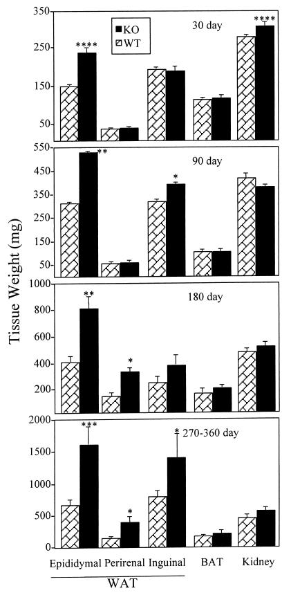
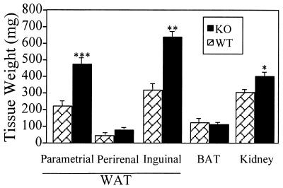
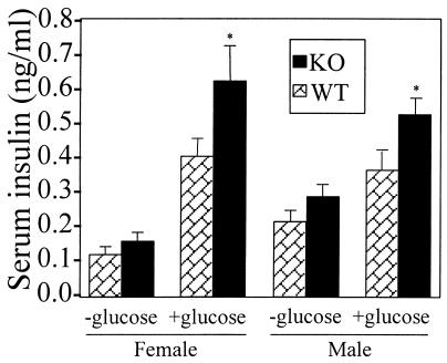
Estrogen regulates the amount of white adipose tissue (WAT) in females, but its role in males and whether WAT effects involve estrogen receptor-alpha (ERalpha) or ERbeta were unclear. We analyzed the role of ERalpha in WAT and brown adipose tissue by comparing these tissues in wild-type (WT) and ERalpha-knockout (alphaERKO) male and female mice. Brown adipose tissue weight was similar in alphaERKO and WT males at all ages. Progressive increases in WAT were seen in alphaERKO males with advancing age. Epididymal, perirenal, and inguinal WAT weighed 139-185% more in alphaERKO than in WT males by 270-360 days of age. Epididymal and perirenal adipocyte size was increased 20% in alphaERKO males. Adipocyte number was 82-168% greater in fat pads of alphaERKO vs. WT males. Compared with WT, 90-day-old alphaERKO females had increases in fat pad weights (54-103%), adipocyte size, and number. Both alphaERKO males and females had insulin resistance and impaired glucose tolerance, similar to humans lacking ERalpha or aromatase. Energy intake was equal in WT and alphaERKO males, indicating that obesity was not induced by hyperphagia. In contrast, energy expenditure was reduced by 11% in alphaERKO compared with WT males, indicating that altered energy expenditure may be important for the observed obesity. In summary, ERalpha absence causes adipocyte hyperplasia and hypertrophy, insulin resistance, and glucose intolerance in both sexes. These results are evidence that estrogen/ERalpha signaling is critical in female and male WAT; obesity in alphaERKO males involves a mechanism of reduced energy expenditure rather than increased energy intake.


Figures

Figure 1
Figure 1
Weights of adipose and other tissues in WT and αERKO males at various ages. Data are expressed as mean ± SEM, and n ≥ 5 for both groups at all ages. Values significantly different from the age-matched WT controls are designated by asterisks above the column: ****, P < 0.001; **, P < 0.01; *, P < 0.05. Note different scales in each panel.
Figure 2
Figure 2
Weights of adipose and other tissues in 90-day-old WT and αERKO females. Data are expressed as mean ± SEM, and n ≥ 4 for both groups. Values significantly different from age-matched WTs are designated by asterisks above the column: ***, P < 0.005; **, P < 0.01; *, P < 0.05.
Figure 3
Figure 3
Blood glucose concentrations in adult male (A) and female (B) WT (n = 8 and 13, respectively) and αERKO (n = 10 and 20, respectively) mice before and after a glucose challenge (2 mg/kg body weight); data are expressed as mean ± SEM. Blood glucose levels were not different before glucose injection in αERKO and WT mice of either sex, but both male and female αERKO mice had a more pronounced and sustained increase in blood glucose after the initial glucose injection; αERKO values in both sexes were significantly (P < 0.05) greater than WTs at other time points (30, 60, and 120 min).
Figure 4
Figure 4
Serum insulin concentrations in adult male and female WT (n = 10 and 8, respectively) and αERKO (n = 10 and 8, respectively) mice 30 min after glucose challenge. Fasted mice were injected i.p. with saline or glucose (2 mg/kg body weight), then blood was collected 30 min later. Data are expressed as mean ± SEM. Basal serum insulin levels in mice not given glucose challenge were 30–35% higher in both αERKO males and females than WT (e.g., 0.11 ± 0.02 and 0.15 ± 0.02 ng/ml in WT and αERKO females, respectively) and showed a trend toward an increase compared with WT controls that did not reach statistical significance in either sex (P = 0.06 and 0.08, respectively). Insulin levels at 30 min after glucose challenge were increased over baseline in all mice and were more than 40% greater in glucose-challenged αERKO than WT males (P < 0.01). Similarly, insulin levels were 50% greater (P < 0.05) in αERKO vs. WT females after glucose challenge.
Figure 5
Figure 5
Body weights (A) and food intake (B) of five male sibling pairs of WT (dashed lines) and αERKO (solid lines) mice from 2–11 months of age. A quadratic function was fit to the data for each group, which indicated significant differences in the linear (body weight) term. Body weights were significantly greater in αERKO compared with WT mice beginning at 3 months of age and subsequently. Despite progressively greater body weight in αERKO mice compared with WT, average food consumption was similar and unchanged over time in the two groups (14.9 ± 0.8 and 14.8 ± 0.8 kcal/day in WT and αERKO, respectively), indicating greater feed efficiency in αERKO mice. SE refers to the overall standard deviation of the error term for each regression analysis.

References

    1. Taubes G. Science. 1998;280:1367–1368. - PubMed
    1. Allison D B, Fontaine K R, Manson J E, Stevens J, VanItallie T B. J Am Med Assoc. 1999;282:1530–1538. - PubMed
    1. Wade G N, Gray J M. Physiol Behav. 1985;22:583–593. - PubMed
    1. Tchernof A, Calles-Escandon J, Sites C K, Poehlman E T. Coron Artery Dis. 1998;9:503–511. - PubMed
    1. Wade G N, Gray J M. Endocrinology. 1978;103:1695–1701. - PubMed

Publication types

LinkOut - more resources