. 2000 Nov;11(11):3751-63.
doi: 10.1091/mbc.11.11.3751.

Sequential activation of ERK and repression of JNK by scatter factor/hepatocyte growth factor in madin-darby canine kidney epithelial cells

Affiliations
Free PMC article

Sequential activation of ERK and repression of JNK by scatter factor/hepatocyte growth factor in madin-darby canine kidney epithelial cells

R Paumelle et al. Mol Biol Cell. 2000 Nov.
Free PMC article

Abstract

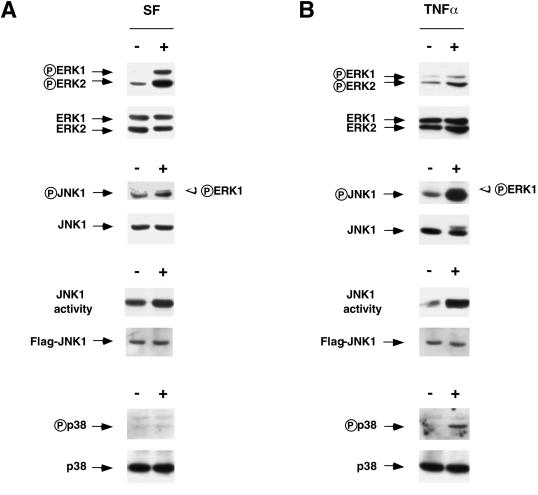
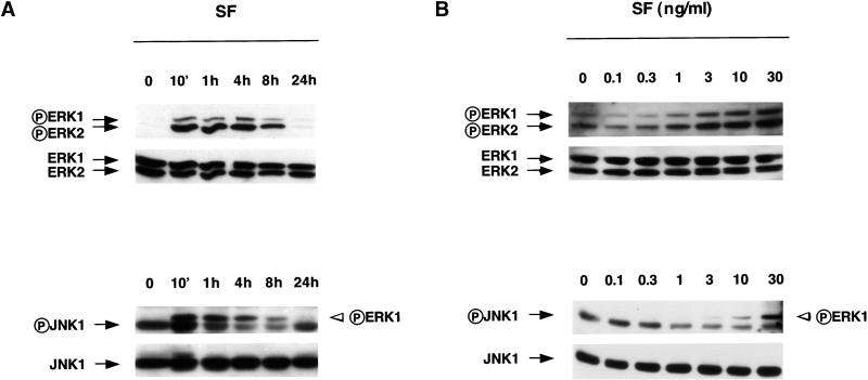
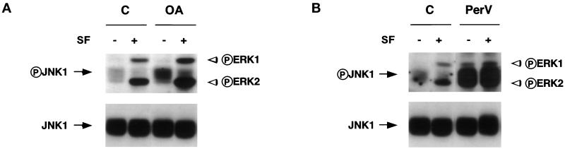
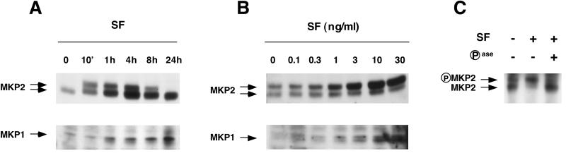
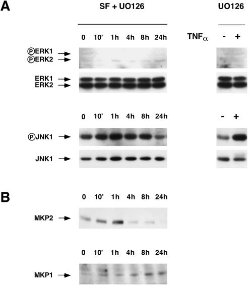
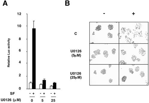
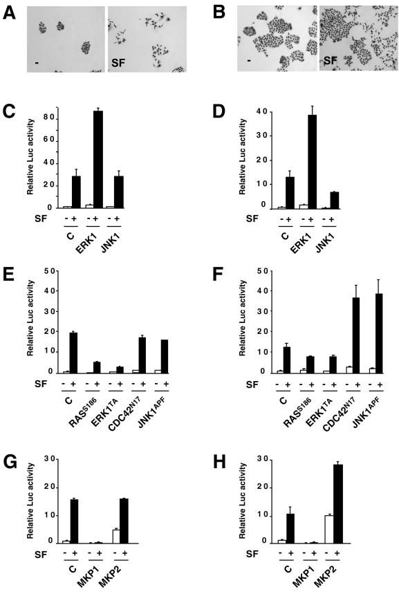
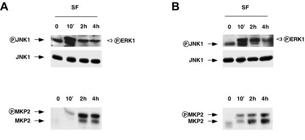
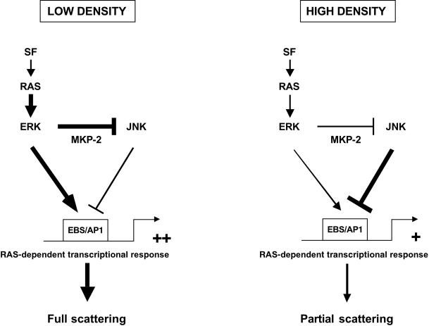
The scattering of Madin-Darby canine kidney (MDCK) epithelial cells by scatter factor/hepatocyte growth factor (SF/HGF) is associated with transcriptional induction of the urokinase gene, which occurs essentially through activation of an EBS/AP1 response element. We have investigated the signal transduction pathways leading to this transcriptional response. We found that SF/HGF induces rapid and sustained phosphorylation of the extracellular signal-regulated kinase (ERK) MAPK while stimulating weakly and then repressing phosphorylation of the JUN N-terminal kinase (JNK) MAPK for several hours. This delayed repression of JNK was preceded by phosphorylation of the MKP2 phosphatase, and both MKP2 induction and JNK dephosphorylation were under the control of MEK, the upstream kinase of ERK. ERK and MKP2 stimulate the EBS/AP1-dependent transcriptional response to SF/HGF, but not JNK, which inhibits this response. We further demonstrated that depending on cell density, the RAS-ERK-MKP2 pathway controls this transrepressing effect of JNK. Together, these data demonstrate that in a sequential manner SF/HGF activates ERK and MKP2, which in turn dephosphorylates JNK. This sequence of events provides a model for efficient cell scattering by SF/HGF at low cell density.


Figures

Figure 1
Figure 1
Short-term effects of SF/HGF and TNF-α on phosphorylation of ERK, JNK, and p38 MAPKs. (A) Cells were treated for 10 min with or without 30 ng/ml SF/HGF. Whole cell lysates were collected, and 20 μg of cell extracts was fractionated by 10% SDS-PAGE and examined by immunoblot analysis with the use of either anti-phospho-MAPK antibodies or their respective anti-MAPK antibodies. Positions of ERK1,2, JNK1, and p38 are shown by arrows, and their phosphorylated forms are indicated by a circled P. JNK1 activity was evaluated by a specific immunoprecipitation/kinase assay with the use of GST-JUN1–79 as a substrate; immunoblot of FLAG-tagged JNK1 with anti-FLAG antibody indicates comparable loading. (B) Cells were treated for 10 min with or without 30 ng/ml TNF-α. Cell extracts and immunoblot analysis were performed as described above. The filters used for examination of phospho-MAPKs were stripped and reprobed with their respective anti-MAPK antibodies. Symbols are as in A. It is worth noting that the anti-phospho-JNK kinase antibody recognized the phosphorylated forms of both JNK1 (left arrow) and ERK1 (right open arrowhead).
Figure 2
Figure 2
Long-term effects of SF/HGF on phosphorylation of ERK and JNK MAPKs. (A) Cells were treated for the indicated times with SF/HGF (30 ng/ml). Cell extracts (20 μg for ERK determination, 30 μg for JNK determination) were fractionated on 10% SDS-PAGE. Immunoblot analysis was performed with the use of the anti-phospho-ERK kinase antibody (top panel) or the anti-phospho-JNK kinase antibody (bottom panel). The filters used for examination of phospho-MAPKs were stripped and reprobed with their respective anti-MAPK antibodies. Positions of ERK1,2 and JNK1 are shown by arrows, and their phosphorylated forms are indicated by a circled P. Middle panel, the anti-phospho-JNK kinase antibody recognized the phosphorylated forms of both JNK1 (left arrow) and ERK1 (right open arrowhead). (B) Cells were treated for 4 h with increasing concentrations of SF/HGF (0.1–30 ng/ml). As described for A, cell extracts and immunoblot analysis were performed with the use of the anti-phospho-ERK kinase antibody (top panel) or the anti-phospho-JNK kinase antibody (bottom panel).
Figure 3
Figure 3
Effects of okadaic acid and pervanadate on SF/HGF-induced JNK dephosphorylation. Cells were pretreated for 30 min with 250 nM okadaic acid (OA; A) or 3 μM pervanadate (PerV; B) and were then treated for 2 h with or without SF/HGF (30 ng/ml). Cell extracts, immunoblot analysis of phosphorylated JNK1, and rehybridization with the anti-JNK1 antibody were performed as described in Figure 2. The band corresponding to JNK1 and its phosphorylated form is large and was also found with the use of a lower exposure of the gel because the 10% SDS-PAGE was particularly well separated.
Figure 4
Figure 4
Long-term effects of SF/HGF on expression of MKP1 and MKP2 phosphatases. (A) Cells were treated for the indicated times with SF/HGF (30 ng/ml). Cell extracts (30 μg) were fractionated by 10% SDS-PAGE, and immunoblot analysis was performed with the use of the anti-MKP2 or anti-MKP1 antibody. MKP1 and MKP2 migrated at their expected sizes (∼42 and 39 kDa, respectively). (B) Cells were treated for 4 h with increasing concentrations of SF/HGF (0.1–30 ng/ml). Cell extracts (30 μg) were fractionated by 10% SDS-PAGE, and immunoblot analysis was performed with the use of the anti-MKP2 or anti-MKP1 antibody. (C) Cells were treated for 10 min with (+) or without (−) SF/HGF (30 ng/ml). Immunoprecipitated MKP2 was incubated (+) or not (−) with alkaline phosphatase (Pase), and MKP2 detection was performed by immunoblot analysis with the anti-MKP2 antibody.
Figure 5
Figure 5
Effects of U0126 on MAPKs and MKP phosphatases induced by SF/HGF. (A) Cells were pretreated with U0126 (25 μM) for 30 min and then treated for the indicated times with 30 ng/ml SF/HGF (SF + U0126) or for 10 min with or without 30 ng/ml TNF-α (right panel). Cell extracts and immunoblot analysis were performed as described in Figure 2 with the use of the anti-phospho-ERK kinase antibody (top panel) or the anti-phospho-JNK kinase antibody (bottom panel). The filters used for examination of phospho-MAPKs were stripped and reprobed with their respective anti-MAPK antibodies. (B) Cell extracts obtained in A were analyzed by immunoblot with the use of the anti-MKP2 or anti-MKP1 antibody.
Figure 6
Figure 6
Effects of U0126 on EBS/AP1-dependent transactivation and cell scattering induced by SF/HGF. (A) Cells were seeded on 12-well plates (30,000) and were transfected with the EBS/AP1-Luc reporter vector. The next day, cells were pretreated with U0126 (5 or 25 μM) for 30 min and then left untreated (white bars) or treated with SF/HGF (10 ng/ml) (black bars), and luciferase activity was measured 24 h later. (B) Cells were seeded on 12-well plates (2500) until they formed colonies. Cells were then treated with U0126 (5 or 25 μM) for 30 min. After this time, SF/HGF (10 ng/ml) was added and the cells were further cultured for 24 h.
Figure 7
Figure 7
Effects of ERK1, JNK1, MKP2, and MKP1 in inducing an EBS/AP1-dependent transcriptional response at different cell densities. (A and B) Scattering. Cells were seeded on 12-well plates (1250 cells [A] and 5000 cells [B]) and cultured for 2 d. The medium was replaced by DMEM–0.5% FCS, and SF/HGF (10 ng/ml) was added (SF) or not (−) for 24 h. (C and D) Effects of ERK1 and JNK1 on SF/HGF-induced transactivation of the EBS/AP1-Luc reporter vector. Cells were seeded on 12-well plates (10,000 cells [C] and 30,000 cells [D]). The next day, they were cotransfected with the EBS/AP1-Luc reporter vector and with expression vectors, either empty (C) or encoding wild-type ERK1 (ERK1) or wild-type JNK1 (JNK1). The following day, cells were left untreated (−) or treated with 10 ng/ml SF/HGF (+), and luciferase activity was measured 24 h later. The usual condition of transfection is obtained by seeding the cells at 30,000 cells per well, which gives a confluence of 50–60% at the end of the experiment. It is worth noting that at the end of the assay, the sizes of the cell islets are comparable between the cell-scattering (A and B) and transactivation (C and D) assays because of cell mortality during the transfection procedure. (E and F) Effects of dominant negative mutants of RAS, ERK1, CDC42, and JNK1 on SF/HGF-induced transactivation of the EBS/AP1-Luc reporter vector. Cells were seeded on 12-well plates (10,000 cells [E] and 30,000 cells [F]). The next day, they were cotransfected with the EBS/AP1-Luc reporter vector and with expression vectors, either empty (C) or encoding dominant negative forms of RAS (RASS186), ERK (ERK1TA), CDC42 (CDC42N17), or JNK (JNK1APF). The following day, cells were treated with SF/HGF (10 ng/ml) (black bars) or not (white bars), and luciferase activity was measured 24 h later. (G and H) Effects of MKP1 and MKP2 on SF/HGF-induced transactivation of the EBS/AP1-Luc reporter vector. Cells were seeded on 12-well plates (10,000 cells [G] and 30,000 cells [H]). The next day, they were cotransfected with the EBS/AP1-Luc reporter vector and with expression vectors, either empty (C) or encoding wild-type MKP1 (MKP1) or MKP2 (MKP2). The following day, cells were treated with SF/HGF (10 ng/ml) (black bars) or not (white bars), and luciferase activity was measured 24 h later.
Figure 8
Figure 8
Effects of SF/HGF on phosphorylation of ERK and JNK and on expression of MKP2 at different cell densities. Cells were seeded on 100-mm plates (100,000 cells [A] and 400,000 cells [B]). The next day, the cells were incubated in DMEM–0.5% FCS. The following day, cells were treated for the indicated times with SF/HGF (30 ng/ml). Cell extracts and immunoblot analysis of phosphorylated JNK1 or MKP2 expression were performed as described in Figure 5. The filters were stripped and reprobed with the anti-JNK antibody.
Figure 9
Figure 9
Model of SF/HGF signaling implicating ERK, MKP2, and JNK in MDCK cells at low and high cell density. See text for details.

References

    1. Auer KL, et al. The Ras/Rac1/Cdc42/SEK/JNK/c-Jun cascade is a key pathway by which agonists stimulate DNA synthesis in primary cultures of rat hepatocytes. Mol Biol Cell. 1998;9:561–573. - PMC - PubMed
    1. Bagrodia S, Dérijard B, Davis RJ, Cerione RA. Cdc42 and PAK-mediated signaling leads to jun kinase and p38 mitogen-activated protein kinase activation. J Biol Chem. 1995;270:27995–27998. - PubMed
    1. Beauchemin N, Kunath T, Robitaille J, Chow B, Turbide C, Daniels E, Veillette A. Association of biliary glycoprotein with protein tyrosine phosphatase SHP-1 in malignant colon epithelial cells. Oncogene. 1997;14:783–790. - PubMed
    1. Brondello JM, Brunet A, Pouyssegur J, McKenzie FR. The dual specificity mitogen-activated protein kinase phosphatase-1 and -2 are induced by the p42/p44MAPK cascade. J Biol Chem. 1997;272:1368–1376. - PubMed
    1. Brondello JM, Pouyssegur J, McKenzie FR. Reduced MAP kinase phosphatase-1 degradation after p42/p44MAPK-dependent phosphorylation. Science. 1999;286:2514–2517. - PubMed

Publication types

MeSH terms

Substances