

Epidermal growth factor receptor distribution during chemotactic responses
- PMID: 11071913
- PMCID: PMC15043
- DOI: 10.1091/mbc.11.11.3873
Epidermal growth factor receptor distribution during chemotactic responses
Abstract
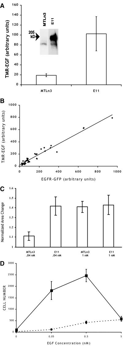
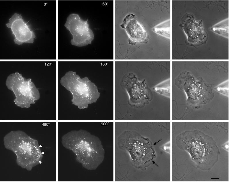
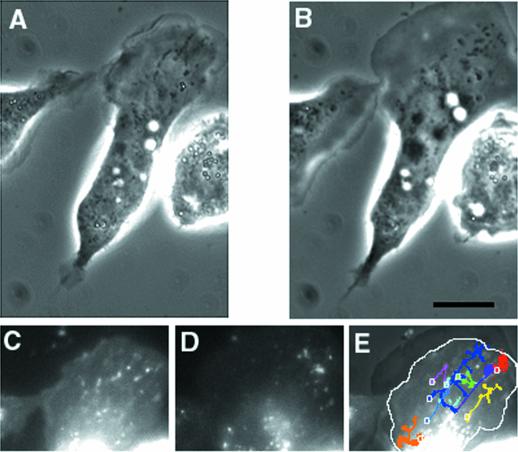
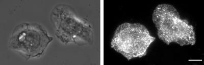
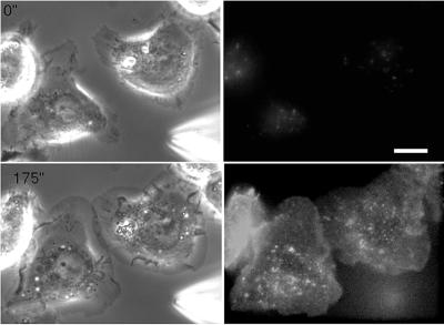
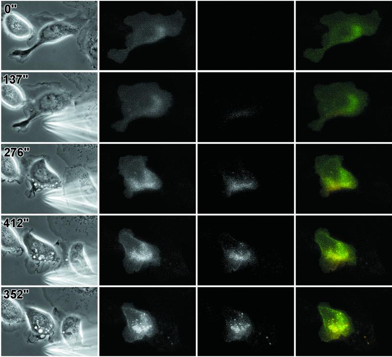
To determine the distribution of the epidermal growth factor (EGF) receptor (EGFR) on the surface of cells responding to EGF as a chemoattractant, an EGFR-green fluorescent protein chimera was expressed in the MTLn3 mammary carcinoma cell line. The chimera was functional and easily visualized on the cell surface. In contrast to other studies indicating that the EGFR might be localized to certain regions of the plasma membrane, we found that the chimera is homogeneously distributed on the plasma membrane and becomes most concentrated in vesicles after endocytosis. In spatial gradients of EGF, endocytosed receptor accumulates on the upgradient side of the cell. Visualization of the binding of fluorescent EGF to cells reveals that the affinity properties of the receptor, together with its expression level on cells, can provide an initial amplification step in spatial gradient sensing.
Figures
References
-
- Bailly M, Condeelis JS, Segall JE. Chemoattractant-induced lamellipod extension. Microsc Res Tech. 1998a;43:433–443. - PubMed
-
- Bailly M, Yan L, Whitesides GM, Condeelis JS, Segall JE. Regulation of protrusion shape and adhesion to the substratum during chemotactic responses of mammalian carcinoma cells. Exp Cell Res. 1998b;241:285–299. - PubMed
-
- Bretscher MS, Aguado-Velasco C. EGF induces recycling membrane to form ruffles. Curr Biol. 1998a;8:721–724. - PubMed
-
- Bretscher MS, Aguado-Velasco C. Membrane traffic during cell locomotion. Curr Opin Cell Biol. 1998b;10:537–541. - PubMed
Publication types
MeSH terms
Substances
Grants and funding
LinkOut - more resources
Full Text Sources
Molecular Biology Databases
Research Materials
Miscellaneous
