. 2000 Nov;11(11):3963-76.
doi: 10.1091/mbc.11.11.3963.

Saccharomyces cerevisiae PTS1 receptor Pex5p interacts with the SH3 domain of the peroxisomal membrane protein Pex13p in an unconventional, non-PXXP-related manner

Affiliations
Free PMC article

Saccharomyces cerevisiae PTS1 receptor Pex5p interacts with the SH3 domain of the peroxisomal membrane protein Pex13p in an unconventional, non-PXXP-related manner

G Bottger et al. Mol Biol Cell. 2000 Nov.
Free PMC article

Abstract

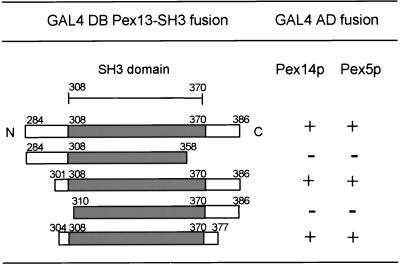
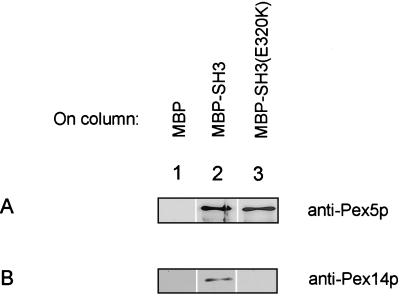
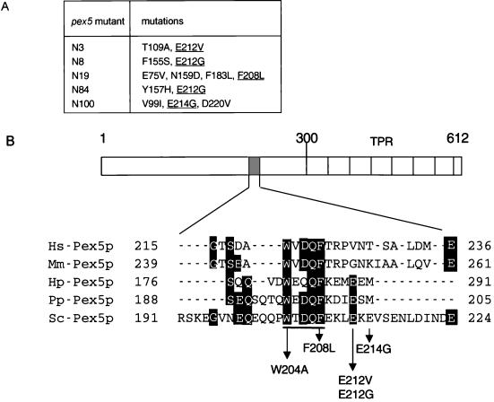
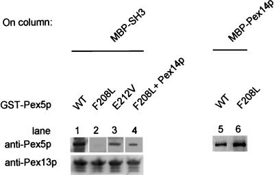
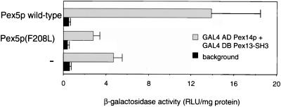
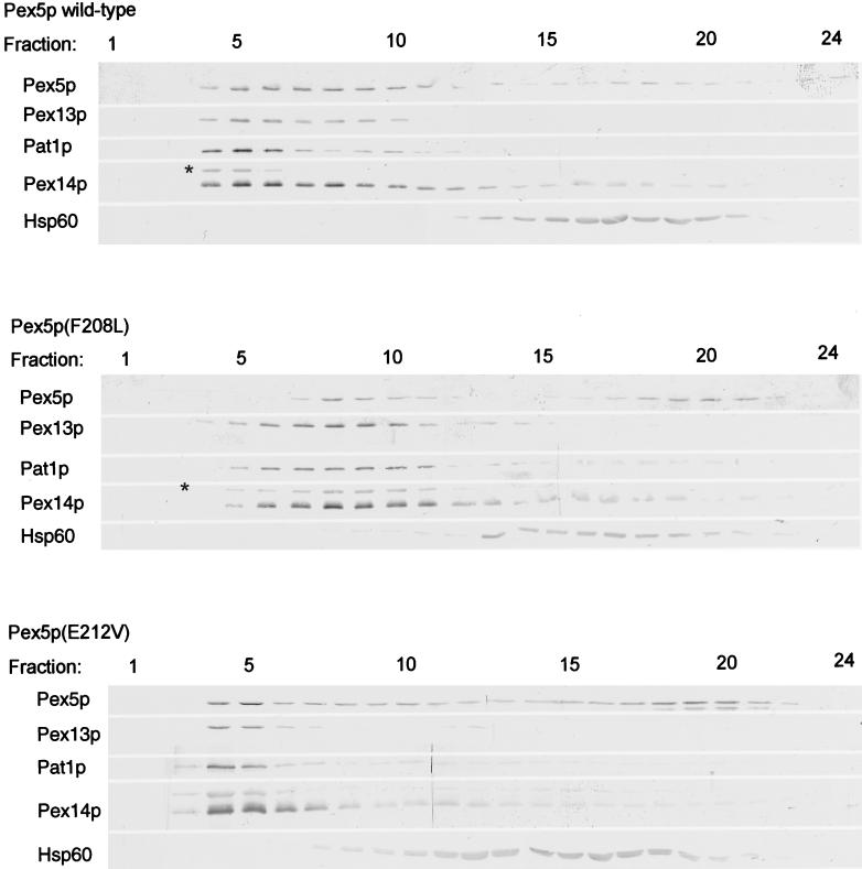
A number of peroxisome-associated proteins have been described that are involved in the import of proteins into peroxisomes, among which is the receptor for peroxisomal targeting signal 1 (PTS1) proteins Pex5p, the integral membrane protein Pex13p, which contains an Src homology 3 (SH3) domain, and the peripheral membrane protein Pex14p. In the yeast Saccharomyces cerevisiae, both Pex5p and Pex14p are able to bind Pex13p via its SH3 domain. Pex14p contains the classical SH3 binding motif PXXP, whereas this sequence is absent in Pex5p. Mutation of the conserved tryptophan in the PXXP binding pocket of Pex13-SH3 abolished interaction with Pex14p, but did not affect interaction with Pex5p, suggesting that Pex14p is the classical SH3 domain ligand and that Pex5p binds the SH3 domain in an alternative way. To identify the SH3 binding site in Pex5p, we screened a randomly mutagenized PEX5 library for loss of interaction with Pex13-SH3. Such mutations were all located in a small region in the N-terminal half of Pex5p. One of the altered residues (F208) was part of the sequence W(204)XXQF(208), that is conserved between Pex5 proteins of different species. Site-directed mutagenesis of Trp204 confirmed the essential role of this motif in recognition of the SH3 domain. The Pex5p mutants could only partially restore PTS1-protein import in pex5Delta cells in vivo. In vitro binding studies showed that these Pex5p mutants failed to interact with Pex13-SH3 in the absence of Pex14p, but regained their ability to bind in the presence of Pex14p, suggesting the formation of a heterotrimeric complex consisting of Pex5p, Pex14p, and Pex13-SH3. In vivo, these Pex5p mutants, like wild-type Pex5p, were still found to be associated with peroxisomes. Taken together, this indicates that in the absence of Pex13-SH3 interaction, other protein(s) is able to bind Pex5p at the peroxisome; Pex14p is a likely candidate for this function.


Figures

Figure 1
Figure 1
Pex13-SH3 domain binds Pex5p and Pex14p in the two-hybrid assay. Truncated versions of Pex13-SH3 fused to GAL4 DB were cotransformed with GAL4 AD fusions of Pex5p or Pex14p into the yeast two-hybrid reporter strain PCY2. Interaction was monitored by determining β-galactosidase activity with a filter lift assay. “+” indicates blue staining of colonies within 1 h, “−” indicates that colonies remained white after incubation overnight.
Figure 2
Figure 2
Pex13-SH3 domain interacts with Pex5p and with Pex14p in vitro. MBP, MBP-SH3(wild-type) and MBP-SH3(E320K) were expressed in E. coli and immobilized on amylose columns. E. coli lysates containing either GST-Pex5p (A) or 6xHisPex14p (B) were then passed over the columns. The columns were washed with five column volumes and bound proteins were eluted with maltose. Eluates were analyzed by SDS-PAGE and Western blotting by using antibodies specific for Pex14p and Pex5p.
Figure 3
Figure 3
Amino acid substitutions in pex5 mutants selected for loss of two-hybrid interaction with Pex13-SH3 are clustered in the region between amino acids 208 and 214. (A) Amino acid substitutions in pex5 mutants selected for loss of interaction with Pex13-SH3. Mutations in or near the conserved W204XDQF208 motif are underlined. (B) Multiple sequence alignment (GeneInspector) of the region in Pex5p important for Pex13-SH3 interaction. Amino acid substitutions in pex5 mutants are indicated. The loss of interaction mutant W204A created by site-directed mutagenesis is also shown. The conserved WXDQF motif is underlined. Hs, Homo sapiens; Mm, Mus musculus; Hp, H. polymorpha; Pp, P. pastoris; Sc, S. cerevisiae.
Figure 4
Figure 4
Pex5p(F208L) and Pex5p(E212V) are disturbed in Pex13-SH3 binding, but maintain interaction with Pex14p in vitro. GST-Pex5p(wild-type) (WT) or mutant GST-Pex5p(F208L or E212V) were expressed in E. coli and passed over a column with immobilized MBP-SH3 (lanes 1–3) or MBP-Pex14p (lanes 5 and 6). Columns were washed, eluted, and analyzed as described below. Pex13-SH3, Pex14p, and Pex5p(F208L) can form a trimeric complex in vitro (lane 4): an E. coli lysate containing 6xHisPex14p was passed over an amylose column with immobilized MBP-SH3. After washing, an E. coli lysate containing GST-Pex5p(F208L) was passed over the column. Proteins were eluted with maltose and eluates were analyzed by SDS-PAGE and Western blotting by using antibodies specific for Pex13-SH3 and Pex5p.
Figure 5
Figure 5
Two-hybrid interaction of Pex13-SH3 and Pex14p is reduced in the absence of wild-type Pex5p. A PCY2 strain harboring a deletion of the PEX5 gene (PCY2 pex5Δ) was transformed with expression constructs encoding either wild-type Pex5p or mutant Pex5p, or with an empty plasmid (−). The two-hybrid interaction of Pex13-SH3 and Pex14p was determined in these different strains by measuring the β-galactosidase activity. Indicated is the mean of two measurements in triplicate cultures ± SD. Background (▪) represents β-galactosidase activity in strains transformed with either GAL4DB-Pex13-SH3 or GAL4AD-Pex14p alone.
Figure 6
Figure 6
Growth characteristics on oleate of wild-type cells overexpressing Pex13p, Pex14p, and Pex5p under the control of the CTA1 promoter. Peroxins were expressed in wild-type cells either separately (A) or in combination (B) as indicated. Transformants are indicated with “+ Pex.” Positive and negative controls for growth are untransformed wild-type cells and the pex13Δ strain, respectively.
Figure 7
Figure 7
Growth on liquid oleate medium of pex5Δ cells (○) expressing wild-type Pex5p (▪), Pex5p(F208L) (▵), Pex5p(E212V) (▴), Pex5p(F208L; E212V; E214G)(●; see inset). Cells were grown to mid-log phase in 0.3% glucose medium and inoculated at OD600 of 0.001 in liquid oleate medium. Growth was followed with time by measuring the optical density at 600 nm (OD600).
Figure 8
Figure 8
Localization of peroxisomal matrix proteins in pex5Δ cells expressing wild-type Pex5p, Pex5p(F208L), or Pex5p(E212V). Subcellular distribution of GFP-SKL visualized by fluorescence microscopy (A). Bar, 10 μm. Subcellular distribution of CTA1 and 3HAD (B) and 3-ketoacyl-CoA thiolase and Mdh3p (C). After subcellular fractionation equivalent volumes of the 600 × g postnuclear supernatant (homogenate [H]), 20,000 × g pellet (P) and 20,000 × g supernatant (S) were analyzed by measuring enzyme activities (B) or by Western blotting (C). Antibodies were directed against the proteins as indicated. Recoveries varied between 90 and 110%.
Figure 9
Figure 9
Pex5p mutants are associated with peroxisomes. 20,000 × g pellet fractions of pex5Δ cells expressing wild-type Pex5p, Pex5p(F208L), or Pex5p(E212V) were loaded on top of a continuous Nycodenz gradient and centrifuged at 29,000 × g in a vertical rotor for 2.5 h. Fractions of 0.5 ml were collected and analyzed by SDS-PAGE and Western blotting with antibodies specific for Pex5p; the peroxisomal membrane markers Pex13p, Pex14p, and Pat1p; and the mitochondrial marker Hsp60. Fraction 1 is the bottom of the gradient. The asterisk indicates a cross-reacting band.

References

    1. Albertini M, Rehling P, Erdmann R, Girzalsky W, Kiel JAKW, Veenhuis M, Kunau W-H. Pex14p, a peroxisomal membrane protein binding both receptors of the two PTS-dependent import pathways. Cell. 1997;89:83–92. - PubMed
    1. Arold S, O'Brien R, Franken P, Strub M-P, Hoh F, Dumas C, Ladbury JE. RT loop flexibility enhances the specificity of Src family SH3 domains for HIV-1 Nef. Biochemistry. 1998;37:14683–14691. - PubMed
    1. Bradford MM. A rapid and sensitive method for quantification of microgram quantities of protein utilizing the principle of protein-dye binding. Anal Biochem. 1976;72:248–254. - PubMed
    1. Brocard C, Kragler F, Simon MM, Schuster T, Hartig A. The tetratricopeptide repeat-domain of the PAS10 protein of Saccharomyces cerevisiae is essential for binding the peroxisomal targeting signal-SKL. Biochem Biophys Res Commun. 1994;204:1016–1022. - PubMed
    1. Brocard C, Lametschwandtner G, Koudelka R, Hartig A. Pex14p is a member of the protein linkage map of Pex5p. EMBO J. 1997;16:5491–5500. - PMC - PubMed

Publication types

MeSH terms

LinkOut - more resources