Review
. 2000 Nov 15;44(2-3):81-95.
doi: 10.1016/s0169-409x(00)00087-9.

Peptide nucleic acids: versatile tools for gene therapy strategies

Affiliations
Review

Peptide nucleic acids: versatile tools for gene therapy strategies

D A Dean. Adv Drug Deliv Rev. .

Abstract

Peptide nucleic acids, or PNAs, are oligonucleotide analogs in which the phosphodiester backbone is replaced with a polyamide structure. First synthesized less than 10 years ago, they have received great attention due to their several favorable properties, including resistance to nuclease and protease digestion, stability in serum and cell extracts, and their high affinity for RNA and single and double-stranded DNA targets. Although initially designed and demonstrated to function as antisense and antigene reagents that inhibit both transcription and translation by steric hindrance, more recent applications have included gene activation by synthetic promoter formation and mutagenesis of chromosomal targets. Most notably for gene delivery, they have been used to specifically label plasmids and act as adapters to link synthetic peptides or ligands to the DNA. Thus, their great potential lies in the ability to attach specific targeting peptides to plasmids to circumvent such barriers to gene transfer as cell-targeting or nuclear localization, thereby increasing the efficacy of gene therapy.


Figures
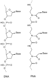
Fig. 1
Fig. 1
Structures of single-stranded DNA and PNA.
Fig. 2
Fig. 2
Structures of duplex and triplex PNA complexes. (A) Duplex PNA–DNA or PNA–RNA complex. The target sequence present in either DNA or RNA is shown as the top strand, while the PNA (bold, italics) is shown as the bottom strand, in the antiparallel orientation. Base-pairing interactions are denoted by dots between the dases. (B) Triplex PNA2–DNA complex. In this case, the DNA target sequence is depicted as the upper DNA strand and the displaced ‘D-loop’ is shown below the triplex structure, bulging away from the complementary DNA strand. The amino-terminal half of the PNA hybridizes to the target sequence using standard Watson–Crick base pairs (WC) and the carboxy-terminal half of the PNA uses Hoogstein base pairs to form the third strand. 8-amino-3,6-dioxaoctanoic acid linkers used to connect the two halves of the PNA are abbreviated as ‘O’.
Fig. 3
Fig. 3
Stability of PNA2–DNA triplex. A fluorescein-labeled 20-mer oligonucleotide (Fl-ODN) was reacted with a triplex-forming rhodamine-labeled PNA (8 base target site; Rh-PNA) for 2 h at 37°C to form a stable ‘PNA-clamp’, and then incubated for at least 3 h with 100–1000-fold molar excess of competitors. Fluorescence resonance energy transfer (FRET) was used to measure the degree of dissociation of PNA from the fluorescein-labeled oligonucleotide. Peak fluorescence intensity of the free Fl-ODN at 520 nm is shown. Thus, 100% of the Fl-ODN is free when unincubated with PNA, whereas 25% of the Fl-ODN is free after the 2 h reaction with the PNA. Values above 25% therefore represent dissociation of the PNA from the Fl-ODN. Competitors included 1000-fold molar excess of salmon sperm DNA or unlabeled target site-containing oligonucleotide (ODN), 350-fold molar excess of histones, or 100-fold molar excess of the DMRIE:DOPE liposomes. The triplex complex was also incubated for 1 h at 58°C with 50 mM Tris–HCl (pH 8.9), 0.5 M NaCl, 10 mM EDTA, and 1% SDS. This Fig. was adapted from data in [11].
Fig. 4
Fig. 4
Use of peptide–PNA conjugates as targeting reagents for plasmids. A generic transgene-expressing plasmid is shown with a triplex-forming PNA–synthetic peptide conjugate bound to its target site at a unique location. Several potential targeting peptides or ligands are listed along with their activities or sites of action. Such PNAs can be used alone or in any combination on the plasmid by varying the number of PNA-binding sites on the plasmid, thus creating a wide variety of non-viral vectors optimized for different aspects of delivery and function.

References

    1. Nielsen PE, Eghoim M, Berg RH, Buchardt O. Sequence-selective recognition of DNA by strand displacement with a thymine-substituted polyamide. Science. 1991;254:1497–1500. - PubMed
    1. Brown SC, Thomson SA, Veal JM, Davis DG. NMIR solution structure of a peptide nucleic acid complexed with RNA. Science. 1994;265:777–780. - PubMed
    1. Wittung P, Nielsen PE, Buchardt O, Eghoim M, Norden B. DNA-like double helix formed by peptide nucleic acid. Nature. 1994;368:561–563. - PubMed
    1. Egholm M, Buchardt O, Christensen L, Bebrens C, Freier SM, Driver DA, Berg RH, Kim SK, Norden B, Nielsen PE. PNA hybridizes to complementary oligonucleotides obeying the Watson–Crick hydrogen-bonding rules. Nature. 1993;365:566–568. - PubMed
    1. Nielsen PE, Egholm M, Buchardt O. Evidence for (PNA)2IDNA triplex structure upon binding of PNA to dsDNA by strand displacement. J Mol Recognit. 1994;7:165–170. - PubMed

Publication types

MeSH terms