. 2000 Nov;157(5):1415-30.
doi: 10.1016/S0002-9440(10)64779-7.

Mechanisms of apoptosis

Affiliations

Mechanisms of apoptosis

J C Reed. Am J Pathol. 2000 Nov.

Abstract

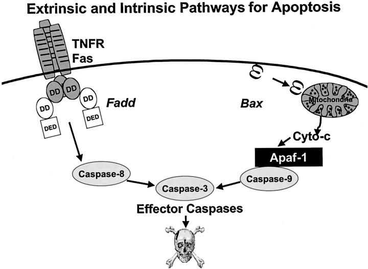
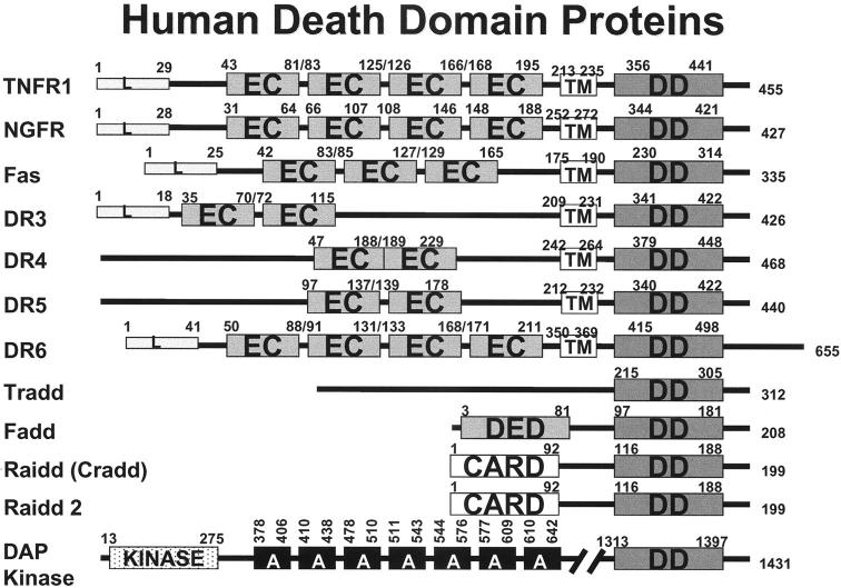
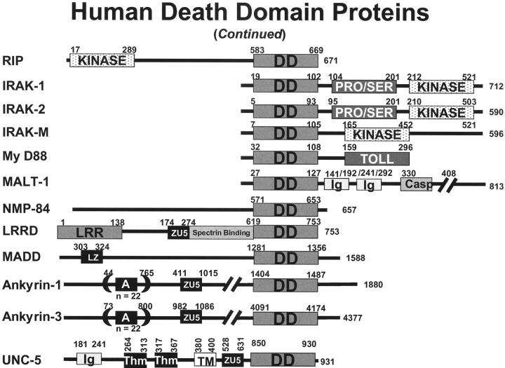
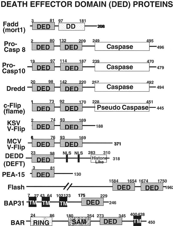
Programmed cell death plays critical roles in a wide variety of physiological processes during fetal development and in adult tissues. In most cases, physiological cell death occurs by apoptosis as opposed to necrosis. Defects in apoptotic cell death regulation contribute to many diseases, including disorders where cell accumulation occurs (cancer, restenosis) or where cell loss ensues (stroke, heart failure, neurodegeneration, AIDS). In recent years, the molecular machinery responsible for apoptosis has been elucidated, revealing a family of intracellular proteases, the caspases, which are responsible directly or indirectly for the morphological and biochemical changes that characterize the phenomenon of apoptosis. Diverse regulators of the caspases have also been discovered, including activators and inhibitors of these cell death proteases. Inputs from signal transduction pathways into the core of the cell death machinery have also been identified, demonstrating ways of linking environmental stimuli to cell death responses or cell survival maintenance. Knowledge of the molecular mechanisms of apoptosis is providing insights into the causes of multiple pathologies where aberrant cell death regulation occurs and is beginning to provide new approaches to the treatment of human diseases.


Figures

Figure 1.
Figure 1.
Pathways for caspase activation. Two of the major pathways for caspase activation in mammalian cells are presented, the extrinsic (left) and intrinsic (right). The extrinsic pathway can be induced by members of the TNF family of cytokine receptors, such as TNFR1 and Fas. These proteins recruit adapter proteins to their cytosolic DDs, including Fadd, which then binds DED-containing pro-caspases, particularly pro-caspase-8. The intrinsic pathway can be induced by release of cytochrome c from mitochondria, induced by various stimuli, including elevations in the levels of pore-forming pro-apoptotic Bcl-2 family proteins such as Bax. In the cytosol, cytochrome c binds and activates Apaf-1, allowing it to bind and activate pro-caspase-9. Active caspase-9 (intrinsic) and caspase-8 (extrinsic) have been shown to directly cleave and activate the effector protease, caspase-3. Other caspases can also become involved in these pathways (not shown); thus, the schematic represents an oversimplification of the events that occur in vivo.
Figure 2.
Figure 2.
Human death domain (DD) family proteins. The domain arrangements of the known members of the human DD family are presented. Numbers represent amino acid residue positions.
Figure 2.
Figure 2.
Human death domain (DD) family proteins. The domain arrangements of the known members of the human DD family are presented. Numbers represent amino acid residue positions.
Figure 3.
Figure 3.
Death effector domain (DED) family proteins. The domain arrangements of some of the known members of the DED family are presented. Numbers represent amino acid residue positions. Included in the diagram are the Drosophila caspase, Dronc, and the viral DED proteins from Kaposi’s sarcoma virus and MCV.
Figure 4.
Figure 4.
Caspase-associated recruitment domain (CARD) family proteins. Non-caspase CARD family proteins are shown. The domain arrangements of the known members of the CARD family are presented. Numbers represent amino acid residue positions. In addition to human proteins, the diagram includes the murine caspase-11 and caspase-12 proteins, C. elegans CED-3 and CED-4 proteins, and the Drosophila Dronc protein. Additional family members in lower organisms are not presented.
Figure 5.
Figure 5.
Human IAP family proteins. The domain arrangements of the known members of the human IAP family are presented. Numbers represent amino acid residue positions. Shown are BIR, nucleotide-binding (NB), ubiquitin-conjugating (Ubc), and RING zinc-finger domains.
Figure 6.
Figure 6.
Bcl-2 family proteins. The domain arrangements of the known members of the Bcl-2 family are presented. The BH1, BH2, BH3, BH4, and transmembrane domains are indicated. In addition to human or murine proteins, included in the diagram are the chicken Nr13 protein, C. elegans proteins CED-9, EGL1, and CeBNIP, and the Drosophila proteins DBok (Drob1, dBorg 1, Debcl). Homologues from viruses, xenopas, and marine sponges are not depicted. Human noxa (APR) has only one BH3 domain.

References

    1. Kerr JF, Wyllie AH, Currie AR: Apoptosis: a basic biological phenomenon with wide-ranging implications in tissue kinetics. Br J Cancer 1972, 26:239-257 - PMC - PubMed
    1. Alnemri ES, Livingston DJ, Nicholson DW, Salvesen G, Thornberry NA, Wong WW, Yuan J: Human ICE/CED-3 protease nomenclature. Cell 1996, 87:171. - PubMed
    1. Thornberry N, Lazebnik Y: Caspases: enemies within. Science 1998, 281:1312-1316 - PubMed
    1. Cryns V, Yuan Y: Proteases to die for. Genes Dev 1999, 13:371 - PubMed
    1. Nicholson DW: ICE/CED3-like proteases as therapeutic targets for the control of inappropriate apoptosis. Nat Biotechnol 1996, 14:297-301 - PubMed

LinkOut - more resources