. 2000 Nov;157(5):1473-83.
doi: 10.1016/S0002-9440(10)64786-4.

Expression of angiopoietin-1, angiopoietin-2, and tie receptors after middle cerebral artery occlusion in the rat

Affiliations

Expression of angiopoietin-1, angiopoietin-2, and tie receptors after middle cerebral artery occlusion in the rat

H Beck et al. Am J Pathol. 2000 Nov.

Abstract

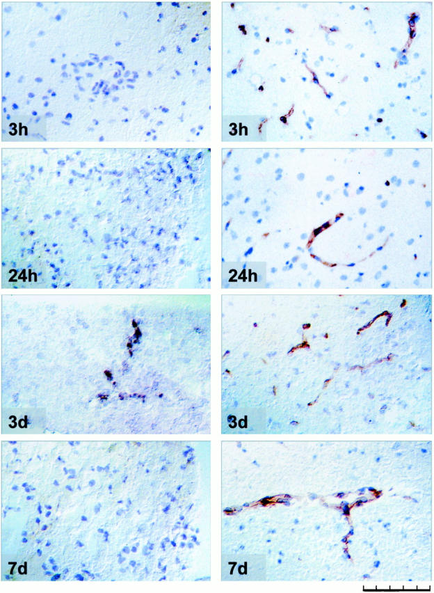
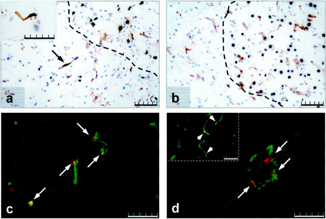
Vascular endothelial growth factor (VEGF), a key regulator of vasculogenesis and embryonic angiogenesis, was recently found to be up-regulated in an animal model of stroke. Unlike VEGF, angiopoietin (Ang)-1 and -2, their receptor tie-2, and the associated receptor tie-1 exert their functions at later stages of vascular development, i.e., during vascular remodeling and maturation. To assess the role of the angiopoietin/tie family in ischemia-triggered angiogenesis we analyzed their temporal and spatial expression pattern after middle cerebral artery occlusion (MCAO) using in situ hybridization and immunohistochemistry. Ang-1 mRNA was constitutively expressed in a subset of glial and neuronal cells with no apparent change in expression after MCAO. Ang-2 mRNA was up-regulated 6 hours after MCAO and was mainly observed in endothelial cell (EC) cord tips in the peri-infarct and infarct area. Up-regulation of both Ang-2 and VEGF coincided with EC proliferation. Interestingly, EC proliferation was preceded by a transient period of EC apoptosis, correlating with a change in VEGF/Ang-2 balance. Our observation of specific stages of vascular regression and growth after MCAO are in agreement with recent findings suggesting a dual role of Ang-2 in blood vessel formation, depending on the availability of VEGF.


Figures

Figure 1.
Figure 1.
H&E-stained rat brain 24 hours after MCAO. I, infarct area; P, peri-infarct area. For analysis, corresponding areas were taken from the ipsilateral and contralateral hemispheres relative to the side of MCAO. Scale bar, 2 mm.
Figure 2.
Figure 2.
In situ hybridization for Ang-2 mRNA in rat brain subjected to MCAO. Figure ▶ shows ipsilateral (left) peri-infarct cortex and corresponding area in the contralateral (right) hemisphere relative to the side of MCAO, except for 24-hour contralateral, where the hippocampus is shown. Time indicates hours after permanent MCAO. Note up-regulation of Ang-2 mRNA in the contralateral hemisphere 12 and 24 hours after MCAO in single cells (arrows), low-expressing large vessel (arrowhead) at 12 hours, and marked Ang-2 induction at 3 days in a large vessel (arrowhead) as well as smaller vessels (arrows). Scale bar, 100 μm.
Figure 3.
Figure 3.
Characterization of Ang-2 mRNA-expressing cell types. Double labeling for Ang-2 mRNA (shown in black) and vWF (a) or GFAP immunostaining (b) in the peri-infarct area 24 hours after MCAO. Immunoreactivity for the proteins is indicated by brown color. a: Ang-2 mRNA-expressing blood vessel. b: Ang-2 mRNA-expressing astrocyte. Scale bar, 20 μm.
Figure 4.
Figure 4.
Tie-1 immunostaining at 3 days MCAO of the ipsilateral peri-infarct (left) area and the corresponding area in the contralateral cortex (right); note the up-regulation of tie-1 protein in peri-infarct vessels. Scale bar, 100 μm.
Figure 5.
Figure 5.
Tie-2 in situ hybridization and immunolocalization following different times after MCAO. Left: Tie-2 mRNA expression. Right: Immunolocalization of tie-2 protein in vessels in the peri-infarct cortical area. Scale bar, 100 μm.
Figure 6.
Figure 6.
Apoptosis versus proliferation in ECs in the peri-infarct area. TUNEL staining combined with lectin staining 12 hours (a) and 24 hours (b) after MCAO. Dotted line in a and b demarcates the infarct area. Arrow in a indicates an apoptotic vessel in the peri-infarct area, insert in a shows a high magnification view of an apoptotic vessel in the contralateral hemisphere at 12 hours. Double immunofluorescence for CD31 (green color) and Ki67 (red color) at 24 hours (c) and 3 days MCAO (d). Arrows in c and d indicate proliferating vessels. Note large proliferating blood vessel in d, insert in d depicts small proliferating vessels. Scale bar, 100 μm (a and b), 50 μm (c and d, insert in a and d).

References

    1. Krupinski J, Kaluza J, Kumar P, Kumar S, Wang JM: Role of angiogenesis in patients with cerebral ischemic stroke. Stroke 1994, 25:1794-1798 - PubMed
    1. Marchal G, Serrati C, Rioux P, Petit-Taboue MC, Viader F, de IS V, Le Doze F, Lochon P, Derlon JM, Orgogozo JM: PET imaging of cerebral perfusion and oxygen consumption in acute ischaemic stroke: relation to outcome. Lancet 1993, 341:925-927 - PubMed
    1. Plate KH, Beck H, Danner S, Allegrini PR, Wiessner C: Cell type specific upregulation of vascular endothelial growth factor in an MCA-occlusion model of cerebral infarct. J Neuropathol Exp Neurol 1999, 58:654-666 - PubMed
    1. Marti HJ, Bernaudin M, Bellail A, Schoch H, Euler M, Petit E, Risau W: Hypoxia-induced vascular endothelial growth factor expression precedes neovascularization after cerebral ischemia. Am J Pathol 2000, 156:965-976 - PMC - PubMed
    1. Risau W: Mechanisms of angiogenesis. Nature 1997, 386:671-674 - PubMed

Publication types

MeSH terms