. 2000 Nov;157(5):1495-510.
doi: 10.1016/S0002-9440(10)64788-8.

Prominent axonopathy and disruption of axonal transport in transgenic mice expressing human apolipoprotein E4 in neurons of brain and spinal cord

Affiliations

Prominent axonopathy and disruption of axonal transport in transgenic mice expressing human apolipoprotein E4 in neurons of brain and spinal cord

I Tesseur et al. Am J Pathol. 2000 Nov.

Abstract

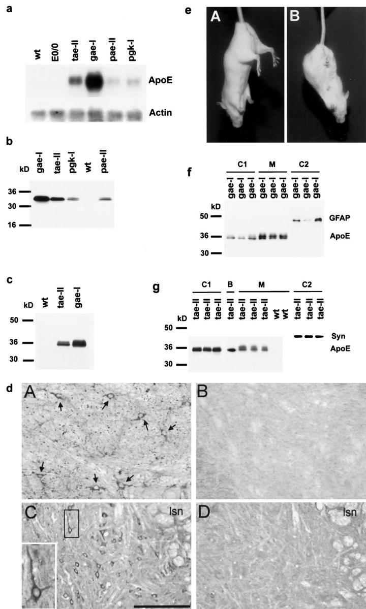
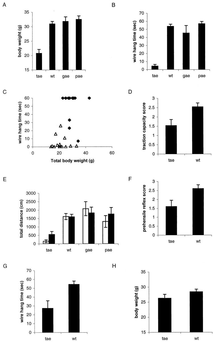
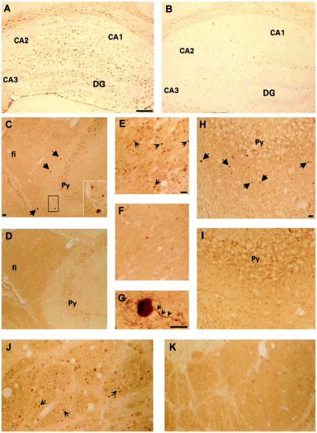
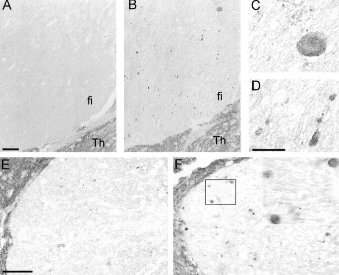
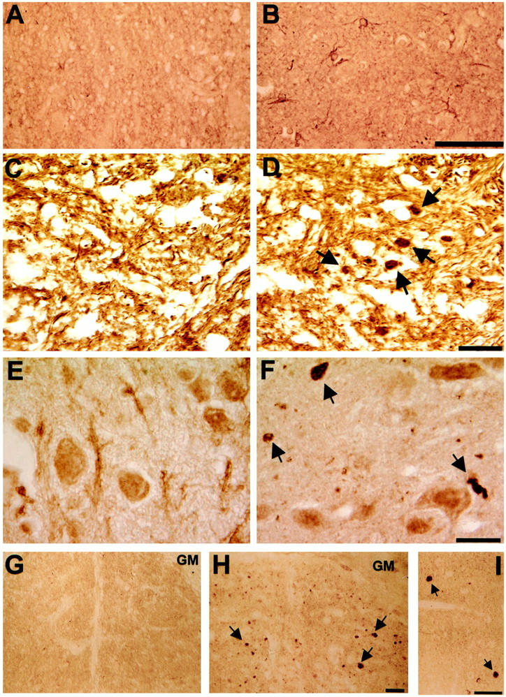
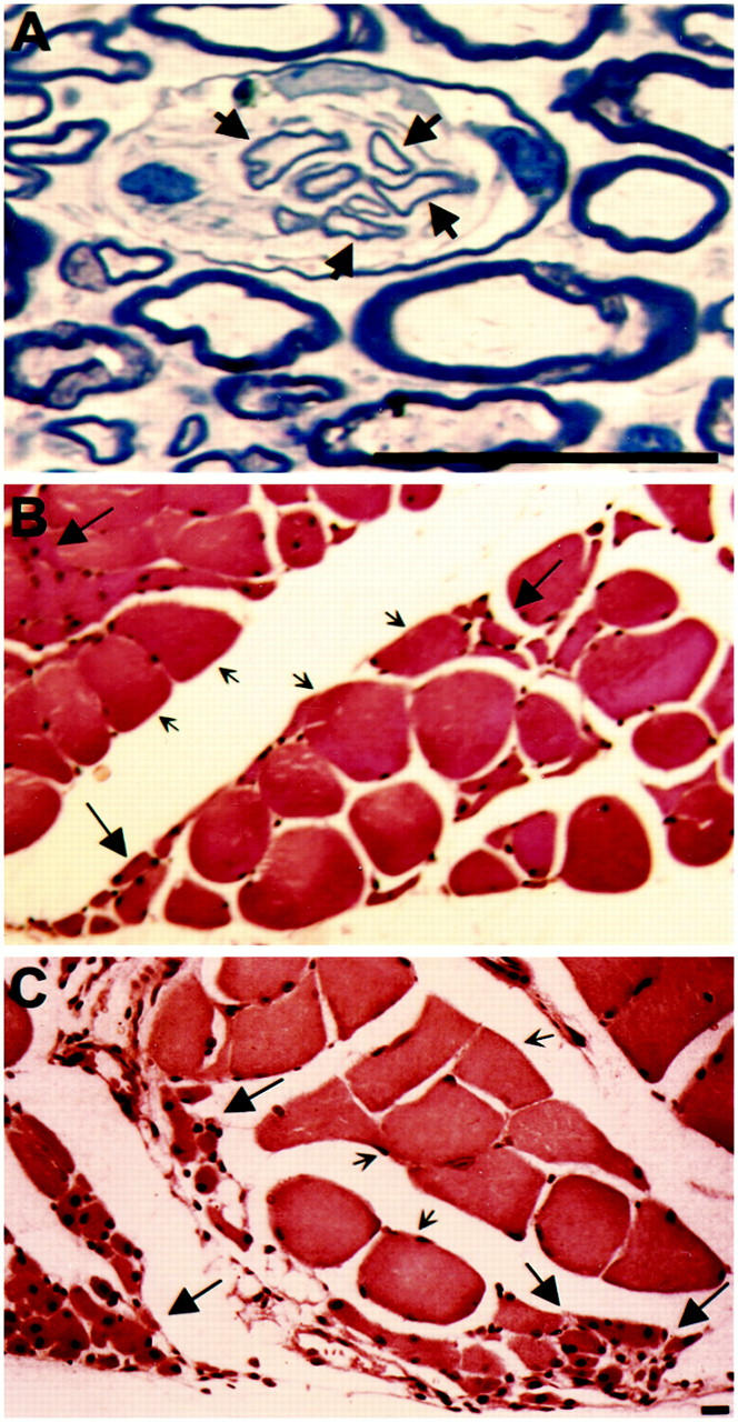
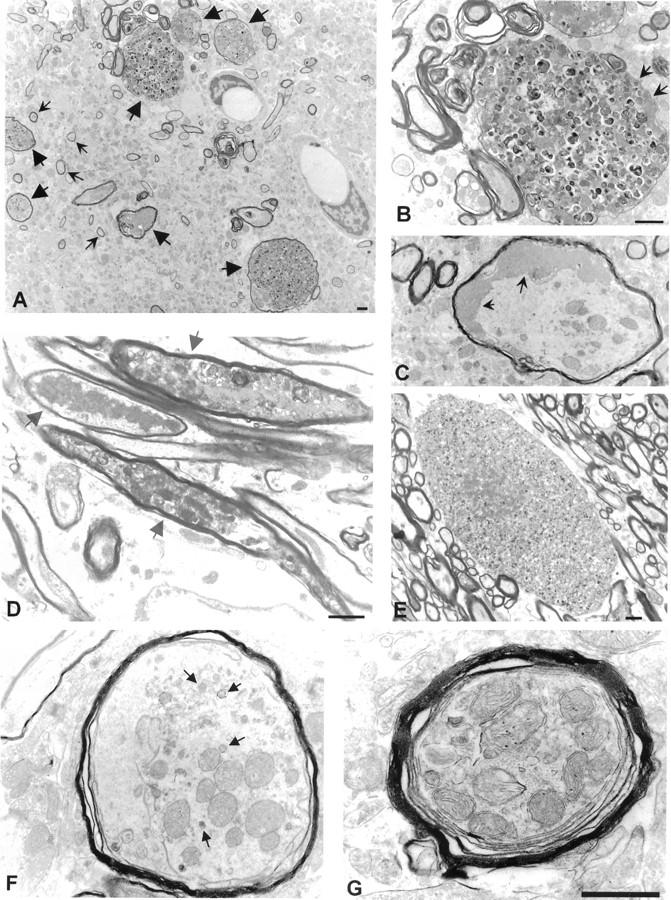
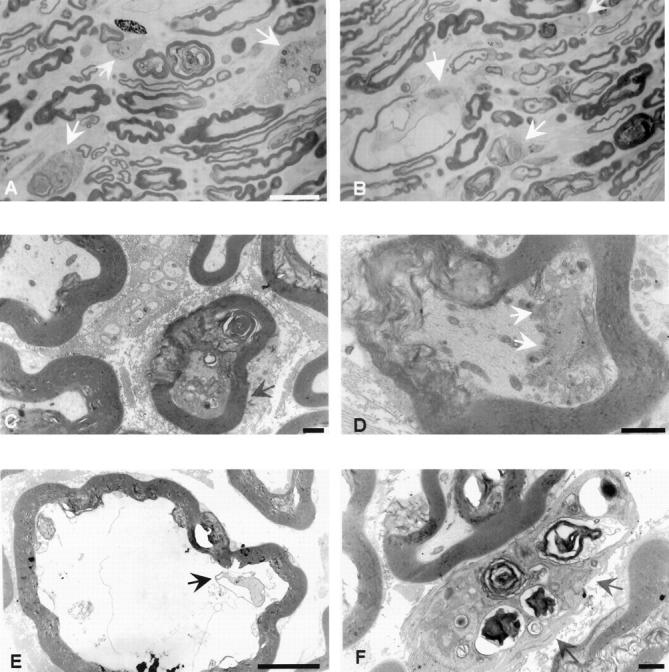
The epsilon 4 allele of the human apolipoprotein E gene (ApoE4) constitutes an important genetic risk factor for Alzheimer's disease. Recent experimental evidence suggests that human ApoE is expressed in neurons, in addition to being synthesized in glial cells. Moreover, brain regions in which neurons express ApoE seem to be most vulnerable to neurofibrillary pathology. The hypothesis that the expression pattern of human ApoE might be important for the pathogenesis of Alzheimer's disease was tested by generating transgenic mice that express human ApoE4 in neurons or in astrocytes of the central nervous system. Transgenic mice expressing human ApoE4 in neurons developed axonal degeneration and gliosis in brain and in spinal cord, resulting in reduced sensorimotor capacities. In these mice, axonal dilatations with accumulation of synaptophysin, neurofilaments, mitochondria, and vesicles were documented, suggesting impairment of axonal transport. In contrast, transgenic mice expressing human ApoE4 in astrocytes remained normal throughout life. These results suggest that expression of human ApoE in neurons of the central nervous system could contribute to impaired axonal transport and axonal degeneration. The possible contribution of hyperphosphorylation of protein Tau to the resulting phenotype is discussed.


Figures

Figure 1.
Figure 1.
Analysis of expression of human ApoE4 mRNA and protein and secretion of human ApoE4 in ApoE4 transgenic mice and abnormal extension reflex in Thy1-ApoE4 transgenic mice. a: Northern blot from spinal cord mRNA of representative mice from the four different transgenic ApoE4 strains and wild-type mice. Total spinal cord RNA (10 μg) was blotted and hybridized for human ApoE4 mRNA and for actin mRNA. b: Western blot of proteins extracted from spinal cord of ApoE4 transgenic and wild-type mice. Bands were detected with a polyclonal anti-human-ApoE antibody. c: Western blot of human ApoE4 in CSF from wild-type, Thy1-ApoE4 (tae-II), and GFAP-ApoE4 (gae-I) transgenic mice. d: Immunohistochemistry for human ApoE4 in spinal cord. Ventral horn of a GFAP-ApoE4 (A) transgenic mouse and wild-type (B) mouse, and dorsal horn of a Thy1-ApoE4 (C) transgenic mouse and wild-type (D) mouse. Scale bar, 50 μm. e: Extension reflex in an 8-month-old wild-type (A) and an age-matched Thy1-ApoE4 (tae-II) transgenic (B) mouse. f: Western blotting of human ApoE4 in primary cortical glial cultures derived form three GFAP-ApoE4 (gae-I) transgenic pups. Human ApoE4 was expressed by cultured glial cells (C1, ApoE) and secreted into the medium (M, ApoE). Reaction with the GFAP antibody identified the cells as glial cells (C2, ApoE). g: Western blotting of human ApoE4 in primary hippocampal neuronal cultures derived from three Thy1-ApoE4 (tae-XIII) transgenic pups. Human ApoE4 was expressed by transgenic neurons (C1, ApoE) and secreted into the medium (M, ApoE), but was not secreted by wild-type neurons. Reaction with the synaptophysin-antibody identified the cells as neurons (C2, Syn). Brain extract (B, ApoE) of a Thy1-ApoE4 transgenic mouse is shown as reference. Abbreviations: wt, wild type; tae-II, Thy1-ApoE4 line 2; gae-I, GFAP-ApoE4 line 1; pae-II, PDGF-ApoE4 line 2; pgk, PGK-ApoE4 line 1.
Figure 2.
Figure 2.
Body weight, parameters of muscle strength, and spontaneous locomotion in ApoE4 transgenic and wild-type mice. A: Body weight of Thy1-ApoE4 (tae, n = 6), GFAP-ApoE4 (gae, n = 5), PDGF-ApoE4 (pae, n = 6), and PGK-ApoE4 (pgk, n = 7) transgenic mice, and wild-type (wt, n = 8) mice (12 to 16 months). B: Wire hang time for Thy1-ApoE4 (tae, n = 6), GFAP-ApoE4 (gae, n = 5), PDGF-ApoE4 (pae, n = 6), and PGK-ApoE4 (pgk, n = 7) transgenic mice and wild-type (wt, n = 8) mice (12 to 16 months), as measured in the grid suspension test. C: Relation between muscle strength, measured in the grid suspension test, and body weight in individual Thy1-ApoE4 mice (tae ▵, n = 15) and wild-type (wt ♦, n = 17) littermates. D: Traction capacity in Thy1-ApoE4 (tae, n = 15) and wild-type (wt, n = 17) mice, 3 months old. E: Spontaneous locomotor activity, measured as distance traveled in an open field area for 10 minutes, and analyzed in two consecutive rounds of 5 minutes each (□ = 1′ to 5′; ▪ = 5′ to 10′). Thy1-ApoE4 (tae, n = 6), GFAP-ApoE4 (gae, n = 5), PDGF-ApoE4 (pae, n = 6), and PGK-ApoE4 (pgk, n = 7) transgenic mice and wild-type (wt, n = 8) mice, 12 to 16 months old, were used. F: Prehensile reflex in Thy1-ApoE4 (tae, n = 15) and wild-type (wt, n = 17) mice, 3 months old. G: Wire hang time for Thy1-ApoE4 (tae, n = 10) transgenic mice and wild-type (wt, n = 13) mice, 3 months old, measured in the grid suspension test. H: Body weight of Thy1-ApoE4 (tae, n = 10) transgenic mice and wild-type (wt, n = 13) mice of 3 months. All results are expressed as means (±SEM).
Figure 3.
Figure 3.
GFAP staining showing reactive astrogliosis in the hippocampus of an 18-month-old Thy1-ApoE4 mouse (A), but not in an age-matched wild-type mouse (B). Ubiquitin-positive dilated axons (arrows) can be seen in the stratum oriens of the hippocampus (C), hippocampal fimbria (E) and corticospinal tract (J) of Thy1-ApoE4 transgenic mice, but not in wild-type littermates (D, F, and K). Note the fine granularity of the white matter of the hippocampal fimbria and corticospinal tract in the Thy1-ApoE4 transgenic mice (E and J), corresponding to slightly dilated ubiquitin-positive (degenerating) axons. Neurofilament-positive dilated axons (arrows) were observed in the stratum oriens of the hippocampus of Thy1-ApoE4 mice (H), but not in wild-type littermates (I). Detail of a neurofilament-positive dilated axon (G, the nondilated part of the axon is indicated by small arrows). Scale bars, 1 mm (A and B); 50 μm (C–K and inset in C). CA1, CA2, CA3, regions of hippocampus; DG, dentate gyrus; Py, pyramidal cells of the hippocampus; fi, fimbria of the hippocampus.
Figure 4.
Figure 4.
Accumulation of synaptophysin in dilated axons of Thy1-ApoE4 transgenic mice. Hippocampal fimbria of wild-type (A) and Thy1-ApoE4 transgenic mice (B) of 18 months. Note the fine granularity in the inclusions (C and D are insets of B), corresponding to vesicular structures. Posterior column of the spinal cord of a wild-type (E) and a PDGF-ApoE4 transgenic (F) mice of 20 months. Scale bars, 50 μm (A, B, E, and F); 20 μm (C, D, and inset of F).
Figure 5.
Figure 5.
GFAP staining showing reactive gliosis in the ventral horn of the spinal cord of an 8-month-old Thy1-ApoE4 mouse (B), but not in a wild-type littermate (A). SMI31 staining showed neurofilament-positive dilated axons in the dorsal horn of the spinal cord in Thy1-ApoE4 transgenic mice (D), but not in wild-type mice (C). Ubiquitin-positive dilated axons (arrows) in the dorsal horn (F) and posterior column of the spinal cord (H and I) in a 12-month-old Thy1-ApoE4 transgenic mouse but not in a wild-type littermate (E and G). Scale bars, 50 μm (A, B, G, and H); 20 μm (C–F); 50 μm (I). GM, gray matter.
Figure 6.
Figure 6.
Semithin section of 12-month-old Thy1-ApoE4 transgenic mouse showing a cluster of small axons with a thin myelin sheath, indicative of regeneration (A, arrows). H&E staining of the quadriceps skeletal muscle of a sensorimotor impaired Thy1-ApoE4 (tae-II) mouse of 12 months (B) and of 18 months (C), showing grouping of atrophic (large arrows) fibers, which increased with age. Fibers with a normal caliber are indicated by small arrows. Scale bars, 25 μm (A); 50 μm (B and C).
Figure 7.
Figure 7.
Ultrastructure of the stratum oriens of the hippocampus and the hippocampal fimbria of 15-month-old Thy1-ApoE4 transgenic mice. A: Overview of the stratum oriens of the hippocampus showing degenerative dilated axons (large arrows). Normal axons are indicated by small arrows. B: Higher magnification of a dilated axon containing numerous electron-dense and multivesicular bodies, surrounded by a thinned myelin sheath (arrows). C: Dilated axon containing electron-dense amorphous material (arrows). D: Degenerating, only slightly dilated axons (arrows) in the hippocampal fimbria, filled with degenerative organelles and amorphous material. E: Hippocampal fimbria with grossly dilated degenerating axon, surrounded by nonaffected axons. The myelin sheath is thin or absent. Scale bar, 1 μm. Scale bar in B also applies to C. Accumulation of mitochondria in dilated, nondegenerative axons in 15-month-old Thy1-ApoE4 transgenic mice (F and G). Note the accumulation of small vesicles (arrows) in F. Scale bar, 2 μm (F and G).
Figure 8.
Figure 8.
Ultrastructure of sciatic nerve of 15-month-old Thy1-ApoE4 transgenic mouse. A: Semithin section showing macrophages (arrows) filled with myelin debris, indicative of Wallerian degeneration. B: Semithin section of a collapsed axon surrounded by a thinned and partially disrupted myelin sheath (large arrow); the smaller arrows indicate macrophages. C: Affected axon with accumulation of mitochondria and amorphous material. D: Higher magnification of an axon partially filled with granular and electron-dense amorphous material (arrows). E: High magnification of an atrophic collapsed axon. F: A macrophage (arrows) filled with myelin debris. Scale bars, 10 μm (A, B, and E); 1 μm (C, D, and F).
Figure 9.
Figure 9.
a: Hyperphosphorylation of protein Tau in spinal cord of 18-month-old Thy1-ApoE4 (tae-XIII) transgenic mice, compared to wild-type (wt) littermates. Four independent Tau-specific monoclonal antibodies (AT8, PHF1, AT180, and TAU5) were used to detect bands. Western blots shown are representative examples. b: Immunohistochemical staining with the phosphorylation-dependent monoclonal antibody AT8, demonstrating rare neurons with strong somatodendritic staining (arrows) in the spinal cord of 12-month-old Thy1-ApoE4 (tae-II) transgenic mice (A and C, arrows). Neurons with strong somatodendritic staining with AT8 were not present in wild-type littermates (B and D). Scale bars, 50 μm (A–D). WM, white matter. c: Cresyl violet staining of the ventral horn of a 12-month-old Thy1-ApoE4 (tae-II) transgenic mouse showing loss of Nissl substance in some neurons (arrows). Scale bar, 50 μm.

Similar articles

Cited by

References

    1. Corder EH, Saunders AM, Strittmatter WJ, Schmechel DE, Gaskell PC, Small GW, Roses AD, Haines JL, Pericak-Vance MA: Gene dose of apolipoprotein E type 4 allele and the risk of Alzheimer’s disease in late onset families [see comments]. Science 1993, 261:921-923 - PubMed
    1. Strittmatter WJ, Saunders AM, Goedert M, Weisgraber KH, Dong LM, Jakes R, Huang DY, Pericak-Vance M, Schmechel D, Roses AD: Isoform-specific interactions of apolipoprotein E with microtubule-associated protein tau: implications for Alzheimer disease. Proc Natl Acad Sci USA 1994, 91:11183-11186 - PMC - PubMed
    1. Olichney JM, Hansen LA, Galasko D, Saitoh T, Hofstetter CR, Katzman R, Thal LJ: The apolipoprotein E epsilon 4 allele is associated with increased neuritic plaques and cerebral amyloid angiopathy in Alzheimer’s disease and Lewy body variant. Neurology 1996, 47:190-196 - PubMed
    1. Nagy Z, Esiri MM, Jobst KA, Johnston C, Litchfield S, Sim E, Smith AD: Influence of the apolipoprotein E genotype on amyloid deposition and neurofibrillary tangle formation in Alzheimer’s disease. Neuroscience 1995, 69:757-761 - PubMed
    1. Gearing M, Mori H, Mirra SS: A beta-peptide length and apolipoprotein E genotype in Alzheimer’s disease. Ann Neurol 1996, 39:395-399 - PubMed

Publication types

MeSH terms

LinkOut - more resources