. 2000 Nov;157(5):1587-95.
doi: 10.1016/S0002-9440(10)64796-7.

Near-haploidy and subsequent polyploidization characterize the progression of peripheral chondrosarcoma

Affiliations

Near-haploidy and subsequent polyploidization characterize the progression of peripheral chondrosarcoma

J V Bovée et al. Am J Pathol. 2000 Nov.

Abstract

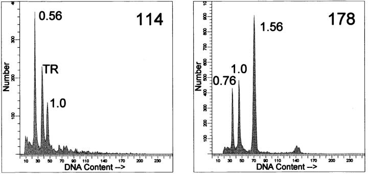
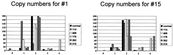
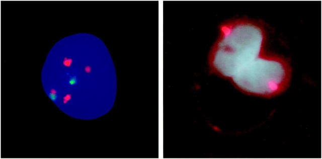
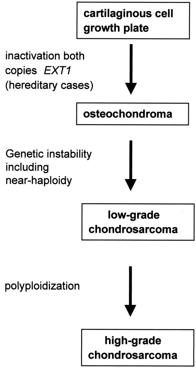
Chondrosarcomas are malignant cartilaginous tumors arising centrally in bone (central chondrosarcoma), or secondarily within the cartilaginous cap of osteochondroma (peripheral chondrosarcoma). We previously used DNA flow cytometry to demonstrate that near-haploidy is relatively frequent in peripheral chondrosarcomas. We performed fluorescence in situ hybridization (FISH) to interphase nuclei using centromeric probes, a genome wide loss of heterozygosity (LOH) analysis, and comparative genomic hybridization on five peripheral chondrosarcomas. We demonstrated near-haploidy in two low-grade tumors with only one copy and LOH of most chromosomes. Few chromosomes are disomic, with retention of heterozygosity and overrepresentation at comparative genomic hybridization. One tumor contains both a near-haploid clone with chromosomes in monosomic and disomic state, and an exactly duplicated clone. Two high-grade tumors clearly demonstrate polyploidization because most chromosomes show LOH and two copies at FISH, whereas few chromosomes have four copies with retention of heterozygosity. Using DNA from a relative, we demonstrate that chromosome loss is random regardless of parental origin. Using FISH on paraffin slides, we exclude near-haploidy to result from meiosis-like division in binucleated cells, characteristic for chondrosarcoma. In conclusion, our results indicate that near-haploidy characterizes the progression from osteochondroma toward low-grade chondrosarcoma. Moreover, further progression toward high-grade chondrosarcoma is characterized by polyploidization.


Figures

Figure 1.
Figure 1.
Examples of flow cytometric DNA histograms from nuclear suspensions. DNA indices are indicated. Left: case 114, demonstrating near-haploidy. TR, trout red blood cells serving as internal standard. Right: case 178 is shown, demonstrating two clones, a hypodiploid clone of 0.76 and a polyploidized clone of 1.56. The trout erythrocytes peak overlaps with the hypodiploid peak and is therefore not shown.
Figure 2.
Figure 2.
The distribution of number of signals for centromeres of chromosome 1 and 15 over 200 counted nuclei in the different tumor samples compared to the normal placenta control. The tumor percentage of case 408 is estimated at maximal 50%. Chromosome 1 in case 408 is estimated at one copy per tumor cell, which is seen in only 57 of 200 nuclei counted. This is however elevated as compared to the placenta control.
Figure 3.
Figure 3.
Left: FISH micrograph showing an example of case 178. Hybridization was simultaneously performed with a probe for chromosome 8 (present in either two or four copies) and chromosome 11 (present in either one or two copies). Indeed we could confirm the presence of two populations. An example of the smallest population, containing four copies of chromosome 8 and two copies of chromosome 11, is shown. Right: FISH on paraffin slides using a sex chromosome (X) centromere probe on a male patient (case 408) is demonstrated. One copy of the X centromere is demonstrated in both nuclei of binucleated cells, indicating that a normal mitosis precedes the formation of these cells, which are highly characteristic for chondrosarcoma. A meiosis-like division in binucleated cells is therefore most probably not the cause of near-haploidy in chondrosarcoma.
Figure 4.
Figure 4.
Multistep genetic model for peripheral cartilaginous tumorigenesis. First, inactivation of both copies of the EXT1 gene in cartilaginous cells of the growth plate is required for osteochondroma formation, as we previously showed by EXT1 germline mutations combined with loss of the remaining wild-type allele in hereditary osteochondromas. If complete inactivation occurs in sporadic cases remains to be investigated. One or more additional genetic alterations, which may involve the EXT-genes, TP53 and RB1 as indicated by a high percentage of LOH at these loci, are then required for peripheral chondrosarcomas to arise within its benign precursor. The process of malignant transformation is genetically represented by chromosomal instability as demonstrated by a high percentage of LOH involving various chromosomes and a broad range in DNA ploidy including near-haploidy. This may be caused by defective cell-cycle checkpoints. In osteochondromas, near-haploidy is absent and can therefore be considered a progression marker toward a low-grade malignant phenotype. Further progression toward high-grade chondrosarcoma is characterized by polyploidization.

Similar articles

Cited by

References

    1. Schmale GA, Conrad EU, Raskind WH: The natural history of hereditary multiple exostoses. J Bone Joint Surg Am 1994, 76A:986-992 - PubMed
    1. Wicklund LC, Pauli RM, Johnston D, Hecht JT: Natural history study of hereditary multiple exostoses. Am J Med Genet 1995, 55:43-46 - PubMed
    1. Springfield DS, Gebhardt MC, McGuire MH: Chondrosarcoma: a review. J Bone Joint Surg Am 1996, 78A:141-149
    1. Mulder JD, Schütte HE, Kroon HM, Taconis WK: Radiologic Atlas of Bone Tumors. Edited by JD Mulder. Amsterdam, Elsevier, 1993
    1. Bovee JVMG, Cleton-Jansen AM, Kuipers-Dijkshoorn N, Van den Broek LJCM, Taminiau AHM, Cornelisse CJ, Hogendoorn PCW: Loss of heterozygosity and DNA ploidy point to a diverging genetic mechanism in the origin of peripheral and central chondrosarcoma. Genes Chromosom Cancer 1999, 26:237-246 - PubMed