

A novel, nuclear pore-associated, widely distributed molecule overexpressed in oncogenesis and development
- PMID: 11073820
- PMCID: PMC1885726
- DOI: 10.1016/S0002-9440(10)64798-0
A novel, nuclear pore-associated, widely distributed molecule overexpressed in oncogenesis and development
Abstract
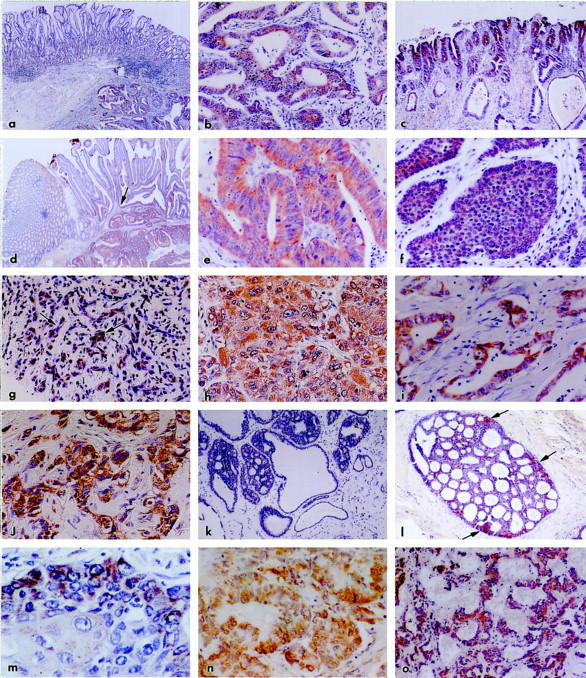
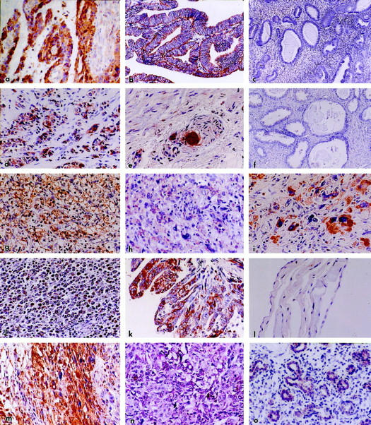
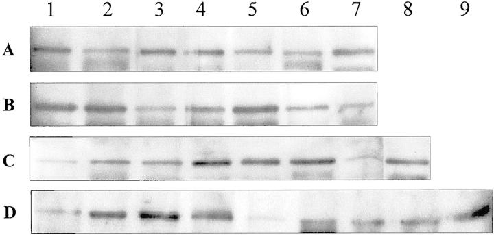
Nuclear pore complexes are large, elaborate macromolecular structures that mediate the bidirectional nucleocytoplasmic traffic. In vertebrates, nuclear pore complexes comprise 50 to 100 proteins termed nucleoporins (Nup). An 88-kd nucleoporin (Nup88) has been recently cloned and characterized, and found to be associated in a dynamic subcomplex with the oncogenic nucleoporin CAN/Nup 214. We have produced a polyclonal antiserum to Nup88, and found that it immunoreacts convincingly in conventional tissue sections of 214 samples of malignant tumors of many types. All carcinomas were stained irrespective of site or line of differentiation; the majority of cases reacted strongly and extensively. In situ carcinomas and highly dysplastic epithelia were similarly reactive. Samples of malignant mesotheliomas, gliomas, sarcomas, and lymphoreticular tumors were also stained. Substantial reactions were also found in certain fetal tissues. Focal reactions were noted in some reactive-proliferative processes. Most benign epithelial and mesenchymal tumors and hyperplasias, and normal adult tissues reacted weakly and sporadically or not at all. Immunoblot analysis of selected samples strongly corroborated those findings. If further substantiated, our findings indicate that Nup88 could be regarded as a selective yet broadly based proliferation marker of potential significance in the histological evaluation and diagnosis of malignant transformation. Its ready applicability on conventional paraffin sections and on cytological preparations may broaden its clinical and investigative significance.
Figures
Similar articles
-
Nup88 (karyoporin) in human malignant neoplasms and dysplasias: correlations of immunostaining of tissue sections, cytologic smears, and immunoblot analysis.Hum Pathol. 2002 May;33(5):536-44. doi: 10.1053/hupa.2002.124785. Hum Pathol. 2002. PMID: 12094380
-
The nuclear pore complex protein Nup88 is overexpressed in tumor cells.Cancer Res. 1999 Nov 1;59(21):5408-11. Cancer Res. 1999. PMID: 10554006
-
The phosphorylated form of connexin43 is up-regulated in breast hyperplasias and carcinomas and in their neoformed capillaries.Hum Pathol. 2005 May;36(5):536-45. doi: 10.1016/j.humpath.2005.03.013. Hum Pathol. 2005. PMID: 15948121
-
Nucleoporins: leaving the nuclear pore complex for a successful mitosis.Cell Signal. 2011 Oct;23(10):1555-62. doi: 10.1016/j.cellsig.2011.05.023. Epub 2011 Jun 13. Cell Signal. 2011. PMID: 21683138 Review.
-
Dynamics of nuclear pore complex organization through the cell cycle.Curr Opin Cell Biol. 2004 Jun;16(3):314-21. doi: 10.1016/j.ceb.2004.04.001. Curr Opin Cell Biol. 2004. PMID: 15145357 Review.
Cited by
-
Overexpressed Nup88 stabilized through interaction with Nup62 promotes NF-κB dependent pathways in cancer.Front Oncol. 2023 Feb 9;13:1095046. doi: 10.3389/fonc.2023.1095046. eCollection 2023. Front Oncol. 2023. PMID: 36845732 Free PMC article.
-
Nucleoporin Nup188 is required for chromosome alignment in mitosis.Cancer Sci. 2013 Jul;104(7):871-9. doi: 10.1111/cas.12159. Epub 2013 May 2. Cancer Sci. 2013. PMID: 23551833 Free PMC article.
-
Aiding and abetting cancer: mRNA export and the nuclear pore.Trends Cell Biol. 2013 Jul;23(7):328-35. doi: 10.1016/j.tcb.2013.03.004. Epub 2013 Apr 10. Trends Cell Biol. 2013. PMID: 23582887 Free PMC article. Review.
-
Role of the nuclear envelope in genome organization and gene expression.Wiley Interdiscip Rev Syst Biol Med. 2011 Mar-Apr;3(2):147-66. doi: 10.1002/wsbm.101. Wiley Interdiscip Rev Syst Biol Med. 2011. PMID: 21305702 Free PMC article. Review.
-
Nucleoporin 88 expression in hepatitis B and C virus-related liver diseases.World J Gastroenterol. 2006 Sep 28;12(36):5870-4. doi: 10.3748/wjg.v12.i36.5870. World J Gastroenterol. 2006. PMID: 17007055 Free PMC article.
References
-
- Yasumoto K, Setoguchi I, Kamei M, Nomoto K, Murakami H, Hasizume S: Cancer-specific binding of mouse Mab vs Candida krusei cytochrome c: an antigen recognized by a cancer-associated human Mab HB4C5. Hum Antibodies Hybridomas 1993, 4:180-186 - PubMed
-
- Martinez N, Alonso A, Moragues MD, Ponton J, Schneider J: The nuclear pore protein complex Nup88 is overexpressed in tumor cells. Cancer Res 1999, 59:5408-5411 - PubMed
-
- Fornerod M, Boer J, van Baal S, Morreau J, Grosveld G: Interactions of cellular proteins with the leukemia-specific fusion protein DEK/CAN and SET/CAN and their normal counterpart, the nucleoporin CAN. Oncogene 1996, 13:1801-1808 - PubMed
Publication types
MeSH terms
Substances
LinkOut - more resources
Full Text Sources
Other Literature Sources
Molecular Biology Databases
