. 2000 Nov;157(5):1661-9.
doi: 10.1016/s0002-9440(10)64803-1.

Keloid-derived fibroblasts are refractory to Fas-mediated apoptosis and neutralization of autocrine transforming growth factor-beta1 can abrogate this resistance

Affiliations

Keloid-derived fibroblasts are refractory to Fas-mediated apoptosis and neutralization of autocrine transforming growth factor-beta1 can abrogate this resistance

T Chodon et al. Am J Pathol. 2000 Nov.

Abstract

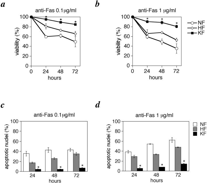
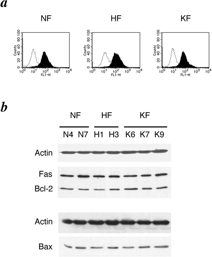
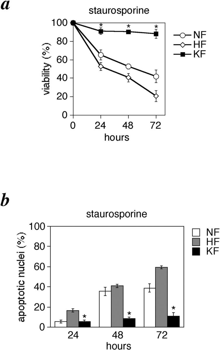
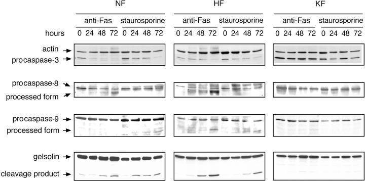
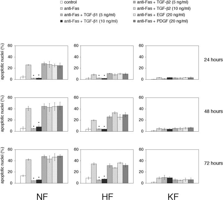
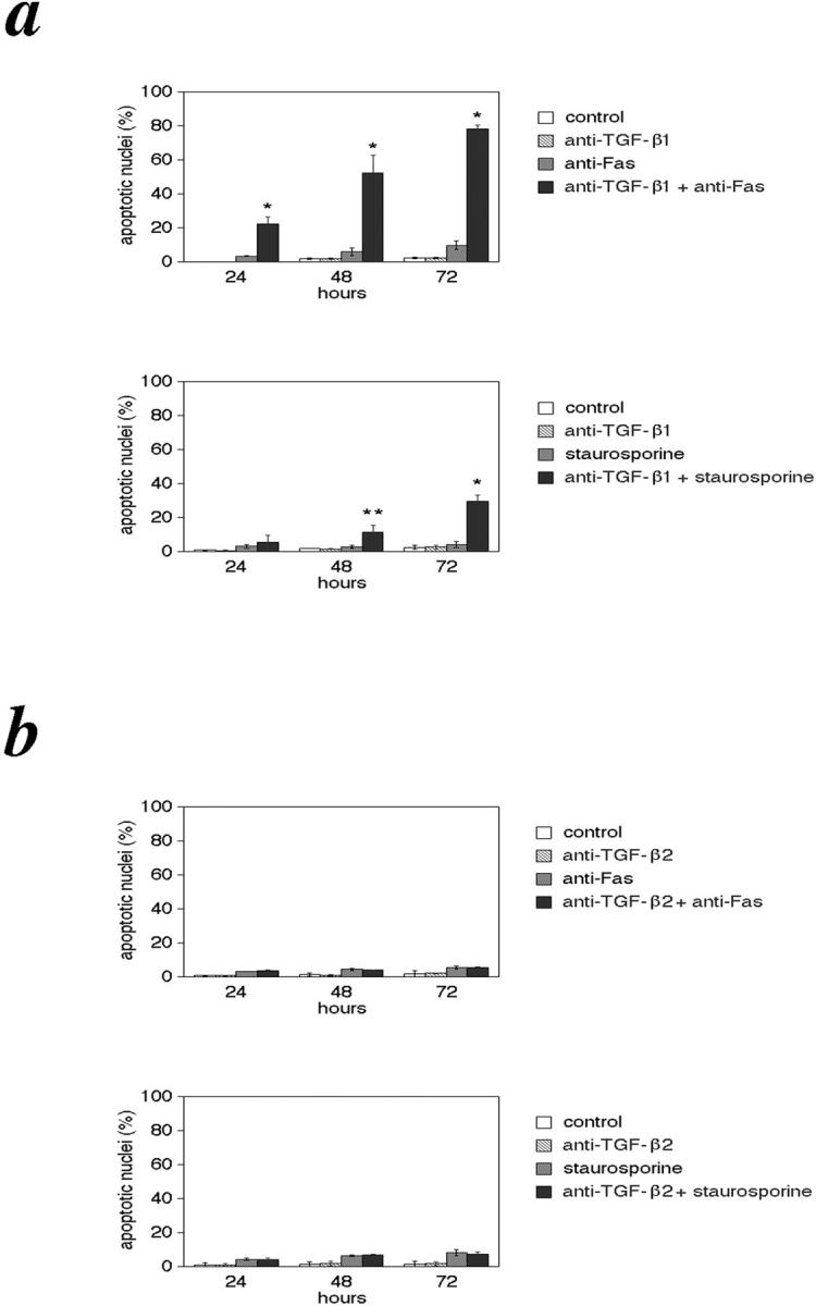
The pathogenesis of keloid remains poorly understood. As no effective therapy for keloid is as yet available, an insight into its pathogenesis may lead to novel approaches. Apoptosis has been found to mediate the decrease in cellularity during the transition between granulation tissue and scar. Here, we report that in contrast to hypertrophic scar-derived and normal skin-derived fibroblasts, keloid-derived fibroblasts are significantly resistant to both Fas-mediated and staurosporine-induced apoptosis. The caspases-3, -8, and -9 were not activated indicating that the block in the apoptotic pathway in keloid is upstream of the caspases. There were no significant differences in the level of expression of Fas, Bcl-2, and Bax between the three groups but addition of transforming growth factor (TGF)-beta1 significantly inhibited Fas-mediated apoptosis in hypertrophic scar-derived and normal skin-derived fibroblasts and neutralization of autocrine TGF-beta1 with anti-TGF-beta1 antibody abrogated the resistance of keloid-derived fibroblasts. Anti-apoptotic activity was not observed with TGF-beta2. This is the first study linking refractory Fas-mediated apoptosis to cellular phenotype in keloids and indicating a pivotal role for the anti-apoptotic effect of TGF-beta1 in this resistance. Hence, it becomes important to treat keloids as a separate entity different from hypertrophic scars and enhancement of Fas-sensitivity could be a promising therapeutic target.


Figures

Figure 1.
Figure 1.
KFs are refractory to Fas-mediated apoptosis. Cells were grown in DMEM and 10% fetal bovine serum and after 24 hours, starved in serum-free medium for 48 hours followed by stimulation with 1 μg/ml of anti-Fas antibody (clone: CH-11; MBL, Japan) for 48 hours. a and b: NFs. c and d: HFs. e and f: KFs. Cellular morphological changes of apoptosis analyzed by phase contrast microscopy. Original magnification, ×200 (a, c, and e). The same fields as that of (a, c, and e) examined by Hoechst 33342 staining (b, d, and f). Note the staining of many condensed nuclei indicative of apoptosis (b and d) that corresponds to the cell rounding seen in (a and c), respectively, and their relative absence in the KFs (e and f).
Figure 2.
Figure 2.
KFs are refractory to Fas-mediated apoptosis. Cells were stimulated with anti-Fas antibody as described. Viability assay by trypan blue exclusion (a and b) and percentage of nuclear condensation assessed by Hoechst staining (c and d) after stimulation with 0.1 and 1 μg/ml of anti-Fas, respectively, for 24, 48, and 72 hours. *, P < 0.01 compared with NFs and HFs (ANOVA with Scheffé’s post hoc tests). Values shown are the mean and SD of 15 independent experiments.
Figure 3.
Figure 3.
Expression of Fas, Bcl-2, and Bax. a: Flow cytometric analysis of cell surface Fas expression on fibroblasts derived from normal skin (NF), hypertrophic scar (HF), and keloid (KF). Cells were incubated with fluorescein isothiocyanate-conjugated goat anti-mouse IgG alone for control (open curve) or with monoclonal anti-Fas antibody followed by incubation with fluorescein isothiocyanate-conjugated goat anti-mouse IgG (filled curve). b: Representative Western blots depicting expression of Fas, Bcl-2, and Bax. To ensure equal loading, each blot was probed for the presence of actin. Here, Fas and Bcl-2 were probed on the same membrane. N4, N7, H1, H3, K6, K7, and K9, are each fibroblasts derived from normal skin, hypertrophic scar, and keloid of different patients.
Figure 4.
Figure 4.
KFs are refractory to staurosporine-induced apoptosis. Cells were grown in DMEM and 10% fetal bovine serum and after 24 hours they were treated with staurosporine (10 nmol/L). Viability assay by trypan blue exclusion (a), and percentage of nuclear condensation assessed by Hoechst staining (b) at 24, 48, and 72 hours of treatment. *, P < 0.01 compared with NFs and HFs (ANOVA with Scheffé’s post hoc tests). Values shown are mean and SD of 15 independent experiments.
Figure 5.
Figure 5.
Caspases are not activated in KFs. After induction of apoptosis with anti-Fas or staurosporine for 24, 48, and 72 hours as described in Materials and Methods, cells were lysed. Cell lysates (40 μg) were loaded onto 10 to 14% polyacrylamide gels and analyzed by Western blotting using monoclonal anti-caspase-3 (top), monoclonal anti-caspase-8 (second row), polyclonal anti-caspase-9 (third row), or monoclonal anti-gelsolin antibody (bottom).
Figure 6.
Figure 6.
TGF-β1 inhibits Fas-mediated apoptosis. Cells were treated with recombinant human TGF-β1 or recombinant human TGF-β2 at a dose of 5 and 10 ng/ml, recombinant human epidermal growth factor or recombinant human platelet-derived growth factor at a dose of 20 ng/ml 6 hours before stimulation with anti-Fas, treated with anti-Fas only or in the absence of both (control) and the percentage of nuclear condensation analyzed by Hoechst staining. *, p < 0.01 compared with the untreated controls and the TGF-β2-, epidermal growth factor-, platelet-derived growth factor-treated groups (ANOVA with Scheffé’s post hoc tests). Values shown are mean and SD of six independent experiments.
Figure 7.
Figure 7.
Neutralizing antibody to TGF-β1 abrogates the resistance of KFs. KFs were treated with monoclonal anti-human TGF-β1 antibody (a) or monoclonal anti-human TGF-β2 antibody (b) alone or together with anti-Fas or staurosporine as described in Materials and Methods and analyzed by Hoechst staining. Control indicates cells not treated with the above mentioned agents. *, P < 0.01; **, P < 0.05 compared with the untreated controls and the anti-TGF-β2 antibody-treated groups (ANOVA with Scheffé’s post hoc tests). Values shown are mean and SD of six independent experiments.

References

    1. Alhady S: Keloids in various races. Plast Reconstr Surg 1969, 44:564-566 - PubMed
    1. Murray JC, Pollack SV, Pinnelli SR: Keloids: a review. J Am Acad Dermatol 1981, 4:461-470 - PubMed
    1. Ladin DA, Garner WL, Smith DJ: Excessive scarring as a consequence of healing. Wound Rep Reg 1995, 3:6-14 - PubMed
    1. Nicoletis C, Bazin S, LeLous M: Clinical and biochemical features of normal, defective and pathologic scars. Clin Plast Surg 1977, 4:347-359 - PubMed
    1. Russell SB, Trupin KM, Rodriguez-Eaton S, Russell JD, Trupin JS: Reduced growth-factor requirement of keloid-derived fibroblasts may account for tumor growth. Proc Natl Acad Sci USA 1988, 85:587-591 - PMC - PubMed

Publication types

MeSH terms