. 2000 Dec;182(23):6724-31.
doi: 10.1128/JB.182.23.6724-6731.2000.

Inducible metabolism of phenolic acids in Pediococcus pentosaceus is encoded by an autoregulated operon which involves a new class of negative transcriptional regulator

Affiliations

Inducible metabolism of phenolic acids in Pediococcus pentosaceus is encoded by an autoregulated operon which involves a new class of negative transcriptional regulator

L Barthelmebs et al. J Bacteriol. 2000 Dec.

Abstract

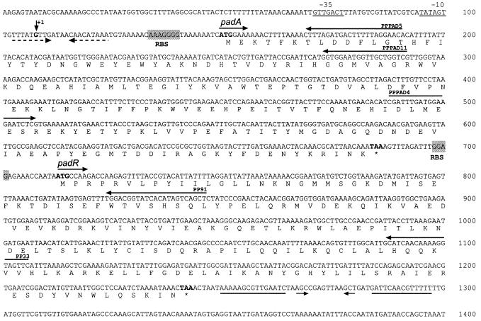
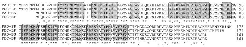
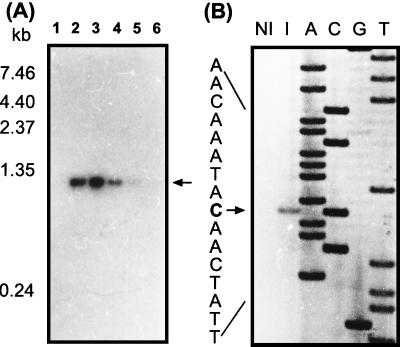
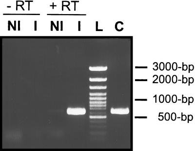
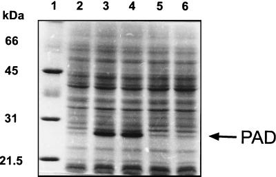
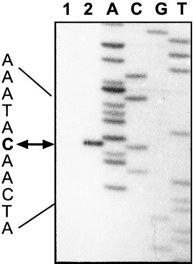
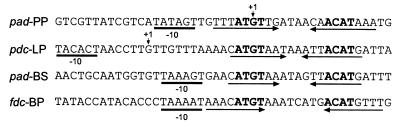
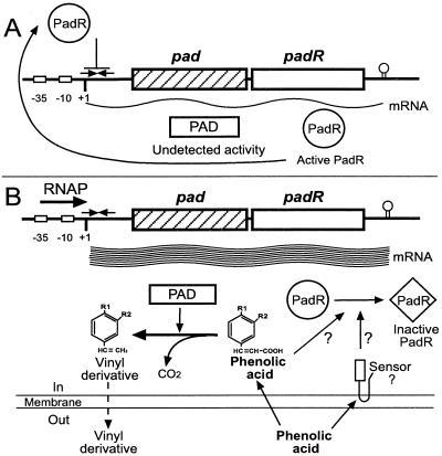
Pediococcus pentosaceus displays a substrate-inducible phenolic acid decarboxylase (PAD) activity on p-coumaric acid. Based on DNA sequence homologies between the three PADs previously cloned, a DNA probe of the Lactobacillus plantarum pdc gene was used to screen a P. pentosaceus genomic library in order to clone the corresponding gene of this bacteria. One clone detected with this probe displayed a low PAD activity. Subcloning of this plasmid insertion allowed us to determine the part of the insert which contains a 534-bp open reading frame (ORF) coding for a 178-amino-acid protein presenting 81.5% of identity with L. plantarum PDC enzyme. This ORF was identified as the padA gene. A second ORF was located just downstream of the padA gene and displayed 37% identity with the product of the Bacillus subtilis yfiO gene. Subcloning, transcriptional analysis, and expression studies with Escherichia coli of these two genes under the padA gene promoter, demonstrated that the genes are organized in an autoregulated bicistronic operonic structure and that the gene located upstream of the padA gene encodes the transcriptional repressor of the padA gene. Transcription of this pad operon in P. pentosaceus is acid phenol dependent.


Figures

FIG. 1
FIG. 1
Physical map of the padAR locus and delineation of subcloned fragments. The parent plasmid pTPADP1 was isolated from the P. pentosaceus genomic library. Restriction sites and primers (small horizontal arrows) used to obtain the different subclones are shown. The ORFs identified by sequencing are indicated by large arrows. PAD activity measured on p-coumaric acid in E. coli is indicated to the right of each subclone (U, micromoles of p-coumaric acid degraded per minute).
FIG. 2
FIG. 2
Nucleotide and deduced amino acid sequences of the pad cluster. The localization and orientation of primers PPPAD5, PPPAD11, and PP91, used to identify the transcriptional start site of padA and padR, and primers PPPAD4 and PP33, used to amplify cDNA from padA mRNA, are indicated by horizontal arrows. The transcriptional start site of the padAR operon, determined by primer extension analysis, is indicated by a vertical arrow, and the corresponding −10 and −35 boxes are underlined. The putative ribosome binding sites (RBS) are shaded. Stop codons are indicated by asterisks. The two convergent arrows located under the sequence indicate the putative rho-independent transcriptional terminator of the padAR operon. The dotted convergent arrows indicate the region of dyad symmetry (inverted repeats), which could be the target of the PadR repressor.
FIG. 3
FIG. 3
Comparison of the deduced amino acid sequence of the padA gene of P. pentosaceus (PAD-PP) with the sequences of B. pumilus FDC (FDC-BP; accession no. X84815), L. plantarum PDC (PDC-LP; accession no. U63827), and B. subtilis PAD (PAD-BS; accession no. AF017117). The sequences were aligned by using the Clustal program. Identical and similar amino acids are indicated by asterisks and dots, respectively. Conserved boxes are shaded. The numbers on the right correspond to the amino acid position in the protein sequence.
FIG. 4
FIG. 4
Transcriptional analysis of the padAR operon. (A) Northern blot analysis with a padA-specific probe of total RNA purified from P. pentosaceus cells harvested after 0 min (lane 1), 5 min (lane 2), 10 min (lane 3), 20 min (lane 4), 40 min (lane 5), and 120 min (lane 6) following the addition of 2.4 mM p-coumaric acid. The arrow indicates the position of the transcript, and molecular size markers are given in the left lane. (B) Mapping of the 5′ end of the padA mRNA by primer extension analysis using primer PPPAD5 with total P. pentosaceus RNA from uninduced cells (NI) and cells induced with 2.4 mM p-coumaric acid (I). The products of reverse transcription were loaded in parallel with DNA sequencing reaction mixtures (lanes A, C, G, and T) initiated with the same primer on padA DNA template. The sequence shown to the left is the complementary strand, and the 5′ end of the padA mRNA is indicated by an arrow.
FIG. 5
FIG. 5
RT-PCR of the padAR region using primers PPPAD4 and PP33 with P. pentosaceus total RNA purified from uninduced cells (NI) and cells induced with 2.4 mM p-coumaric acid (I). Negative controls with no RT (−RT) included are shown on the left. Classical PCR using the same primers and with P. pentosaceus chromosomal DNA added as a positive control is shown in lane C. The 100-bp DNA Ladder Plus (MBI Fermentas, Amherst, Mass.) was used as a molecular weight marker (L).
FIG. 6
FIG. 6
SDS-PAGE of crude cell extracts from E. coli TG1 carrying various subclones of the padAR locus. Lanes: 1, molecular mass standards (SDS-PAGE standards; Bio-Rad); 2, crude extract from E. coli TG1(pJDC9); 3, crude extract from E. coli TG1(pJPADP6); 4, crude extract from E. coli TG1(pJPADP7); 5, crude extract from E. coli TG1(pJPADP8); 6, crude extract from E. coli TG1(pJPADP9). Molecular size markers are indicated on the left.
FIG. 7
FIG. 7
Mapping of the 5′ end of padA mRNA by primer extension with primer PPPAD5 with total RNA from E. coli TG1(pJPADP9) (lane 1) and TG1(pJPADP6) (lane 2). The RT product from E. coli TG1(pJPADP6) was diluted 20-fold before loading. The products of reverse transcription reactions were loaded in parallel with DNA sequencing reaction mixtures (lanes A, C, G, and T) initiated with the same primer on the padA DNA template. The arrow indicates the 5′ end of the padA mRNA.
FIG. 8
FIG. 8
Comparison of the promoter sequence from the padAR operon of P. pentosaceus (pad-PP) with the promoter sequences of the L. plantarum pdc gene (pdc-LP), B. subtilis pad gene (pad-BS), and B. pumilus fdc gene (fdc-BP). The putative −10 boxes are underlined. The transcription start sites are indicated by a vertical arrow, when determined. Convergent arrows indicate regions of dyad symmetry, and the conserved motif is highlighted in boldface.
FIG. 9
FIG. 9
Model for the transcriptional regulation of the padAR operon in P. pentosaceus. (A) In the absence of phenolic acid, the level of pad transcripts is low and could only be detected by RT-PCR, while PAD activity remained undetectable. (B) Addition of phenolic acid causes inactivation of the PadR repressor, possibly through an additional effector or sensor which mediates the conformational change or modulation of PadR. This allows transcription of the padAR operon and synthesis of the PAD enzyme. Toxic phenolic acids are decarboxylated into vinyl derivatives, which are less toxic and can diffuse outside the cell. Exhaustion of phenolic acids results in PadR switching to its active form and repressing padAR transcription. For p-coumaric acid, R1 = OH and R2 = H.

References

    1. Adler K, Beyreuther K, Geisler N, Gronenborn B, Klemm A, Muller-Hill B, Pfahl M, Schmitz A. How lac repressor binds to DNA. Nature. 1972;237:322–327. - PubMed
    1. Ajdic D, Ferretti J J. Transcriptional regulation of the Streptococcus mutans gal operon by the GalR repressor. J Bacteriol. 1998;180:5727–5732. - PMC - PubMed
    1. Alekshun M N, Levy S B. The mar regulon: multiple resistance to antibiotics and other toxic chemicals. Trends Microbiol. 1999;7:410–413. - PubMed
    1. Barthelmebs L, Diviès C, Cavin J-F. Knockout of the p-coumarate decarboxylase gene from Lactobacillus plantarum reveals the existence of two other inducible enzymatic activities involved in phenolic acid metabolism. Appl Environ Microbiol. 2000;66:3368–3375. - PMC - PubMed
    1. Borneman W S, Akin D E, VanEseltine W P. Effect of phenolic monomers on ruminal bacteria. Appl Environ Microbiol. 1986;52:1331–1339. - PMC - PubMed

Publication types

MeSH terms

Associated data

LinkOut - more resources