. 2000 Dec;20(23):8793-802.
doi: 10.1128/MCB.20.23.8793-8802.2000.

Association with Ets-1 causes ligand- and AF2-independent activation of nuclear receptors

Affiliations

Association with Ets-1 causes ligand- and AF2-independent activation of nuclear receptors

R M Tolón et al. Mol Cell Biol. 2000 Dec.

Abstract

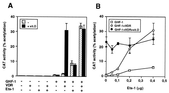
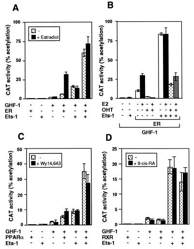
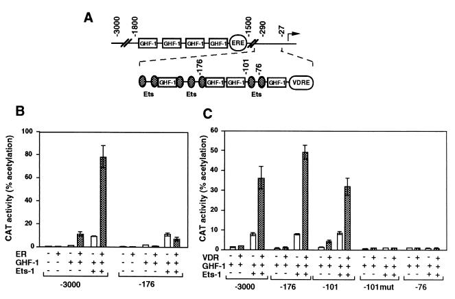
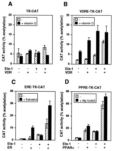
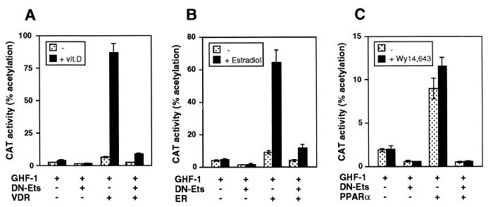
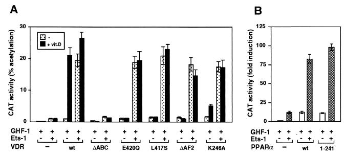
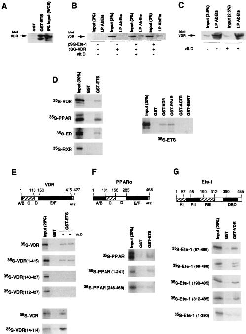
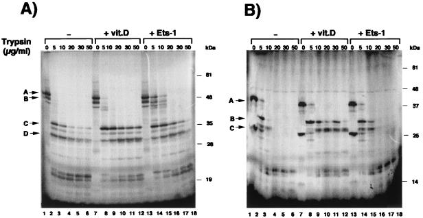
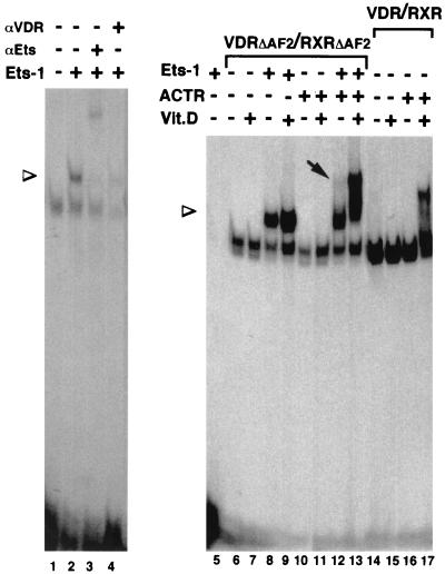
The vitamin D receptor (VDR) normally functions as a ligand-dependent transcriptional activator. Here we show that, in the presence of Ets-1, VDR stimulates the prolactin promoter in a ligand-independent manner, behaving as a constitutive activator. Mutations in the AF2 domain abolish vitamin D-dependent transactivation but do not affect constitutive activation by Ets-1. Therefore, in contrast with the actions of vitamin D, activation by Ets-1 is independent of the AF2 domain. Ets-1 also conferred a ligand-independent activation to the estrogen receptor and to peroxisome proliferator-activated receptor alpha. In addition, Ets-1 cooperated with the unliganded receptors to stimulate the activity of reporter constructs containing consensus response elements fused to the thymidine kinase promoter. There is a direct interaction of the receptors with Ets-1 which requires the DNA binding domains of both proteins. Interaction with Ets-1 induces a conformational change in VDR which can be detected by an increased resistance to proteolytic digestion. Furthermore, a retinoid X receptor-VDR heterodimer in which both receptors lack the core C-terminal AF2 domain can recruit coactivators in the presence, but not in the absence, of Ets-1. This suggests that Ets-1 induces a conformational change in the receptor which creates an active interaction surface with coactivators even in the AF2-defective mutants. These results demonstrate the existence of a novel mechanism, alternative to ligand binding, which can convert an unliganded receptor from an inactive state into a competent transcriptional activator.


Figures

FIG. 1
FIG. 1
Ligand-independent activation of nuclear receptors by Ets-1. (A) HeLa cells were cotransfected with −3000Prl-CAT (5 μg) and vectors for GHF-1 (0.4 μg), VDR (2.5 μg), and/or Ets-1 (0.5 μg). (B) The reporter plasmid was cotransfected with GHF-1 and VDR in the presence of the indicated amounts of Ets-1. CAT activity was determined in untreated cells and cells treated with vitamin D for 48 h.
FIG. 2
FIG. 2
Ets-1 causes constitutive activation of ERα and PPARα. (A) Cells were transfected with −3000Prl-CAT (5 μg) and vectors for GHF-1 (0.4 μg) and Ets-1 (0.5 μg) alone or in combination with 2.5 μg of ERα. CAT activity was determined in cells treated in the presence and absence of estradiol (1 μM) for 48 h. (B) cells were transfected with the same vectors and treated with 10 nM estradiol (E2) and/or 1 μM antagonist OHT. (C and D) The reporter plasmid was cotransfected with the same amounts of GHF-1 and Ets-1 together with 5 μg of PPARα (C) or 1 μg RXR (D) vectors. CAT activity was determined in untreated cells and in cells treated with Wy14,643 or 9-cis-retinoic acid.
FIG. 3
FIG. 3
Influence of Ets-1 on different prolactin promoter fragments. (A) Schematic representation of the rat prolactin 5′-flanking region showing the structure of the distal enhancer (bp −1500 to −1800) and the proximal promoter region. Binding sites for GHF-1 and Ets factors, as well as ERE and VDRE are depicted. (B and C) Cells were transfected with 5 μg of reporter CAT constructs and 0.5 μg of Ets-1 alone or in combination with unliganded ER (B) or VDR (C). The reporter plasmids have progressive deletions of the prolactin promoter (from bp −3000 to −76), and in the −101mut construct the Ets binding sites have been mutated (4). CAT activities were determined 48 h after transfection.
FIG. 4
FIG. 4
Ets increases activity of unliganded receptors in other promoter constructs. HeLa cells were transfected with 5 μg of a reporter CAT plasmid under control of the TK promoter (TK-CAT) (A) or with the same plasmid containing a consensus VDRE, PPRE, or ERE. These plasmids were cotransfected with 0.5 μg of Ets-1 and 2.5 μg of expression vectors for VDR (A and B), ERα (C), or PPARα (D). CAT activity was determined in cells incubated with or without vitamin D, estradiol, or Wy14,643, respectively.
FIG. 5
FIG. 5
A dominant-negative Ets inhibits receptor-mediated stimulation. Cells were transfected with −3000Prl-CAT (5 μg) and GHF-1 (0.4 μg), alone or in combination with VDR (A), ERα (B), or PPARα (C). Five micrograms of a vector encoding a dominant-negative Ets vector (DN-Ets) was cotransfected as indicated. Activity was determined in untreated cells and cells treated with vitamin D, estradiol, or Wy14,643.
FIG. 6
FIG. 6
Ets-1 confers activation to AF2-defective VDR mutants. (A) −3000Prl-CAT was transfected into HeLa cells together with vectors encoding GHF-1 (0.4 μg), Ets-1 (0.5 μg), and wild-type VDR (wt) or the VDR mutants indicated (2.5 μg each). VDR-ΔAF2 lacks the C-terminal helix 12 of the LBD, and ΔABC lacks the 110 N-terminal VDR residues. Two different point mutations in helix 12 (L417S and E420Q), as well as mutation K246A in helix 3, were also used. CAT activity was determined in untreated cells and in cells incubated with vitamin D. (B) The Prl-CAT plasmid was cotransfected with an expression vector for the native PPARα (wt) or for PPARα(1–241), which lacks the LBD. CAT activities were determined 48 h later.
FIG. 7
FIG. 7
Expression of coactivators potentiates Ets-mediated constitutive activity of VDR. The prolactin reporter plasmid −3000Prl-CAT and expression vectors for GHF-1 (0.4 μg), VDR (2.5 μg), and Ets-1 (0.5 μg) were transfected alone or in combination with 2 μg of vectors for the coactivators SRC-1 and CBP, as indicated. CAT activity was determined in cells treated for 48 h in the presence and absence of vitamin D.
FIG. 8
FIG. 8
Interaction of Ets-1 with nuclear receptors. (A) GST or GST-ETS was incubated with 900 μg of WCE, and the VDR bound was analyzed by Western blotting. The input represents 5% of proteins used. (B) HeLa cells were transfected with an empty vector or with vectors encoding Ets-1 and/or VDR. Immunoprecipitates with the anti-Ets antibody (AbEts) from cells treated in the presence and absence of vitamin D were subjected to Western analysis with the VDR antibody together with 3% of the WCE used (input). (C) VDR was detected by Western analysis in immunoprecipitates from untreated and vitamin D-treated GH4C1 cells. Input represents 2.5% (125 μg) of the WCE used. (D, left) Pull-down assays were performed with GST alone or GST-ETS and different in vitro-translated 35S-labeled receptors. (Right) In vitro-translated Ets-1 was used in pull-down experiments with GST-fused VDR and PPARα as well as with the receptor-interacting domains of the receptor coactivator ACTR and the corepressor SMRT. The inputs represent 20% of the proteins used. (E and F) Representation of VDR and PPARα, showing the different functional domains. The indicated 35S-VDR and PPARα deletion mutants were used in pull-down assays with GST and GST-ETS. The pull-down assays with labeled VDR were performed in the presence and absence of vitamin D (1 μM). (G) Schematic representation of the p68 Ets-1 protein. RI and RIII, transcriptional activation domains; RII, regulatory domain; DBD, DNA binding ETS domain. The 35S-Ets-1 fragments indicated were used in pull-down assays with GST or GST-VDR.
FIG. 9
FIG. 9
Ets-1 induces a conformational change in VDR. (A) 35S-VDR preincubated with GST (lanes 1 to 6), GST–Ets-1 (lanes 13 to 18), or vitamin D (lanes 7 to 12) was digested with increasing concentrations of trypsin and analyzed by SDS-PAGE and autoradiography. Arrows A and B, undigested VDR; arrows C and D, resistant protein fragments. (B) The 35S-VDR fragment spanning residues 112 to 427 was used in a similar experiment. Arrow A, mobility of the undigested VDR fragment; arrows B and C, main fragments resistant to proteolysis.
FIG. 10
FIG. 10
Association with Ets-1 allows an AF2-independent recruitment of coactivators. Shown are gel retardation assays with the VDRE oligonucleotide and in vitro-translated VDR and/or RXR. Receptors VDR(1–415) (VDRΔAF2) and RXR(1–445) (RXRΔAF2), lacking helix 12, as well as wild-type receptors (lanes 14 to 17) were used. The assays were performed in the presence and absence of recombinant Ets-1 (300 ng) and/or the p160 coactivator ACTR (600 ng). When indicated, vitamin D (100 nM) was present in the binding reaction mixtures. The presence of VDR and Ets-1 in the complexes was analyzed by incubation with 1 μl of specific antibodies (αVDR and αEts, respectively). Arrowheads, mobilities of the supershifted complexes containing the receptor heterodimer and Ets-1; arrow, appearance of a retarded complex with the RXRΔAF2-VDRΔAF2 heterodimer in the presence of ACTR.

References

    1. Bevan C L, Hoare S, Claessens F, Heery D M, Parker M G. The AF1 and AF2 domains of the androgen receptor interact with distinct regions of SRC-1. Mol Cell Biol. 1999;19:8383–8392. - PMC - PubMed
    1. Bradford A P, Wasylyk C, Wasylyk B, Gutierrez-Hartmann A. Interaction of Ets-1 and the POU-homeodomain protein GHF-1/Pit-1 reconstitutes pituitary-specific gene expression. Mol Cell Biol. 1997;17:1065–1074. - PMC - PubMed
    1. Bunone G, Briand P-A, Miksicek R J, Picard D. Activation of the unliganded estrogen receptor by EGF involves the MAPK pathway and direct phosphorylation. EMBO J. 1996;15:2174–2183. - PMC - PubMed
    1. Castillo A I, Tolon R M, Aranda A. Insulin-like growth factor-1 stimulates rat prolactin gene expression by a Ras, ETS and phosphatidylinositol 3-kinase dependent mechanism. Oncogene. 1998;16:1981–1991. - PubMed
    1. Castillo A I, Jiménez-Lara A M, Tolon R M, Aranda A. Synergistic activation of the prolactin promoter by vitamin D and GHF-1: role of the coactivators CBP and SRC-1. Mol Endocrinol. 1999;13:1141–1154. - PubMed

Publication types

MeSH terms

LinkOut - more resources