Comparative Study
. 2000 Dec;20(23):8879-88.
doi: 10.1128/MCB.20.23.8879-8888.2000.

A specificity and targeting subunit of a human SWI/SNF family-related chromatin-remodeling complex

Affiliations
Comparative Study

A specificity and targeting subunit of a human SWI/SNF family-related chromatin-remodeling complex

Z Nie et al. Mol Cell Biol. 2000 Dec.

Abstract

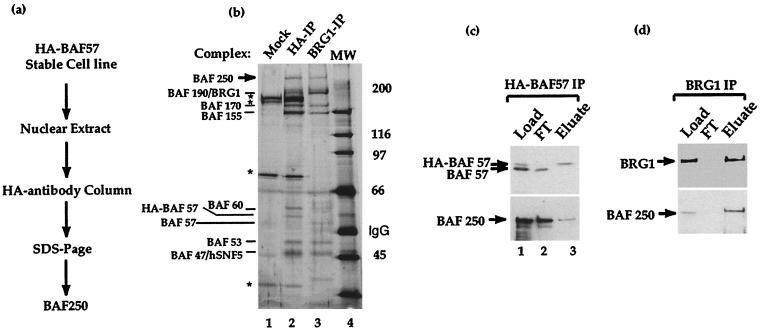
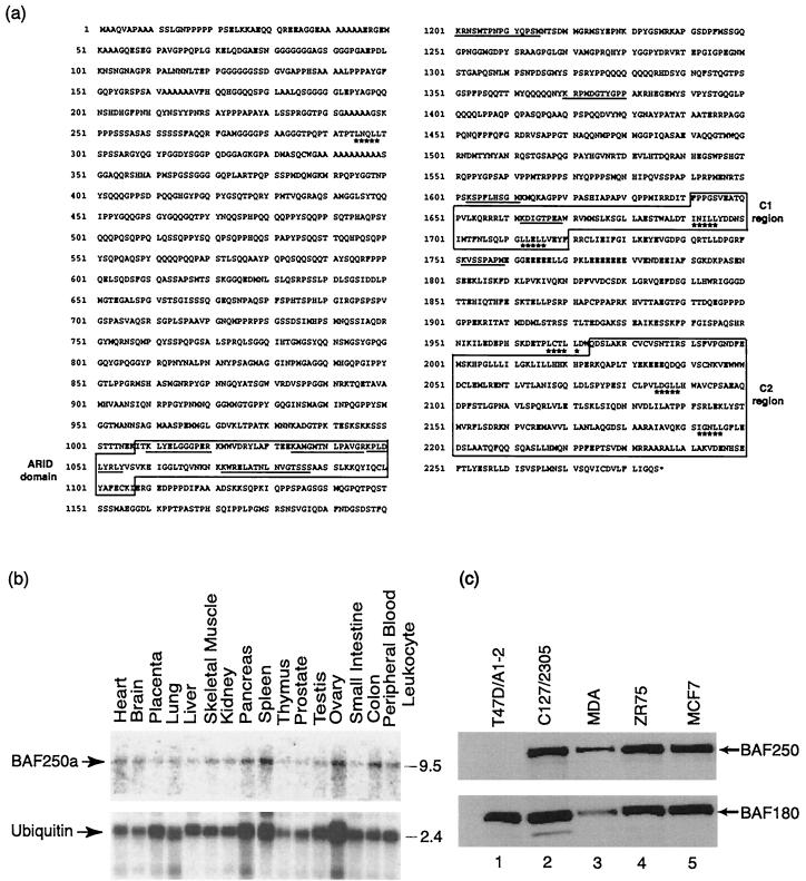
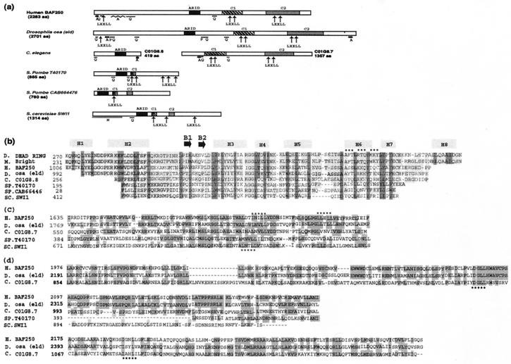
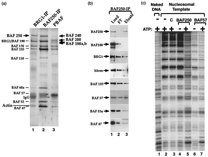
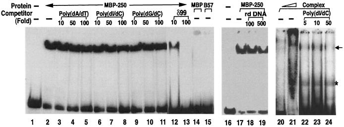
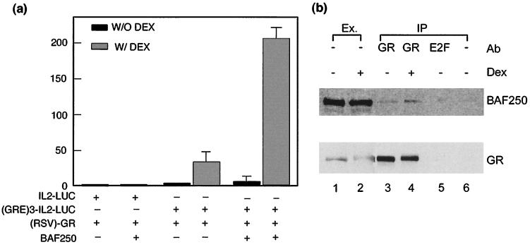
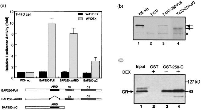
The SWI/SNF family of chromatin-remodeling complexes facilitates gene activation by assisting transcription machinery to gain access to targets in chromatin. This family includes BAF (also called hSWI/SNF-A) and PBAF (hSWI/SNF-B) from humans and SWI/SNF and Rsc from Saccharomyces cerevisiae. However, the relationship between the human and yeast complexes is unclear because all human subunits published to date are similar to those of both yeast SWI/SNF and Rsc. Also, the two human complexes have many identical subunits, making it difficult to distinguish their structures or functions. Here we describe the cloning and characterization of BAF250, a subunit present in human BAF but not PBAF. BAF250 contains structural motifs conserved in yeast SWI1 but not in any Rsc components, suggesting that BAF is related to SWI/SNF. BAF250 is also a homolog of the Drosophila melanogaster Osa protein, which has been shown to interact with a SWI/SNF-like complex in flies. BAF250 possesses at least two conserved domains that could be important for its function. First, it has an AT-rich DNA interaction-type DNA-binding domain, which can specifically bind a DNA sequence known to be recognized by a SWI/SNF family-related complex at the beta-globin locus. Second, BAF250 stimulates glucocorticoid receptor-dependent transcriptional activation, and the stimulation is sharply reduced when the C-terminal region of BAF250 is deleted. This region of BAF250 is capable of interacting directly with the glucocorticoid receptor in vitro. Our data suggest that BAF250 confers specificity to the human BAF complex and may recruit the complex to its targets through either protein-DNA or protein-protein interactions.


Figures

FIG. 1
FIG. 1
Purification of BAF250, the signature subunit of human SWI/SNF family-related BAF complex. (a) Schematic diagram of the purification procedure for BAF250. (b) Silver-stained SDS-polyacrylamide gel of human SWI/SNF family-related BAF complex purified by an antibody against HA-tagged BAF57 (lane 2). The complex purified by anti-BRG1 antibody was used as a control (lane 3). The polypeptides bound to both antibody columns are indicated. Several contaminating polypeptides (∗) were present in the preparation, as shown by a mock purification using the parent cell line that lacks the tagged BAF57 (lane 1). (c and d) Immunoblot of the load, flowthrough (FT), and eluate fractions from the anti-HA-tagged BAF57 (c) or anti-BRG1 antibody (d) column.
FIG. 2
FIG. 2
BAF250 protein and its expression pattern. (a) Predicted amino acid sequence of BAF250. The underlined sequences indicate the nine peptides obtained from microsequencing the BAF250 protein. Boxed regions, ARID domain, the C1 region, and the C2 region, which are homologous to those of Drosophila Osa. Starred amino acid residues, predicted LXXLL motifs. (b) Northern blot analysis of RNA from different human tissues probed with BAF250 (top) or ubiquitin (bottom). Each tissue is indicated at the top. The molecular size markers (right) are in kilobases. (c) Immunoblot analysis of BAF250 in several human and mouse breast cancer cell lines, as indicated at the top. The analysis of BAF180 is also shown for comparison.
FIG. 3
FIG. 3
Several regions of BAF250 are conserved in Drosophila Osa and yeast SWI1. (a) Schematic representation of BAF250 and its orthologs from several organisms. Each box represents a conserved region. Arrows, predicted LXXLL motifs. Regions rich in glutamines (Q), prolines (P), alanines (A), and asparagines (N) are underlined. Note that the two predicted C. elegans ORFs are next to each other on the chromosome and could be a single gene. (b) Alignment of the ARID domain of BAF250 with related sequences from other proteins. Proteins are listed in order of the similarity of sequences in them to those of the ARID domain of BAF250. Dark shading, residues conserved in different α-helices (H1 to H8) and β-sheets (B1 and B2) according to solution structure of the ARID domain of Dead ringer (24); light shading, residues identical in more than three proteins; stars, amino acid residues interacting with DNA. Abbreviations: D, Drosophila; M, mouse; H, human; C, C. elegans; SP, S. pombe; SC, S. cerevisiae. The ARID domains from Dead ringer and Bright can bind specific DNA sequences. (c and d) Alignment of C1 and C2 regions of BAF250 and its orthologs. Shaded amino acid residues are conserved in two or more proteins. Stars, LXXLL motifs. Note that the conserved regions from yeast genes are shorter than those from other species.
FIG. 4
FIG. 4
BAF250 distinguishes human BAF complex from PBAF. (a) Silver-stained SDS-PAGE gel of human BAF complex purified by anti-BRG1 (lane 1) or anti-BAF250 antibody (lane 2). The PBAF complex is shown as a comparison (lane 3). Lines, subunits shared by both complexes; arrows, components unique to each complex. (b) Western blotting of the load, flowthrough (FT), and eluate fractions from the BAF250 antibody column. (c) Autoradiograph showing the mononucleosome disruption activity by human BAF complex purified with a BAF250 antibody (lanes 4 and 5). The results for the BAF complex purified with a BAF57 antibody are also shown for comparison (lanes 6 and 7). The templates and the complexes used in each reaction are shown at the top. The presence (+) or absence (−) of ATP is indicated. C, control in which antibody beads without the complex loaded was tested.
FIG. 5
FIG. 5
The ARID domain of BAF250 specifically binds the pyrimidine-rich element from the β-globin loci. (a) Autoradiograph showing the results of a gel mobility shift assay for the recombinant BAF250 ARID domain and other proteins as indicated. A DNA fragment containing the pyrimidine-rich element (δ99) located between fetal γ-globin and adult β-globin genes was used as a probe (31). A recombinant protein containing the ARID domain of BAF250 fused to MBP (MBP-250) or MBP alone was tested. B57, HMG domain of BAF57, which can bind the four-way junction DNA (44). Several types of unlabeled DNA were used as competitors (lanes 3 to 13). Rd DNA, 41-bp DNA fragment from the mouse rhodopsin promoter region. The BAF complex purified with HA antibody was also analyzed (lanes 20 to 24). Arrow, specific complex formed between the complex and the probe. A contaminant band (∗) which appears to be derived from preparation of the probe was detected.
FIG. 6
FIG. 6
BAF250 facilitates glucocorticoid receptor-dependent transcriptional activation. (a) Graph showing that BAF250 enhances glucocorticoid receptor-mediated gene expression in transient cotransfection assays. The assays were performed in the presence (W/ DEX) or absence (W/O DEX) of DEX (10−7 M). The luciferase reporter plasmids contained either interleukin-2 minimal promoter (IL2-LUC) or the same promoter fused with three GR binding sites upstream [(GRE)3-IL2-LUC]. They were cotransfected with expression vectors for GR (RSV-GR) and BAF250 into T47D cells that lacked endogenous BAF250. An empty expression vector was used as a negative control for BAF250 (bars with − for BAF250). The graph represents the averages of luciferase activities from three independent assays. (b) Immunoblot analysis of the immunoprecipitates obtained with an anti-GR antibody (Ab) from lysates of C127/2305 cells. Cells were either untreated (lanes 1 and 3) or treated with DEX for 1 h (lanes 2 and 4). Immunoprecipitation (IP) with an anti-GR antibody (lanes 3 and 4), an anti-E2F1 nonspecific antibody (lane 5), or no antibody (lane 6) indicates that the interaction between GR and BAF250 is specific. The nuclear extract (Ex.; lanes 1 and 2) was used as the control.
FIG. 7
FIG. 7
The C-terminal region of BAF250 directly interacts with GR. (a) Graph showing that deletion of the C-terminal region of BAF250 decreases its ability to facilitate GR-dependent gene activation. The assays were performed in the presence (shaded bars) or absence (black bars) of DEX (10−7 M). Schematic diagrams of the full-length BAF250 and deletion mutants are shown below. (b) Immunoblot analysis of extract from T47D cells transfected with full-length BAF250 or a C-terminal deletion mutant. The arrows mark each protein. A polypeptide cross-reactive with the antibody (∗) was used as a loading control. The nuclear extract from KB cells was the positive control (lane 1). (c) Autoradiograph showing that the C-terminal region of BAF250 directly interacts with GR in a GST pull-down assay. Either GST alone or GST fused to the C-terminal region of BAF250 (amino acid residues 1670 to 2137) was used. The presence or absence of DEX (10−7 M) is indicated. The 35S-labeled GR protein was produced using an in vitro transcription and translation system.

References

    1. Armstrong J A, Bieker J J, Emerson B M. A SWI/SNF-related chromatin remodeling complex, E-RC1, is required for tissue-specific transcriptional regulation by EKLF in vitro. Cell. 1998;95:93–104. - PubMed
    1. Bourachot B, Yaniv M, Muchardt C. The activity of mammalian brm/SNF2α is dependent on a high-mobility-group protein I/Y-like DNA binding domain. Mol Cell Biol. 1999;19:3931–3939. - PMC - PubMed
    1. Cairns B R, Erdjument-Bromage H, Tempst P, Winston F, Kornberg R D. Two actin-related proteins are shared functional components of the chromatin-remodeling complexes RSC and SWI/SNF. Mol Cell. 1998;2:639–651. - PubMed
    1. Cairns B R, Henry N L, Kornberg R D. TFG/TAF30/ANC1, a component of the yeast SWI/SNF complex that is similar to the leukemogenic proteins ENL and AF-9. Mol Cell Biol. 1996;16:3308–3316. - PMC - PubMed
    1. Cairns B R, Kim Y J, Sayre M H, Laurent B C, Kornberg R D. A multisubunit complex containing the SWI1/ADR6, SWI2/SNF2, SWI3, SNF5, and SNF6 gene products isolated from yeast. Proc Natl Acad Sci USA. 1994;91:1950–1954. - PMC - PubMed

Publication types

Associated data